
Rationale and Objectives
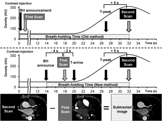
Subtraction coronary computed tomography (CT) angiography (CCTA), which enables the removal of calcium and coronary stents from CCTA images, has been clinically introduced on a second-generation 320-row CT scanner. However, this technique for clinical use is not optimized. The long breath-holding time for two data acquisitions, which causes image misregistration and patient’s discomfort, may limit the clinical availability of this subtraction technique.
Materials and Methods
This study received approval from the institutional review board; prior informed consent to participate was obtained from all patients. We performed subtraction CCTA of five patients using the test injection method and optimized the interval time between the first (pulmonary-arterial phase) and the second (coronary-arterial phase) scans to achieve robust subtraction. The patients’ breath-holding times were recorded. We compared breath-holding times between our new protocol and previous study’s protocol (estimated).
Results
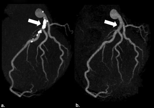
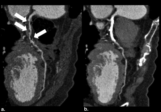
Mean breath-holding time in our new protocol was 18.3 ± 3.4 seconds and that in previous protocol was 29.8 ± 3.6 seconds (difference in mean breath-holding time was 11.5 seconds). Misregistration artifacts were not shown in final subtraction CCTA images. These images improved luminal visualization in the calcified lesion.
Conclusions
Our test injection protocol can shorten the breath-holding time, which is helpful for successful subtraction CCTA imaging, potentially resulting in an increase of subtraction CCTA examinations in many institutions.
A blooming artifact due to the presence of vascular calcium and/or coronary stents affects the evaluation of luminal stenosis by coronary computed tomography (CT) angiography (CCTA) . The appropriate use criteria 2010 reported that the usefulness of CCTA for coronary artery disease (CAD) detection is considered “uncertain” in patients with a calcium score of >400 Agatston units . The diagnosis of CAD in patients with a high calcium score on CCTA images remains challenging.
Recently, subtraction CCTA, a state-of-the-art technique that allows calcium to be subtracted from CCTA images, was clinically introduced on a second-generation 320-row CT scanner. Yoshioka et al. and Tanaka et al. assessed the clinical utility of subtraction CCTA and concluded that coronary calcium subtraction can improve the evaluation of calcified coronary segments in patients with suspected CAD.
Get Radiology Tree app to read full this article<
Materials and methods
Get Radiology Tree app to read full this article<
Get Radiology Tree app to read full this article<
Get Radiology Tree app to read full this article<
Get Radiology Tree app to read full this article<
Get Radiology Tree app to read full this article<
Get Radiology Tree app to read full this article<
Results
Get Radiology Tree app to read full this article<
Get Radiology Tree app to read full this article<
Discussion
Get Radiology Tree app to read full this article<
Get Radiology Tree app to read full this article<
Conclusions
Get Radiology Tree app to read full this article<
Get Radiology Tree app to read full this article<
References
1. Vavere A.L., Arbab-Zadeh A., Rochitte C.E., et. al.: Coronary artery stenoses: accuracy of 64-detector row CT angiography in segments with mild, moderate, or severe calcification—a subanalysis of the CORE-64 trial. Radiology 2011; 261: pp. 100-108.
2. Taylor A.J., Cerqueira M., Hodgson J.M., et. al.: ACCF/SCCT/ACR/AHA/ASE/ASNC/NASCI/SCAI/SCMR 2010 Appropriate use criteria for cardiac computed tomography. A report of the American College of Cardiology Foundation Appropriate Use Criteria Task Force, the Society of Cardiovascular Computed Tomography, the American College of Radiology, the American Heart Association, the American Society of Echocardiography, the American Society of Nuclear Cardiology, the North American Society for Cardiovascular Imaging, the Society for Cardiovascular Angiography and Interventions, and the Society for Cardiovascular Magnetic Resonance. Circulation 2010; 122: pp. e525-e555.
3. Yoshioka K., Tanaka R., Muranaka K.: Subtraction coronary CT angiography for calcified lesions. Cardiol Clin 2012; 30: pp. 93-102.
4. Tanaka R., Yoshioka K., Muranaka K., et. al.: Improved evaluation of calcified segments on coronary CT angiography: a feasibility study of coronary calcium subtraction. Int J Cardiovasc Imaging 2013; 29: pp. 75-81.
5. Kidoh M., Nakaura T., Nakamura S., et. al.: Novel contrast-injection protocol for coronary computed tomographic angiography: contrast-injection protocol customized according to the patient’s time-attenuation response. Heart Vessels 2014; 29: pp. 149-155.
6. Kawaguchi N., Kurata A., Kido T., et. al.: Optimization of coronary attenuation in coronary computed tomography angiography using diluted contrast material. Circ J 2014; 78: pp. 662-670.