
Rationale and Objectives
To evaluate the association of prognostic factors and subtypes of breast cancer with perfusion parameters in dynamic contrast-enhanced magnetic resonance imaging and apparent diffusion coefficient (ADC) values in diffusion-weighted magnetic resonance imaging.
Materials and Methods
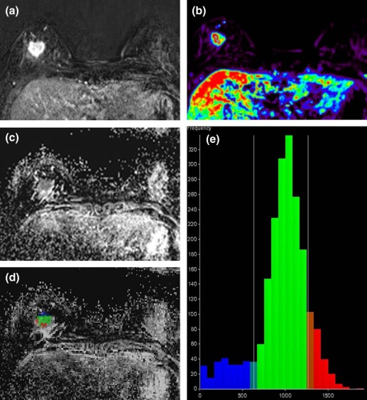
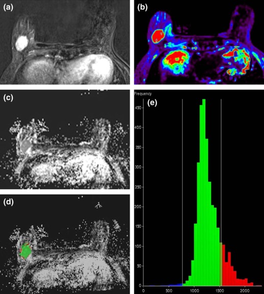
Quantitative perfusion parameters (constant of transfer from plasma to interstitium, constant of transfer from the interstitium to the plasma, extravascular/extracellular volume per unit of volume of tissue [v e ], and initial area under the concentration curve [ i AUC]) and ADC values in the entire tumor volume of 52 invasive ductal carcinomas were obtained using histogram analysis. Four measures (25th percentile, mean, median, 75th percentile) were calculated for each parameter and the ADC value. Associations of perfusion parameters and ADC values with prognostic factors and tumor subtypes were analyzed.
Results
Among perfusion parameters, i AUC mean and i AUC median were greater in tumors larger than 2 cm (8.23 ± 2.33, 8.64 ± 2.67 × 10 4 ) than in those smaller than 2 cm (6.99 ± 1.92, 7.04 ± 2.15 × 10 4 ; P = 0.046, 0.023). V e median was higher in tumors with progesterone receptor (PR) positivity (0.54 ± 0.18) than in those with PR negativity (0.44 ± 0.1, P = 0.041). There were higher ADC mean and ADC median in tumors with human epidermal growth factor receptor 2 (HER2) positivity (1.306 and 1.278 × 10 −3 mm 2 /s) than in those with HER2 negativity (1.078 and 1.053 × 10 −3 mm 2 /s; P = 0.012 and 0.020). Higher ADC mean and ADC median were observed in HER2-enriched type (1.404 and 1.378 × 10 −3 mm 2 /s) than in luminal type (1.096 and 1.073 × 10 −3 mm 2 /s; P = 0.030 and 0.045).
Conclusions
Among perfusion parameters, i AUC was associated with tumor size and v e median was associated with PR positivity. Mean and median ADC values showed positive correlation with HER2-positive and HER2-enriched tumors.
Introduction
Biopsy specimens are required for analysis of conventional prognostic factors such as tumor size, axillary lymph node status, histologic grade, and molecular marker expression . The availability of magnetic resonance imaging (MRI) has prompted efforts to develop noninvasive MRI-based biomarkers to predict the prognosis of breast cancers using techniques such as diffusion-weighted imaging (DWI) and dynamic contrast-enhanced (DCE) MRI. Various apparent diffusion coefficient (ADC) parameters derived from DWI and perfusion parameters obtained from DCE-MRI have been associated with several prognostic factors , and recent studies have also reported correlations between pharmacokinetic parameters of breast DCE-MRI and prognostic factors, suggesting poorer prognosis in tumors with higher constant of transfer from plasma to interstitium (K trans ) and constant of transfer from the interstitium to the plasma (k ep ) values or lower extravascular/extracellular volume per unit of volume of tissue (v e ) values . However, to our knowledge, there have been no studies so far that have analyzed both perfusion parameters and ADC values in the entire tumor volume for simultaneous correlation of prognostic factors or subtypes. The purpose of our study was to investigate the association of prognostic factors and tumor subtypes in patients diagnosed with breast cancer to both MR perfusion parameters and ADC values. In addition, we compared histogram analysis and region of interest (ROI) analysis for the prediction of prognosis of breast cancer.
Materials and Methods
Patients
Institutional review board approval was obtained for this retrospective study, and informed consent was waived. Between February 2012 and March 2013, 81 consecutive breast cancer patients diagnosed by percutaneous biopsy underwent DCE-MRI and DWI on a 3T MRI system for preoperative evaluation. Among them, a total of 29 patients were excluded because they had received preoperative neoadjuvant chemotherapy ( n = 12), had histologic types other than invasive ductal carcinoma (IDC) ( n = 11), or due to processing software failure ( n = 6). Finally, a total of 52 masses from 52 patients (mean age 54.8 years, range 36–72 years) were included in the analysis of perfusion parameters and ADC values.
MRI Acquisition
Get Radiology Tree app to read full this article<
Imaging Analysis
Get Radiology Tree app to read full this article<
Perfusion Parameters
Get Radiology Tree app to read full this article<

Get Radiology Tree app to read full this article<
ADC Values
Get Radiology Tree app to read full this article<
Histopathologic Analysis
Get Radiology Tree app to read full this article<
Statistical Analysis
Get Radiology Tree app to read full this article<
Results
Histopathologic Results
Get Radiology Tree app to read full this article<
Association Between Perfusion Parameters and Prognostic Factors
Get Radiology Tree app to read full this article<
Get Radiology Tree app to read full this article<
Table 1
Association of Perfusion Parameters with Prognostic Factors in ROI Analysis
n K trans Mean (min −1 )P Value v e Mean_P_ Value k ep Mean (min −1 )P Value i AUC Mean (×10 4 )P Value Size (cm) ≤2 23 0.51 ± 0.2 0.417 0.67 ± 0.23 0.461 1.25 ± 0.93 0.253 11.73 ± 3.51 0.261 >2 29 0.58 ± 0.31 0.61 ± 0.19 1.37 ± 0.77 12.42 ± 2.95 Lymph node metastasis Negative 32 0.57 ± 0.3 0.955 0.65 ± 0.22 0.843 1.37 ± 0.96 >0.999 11.70 ± 3.47 0.263 Positive 20 0.52 ± 0.2 0.63 ± 0.19 1.22 ± 0.61 12.78 ± 2.66 Histologic grade Nonhigh (grades 1,2) 35 0.54 ± 0.23 0.961 0.63 ± 0.21 0.653 1.23 ± 0.65 >0.999 12.29 ± 3.15 0.726 High (grade 3) 17 0.57 ± 0.34 0.65 ± 0.21 1.5 ± 1.13 11.75 ± 3.357 Estrogen receptor Negative 14 0.5 ± 0.16 0.718 0.57 ± 0.18 0.134 1.38 ± 0.83 0.789 12.51 ± 2.39 0.599 Positive 38 0.56 ± 0.3 0.67 ± 0.22 1.29 ± 0.85 11.97 ± 3.46 Progesterone receptor Negative 20 0.47 ± 0.16 0.232 0.55 ± 0.16 0.024 \* 1.4 ± 0.8 0.240 12.10 ± 2.19 0.858 Positive 32 0.59 ± 0.31 0.69 ± 0.22 1.26 ± 0.87 12.13 ± 3.73 Human epidermal growth factor receptor 2 Negative 39 0.57 ± 0.28 0.291 0.66 ± 0.2 0.190 1.35 ± 0.86 0.743 12.41 ± 2.63 0.735 Positive 13 0.47 ± 0.19 0.59 ± 0.22 1.22 ± 0.79 11.22 ± 4.52 Ki-67 (%) cutoff 14% Negative 16 0.54 ± 0.27 0.858 0.68 ± 0.21 0.226 1.21 ± 0.82 0.303 13.44 ± 3.04 0.076 Positive 36 0.55 ± 0.27 0.62 ± 0.21 1.36 ± 0.85 11.52 ± 3.13 Tumor subtype Luminal 39 0.57 ± 0.29 0.842 0.67 ± 0.21 0.206 1.31 ± 0.85 0.860 12.06 ± 3.47 0.564 Triple negative 9 0.5 ± 0.18 0.59 ± 0.2 1.34 ± 0.98 11.73 ± 2.11 HER2 enriched 4 0.46 ± 0.14 0.48 ± 0.05 1.26 ± 0.42 13.48 ± 2.57
i AUC, initial area under the concentration curve; k ep , constant of transfer from the interstitium to the plasma; K trans , constant of transfer from plasma to interstitium; SD, standard deviation; v e , extravascular/extracellular volume per unit of volume of tissue.
Data are presented as mean ± standard deviation.
Get Radiology Tree app to read full this article<
Get Radiology Tree app to read full this article<
Get Radiology Tree app to read full this article<
Table 2
Association of Perfusion Parameters with Prognostic Factors in Histogram Analysis
K trans 25th_P_ Value K trans Mean_P_ Value K trans Median_P_ Value K trans 75th_P_ Value v e 25th_P_ Value v e Mean_P_ Value v e Median_P_ Value v e 75th_P_ Value Size (cm) ≤2 0.1 ± 0.03 0.113 0.24 ± 0.11 0.126 0.23 ± 0.13 0.136 0.38 ± 0.2 0.191 0.3 ± 0.11 0.071 0.47 ± 0.12 0.868 0.5 ± 0.18 0.7330 0.63 ± 0.2 0.861 >2 0.15 ± 0.1 0.3 ± 0.16 0.3 ± 0.17 0.45 ± 0.25 0.36 ± 0.1 0.47 ± 0.12 0.5 ± 0.15 0.59 ± 0.15 Lymph node metastasis Negative 0.12 ± 0.08 0.670 0.28 ± 0.16 0.605 0.27 ± 0.16 0.435 0.42 ± 0.25 0.413 0.33 ± 0.12 0.913 0.48 ± 0.12 0.372 0.52 ± 0.18 0.5410 0.63 ± 0.19 0.560 Positive 0.13 ± 0.08 0.27 ± 0.13 0.27 ± 0.14 0.42 ± 0.19 0.34 ± 0.1 0.45 ± 0.1 0.47 ± 0.11 0.58 ± 0.14 Histologic grade Nonhigh (grades 1,2) 0.11 ± 0.07 0.149 0.26 ± 0.13 0.191 0.25 ± 0.15 0.054 0.41 ± 0.22 0.598 0.32 ± 0.12 0.222 0.46 ± 0.11 0.459 0.5 ± 0.16 0.5520 0.6 ± 0.18 0.711 High (grade 3) 0.16 ± 0.1 0.31 ± 0.17 0.31 ± 0.16 0.45 ± 0.26 0.36 ± 0.08 0.48 ± 0.12 0.51 ± 0.16 0.61 ± 0.17 Estrogen receptor Negative 0.1 ± 0.05 0.152 0.23 ± 0.07 0.298 0.22 ± 0.07 0.190 0.36 ± 0.11 0.370 0.33 ± 0.1 0.726 0.43 ± 0.11 0.078 0.44 ± 0.11 0.0510 0.54 ± 0.13 0.068 Positive 0.14 ± 0.09 0.29 ± 0.16 0.29 ± 0.17 0.44 ± 0.26 0.34 ± 0.11 0.48 ± 0.11 0.52 ± 0.17 0.63 ± 0.18 Progesterone receptor Negative 0.1 ± 0.05 0.255 0.23 ± 0.06 0.288 0.22 ± 0.07 0.263 0.36 ± 0.11 0.323 0.33 ± 0.09 0.606 0.43 ± 0.1 0.054 0.44 ± 0.1 0.041 \* 0.54 ± 0.12 0.038 \* Positive 0.14 ± 0.09 0.3 ± 0.17 0.3 ± 0.18 0.46 ± 0.28 0.34 ± 0.12 0.49 ± 0.12 0.54 ± 0.18 0.65 ± 0.19 Human epidermal growth factor receptor 2 Negative 0.12 ± 0.08 0.583 0.28 ± 0.15 0.583 0.28 ± 0.16 0.849 0.44 ± 0.24 0.512 0.35 ± 0.11 0.156 0.48 ± 0.12 0.398 0.5 ± 0.13 0.3000 0.62 ± 0.17 0.228 Positive 0.13 ± 0.08 0.25 ± 0.11 0.25 ± 0.12 0.38 ± 0.18 0.3 ± 0.1 0.45 ± 0.11 0.5 ± 0.23 0.57 ± 0.2 Ki-67 Negative 0.11 ± 0.08 0.422 0.27 ± 0.17 0.388 0.27 ± 0.2 0.346 0.42 ± 0.28 0.326 0.32 ± 0.1 0.479 0.47 ± 0.11 0.656 0.51 ± 0.14 0.5720 0.64 ± 0.19 0.263 Positive 0.13 ± 0.08 0.28 ± 0.13 0.27 ± 0.13 0.42 ± 0.21 0.34 ± 0.11 0.47 ± 0.12 0.5 ± 0.17 0.59 ± 0.17 Subtype Luminal 0.14 ± 0.09 0.179 0.29 ± 0.16 0.246 0.29 ± 0.17 0.153 0.45 ± 0.26 0.375 0.34 ± 0.11 0.590 0.48 ± 0.11 0.098 0.52 ± 0.17 0.0700 0.63 ± 0.18 0.092 Triple negative 0.1 ± 0.05 0.23 ± 0.06 0.22 ± 0.06 0.35 ± 0.09 0.34 ± 0.12 0.46 ± 0.13 0.47 ± 0.13 0.58 ± 0.16 HER2 enriched 0.08 ± 0.01 0.2 ± 0.04 0.17 ± 0.03 0.31 ± 0.09 0.28 ± 0.05 0.37 ± 0.05 0.38 ± 0.05 0.47 ± 0.05
k ep 25th_P_ Value k ep Mean_P_ Value k ep Median_P_ Value k ep 75th_P_ Value i AUC 25th (×10 4 )P Value i AUC Mean (×10 4 )P Value i AUC Median (×10 4 )P Value i AUC 75th (×10 4 )P Value Size (cm) ≤2 0.26 ± 0.12 0.261 0.57 ± 0.31 0.302 0.5 ± 0.24 0.231 0.79 ± 0.48 0.238 4.23 ± 1.30 0.041 \* 6.99 ± 1.92 0.046 \* 7.04 ± 2.15 0.023 \* 9.70 ± 3.04 0.103 >2 0.35 ± 0.22 0.68 ± 0.36 0.62 ± 0.32 0.93 ± 0.47 5.64 ± 2.44 8.23 ± 2.33 8.64 ± 2.67 11.02 ± 2.67 Lymph node metastasis Negative 0.28 ± 0.19 0.150 0.62 ± 0.38 0.232 0.54 ± 0.31 0.198 0.85 ± 0.54 0.211 4.76 ± 1.85 0.458 7.43 ± 2.19 0.297 7.62 ± 2.51 0.270 10.10 ± 3.08 0.296 Positive 0.35 ± 0.18 0.65 ± 0.28 0.6 ± 0.25 0.9 ± 0.37 5.40 ± 2.50 8.10 ± 2.27 8.43 ± 2.62 10.97 ± 2.53 Histologic grade Nonhigh (grades 1,2) 0.28 ± 0.16 0.413 0.59 ± 0.31 0.339 0.54 ± 0.27 0.320 0.84 ± 0.47 0.483 4.83 ± 2.08 0.234 7.58 ± 2.14 0.622 7.70 ± 2.41 0.356 10.37 ± 2.81 0.802 High (grade 3) 0.35 ± 0.24 0.7 ± 0.4 0.63 ± 0.33 0.93 ± 0.49 5.37 ± 2.22 7.91 ± 2.45 8.41 ± 2.86 10.58 ± 3.13 Estrogen receptor Negative 0.26 ± 0.1 0.370 0.58 ± 0.18 0.877 0.53 ± 0.16 0.893 0.8 ± 0.24 0.975 4.99 ± 1.43 0.643 7.92 ± 1.40 0.560 8.25 ± 1.57 0.488 10.92 ± 1.88 0.370 Positive 0.32 ± 0.21 0.65 ± 0.39 0.58 ± 0.33 0.89 ± 0.54 5.02 ± 2.34 7.60 ± 2.47 7.82 ± 2.85 10.26 ± 3.18 Progesterone receptor Negative 0.28 ± 0.12 0.800 0.62 ± 0.2 0.247 0.54 ± 0.14 0.714 0.84 ± 0.23 0.314 4.96 ± 1.26 0.529 7.79 ± 1.36 0.765 8.15 ± 1.70 0.596 10.68 ± 1.86 0.590 Positive 0.32 ± 0.22 0.64 ± 0.41 0.59 ± 0.36 0.89 ± 0.58 5.04 ± 2.54 7.62 ± 2.65 7.80 ± 2.99 10.28 ± 3.40 Human epidermal growth factor receptor 2 Negative 0.32 ± 0.19 0.540 0.64 ± 0.35 0.899 0.59 ± 0.3 0.597 0.89 ± 0.5 0.816 4.99 ± 2.12 0.657 7.75 ± 1.96 0.713 7.95 ± 2.35 0.920 10.57 ± 2.40 0.667 Positive 0.27 ± 0.18 0.59 ± 0.33 0.5 ± 0.27 0.79 ± 0.41 5.07 ± 2.21 7.48 ± 2.97 7.87 ± 3.23 10.04 ± 4.12 Ki-67 Negative 0.3 ± 0.2 0.684 0.63 ± 0.39 0.656 0.57 ± 0.34 0.559 0.89 ± 0.61 0.599 4.96 ± 2.29 0.714 8.06 ± 2.11 0.421 8.11 ± 2.47 0.744 11.13 ± 2.70 0.256 Positive 0.31 ± 0.18 0.63 ± 0.32 0.57 ± 0.27 0.86 ± 0.41 5.03 ± 2.07 7.52 ± 2.29 7.85 ± 2.63 10.13 ± 2.95 Subtype Luminal 0.33 ± 0.21 0.304 0.66 ± 0.39 0.793 0.59 ± 0.33 0.734 0.91 ± 0.54 0.7560 5.11 ± 2.38 0.979 7.68 ± 2.49 0.763 7.92 ± 2.89 0.874 10.35 ± 3.20 0.548 Triple negative 0.23 ± 0.08 0.52 ± 0.13 0.49 ± 0.11 0.73 ± 0.17 4.65 ± 1.07 7.39 ± 0.95 7.73 ± 1.04 10.12 ± 1.41 HER2 enriched 0.27 ± 0.04 0.59 ± 0.08 0.51 ± 0.05 0.82 ± 0.15 4.81 ± 0.90 8.39 ± 1.45 8.53 ± 1.55 11.95 ± 1.9
i AUC, initial area under the concentration curve; k ep , constant of transfer from the interstitium to the plasma; K trans , constant of transfer from plasma to interstitium; SD, standard deviation; v e , extravascular/extracellular volume per unit of volume of tissue.
Data are presented as mean ± SD.
Get Radiology Tree app to read full this article<
Get Radiology Tree app to read full this article<
Association Between ADC Values and Prognostic Factors
Get Radiology Tree app to read full this article<
Get Radiology Tree app to read full this article<
Table 3
Association of Diffusion Parameters with Prognostic Factors
n ADC Mean (ROI) (10 −3 mm 2 /s)P Value ADC 25th (VOI) (10 −3 mm 2 /s)P Value ADC Mean (VOI) (10 −3 mm 2 /s)P Value ADC Median (VOI) (10 −3 mm 2 /s)P Value ADC 75th (VOI) (10 −3 mm 2 /s)P Value Size (cm) ≤2 23 0.887 ± 0.167 0.196 0.853 ± 0.265 0.101 1.066 ± 0.261 0.071 1.054 ± 0.276 0.156 1.273 ± 0.283 0.130 >2 29 0.835 ± 0.118 0.961 ± 0.266 1.190 ± 0.289 1.154 ± 0.292 1.406 ± 0.329 Lymph node metastasis Negative 32 0.893 ± 0.137 0.027 \\ 0.928 ± 0.265 0.579 1.132 ± 0.272 0.814 1.115 ± 0.280 0.873 1.328 ± 0.294 0.588 Positive 20 0.804 ± 0.138 0.890 ± 0.278 1.142 ± 0.302 1.101 ± 0.305 1.377 ± 0.349 Histologic grade Nonhigh (grades 1,2) 35 0.875 ± 0.152 0.236 0.875 ± 0.298 0.320 1.092 ± 0.303 0.178 1.065 ± 0.307 0.258 1.299 ± 0.331 0.115 High (grade 3) 17 0.824 ± 0.117 0.992 ± 0.177 1.225 ± 0.210 1.202 ± 0.220 1.445 ± 0.255 Estrogen receptor Negative 14 0.863 ± 0.147 0.968 1.019 ± 0.255 0.018 \\ 1.261 ± 0.260 0.011 \\ 1.223 ± 0.270 0.024 \\ 1.482 ± 0.292 0.059 Positive 38 0.843 ± 0.134 0.874 ± 0.266 1.089 ± 0.278 1.068 ± 0.285 1.297 ± 0.310 Progesterone receptor Negative 20 0.827 ± 0.118 0.220 1.001 ± 0.231 0.031 \\ 1.240 ± 0.252 0.018 \\ 1.212 ± 0.270 0.028 \\ 1.466 ± 0.297 0.029 \\ Positive 32 0.878 ± 0.155 0.858 ± 0.279 1.070 ± 0.282 1.046 ± 0.284 1.272 ± 0.305 Human epidermal growth factor receptor 2 Negative 39 0.863 ± 0.147 0.663 0.859 ± 0.267 0.011 \\ 1.079 ± 0.276 0.012 \\ 1.053 ± 0.277 0.020 \\ 1.287 ± 0.308 0.016 \\ Positive 13 0.843 ± 0.134 1.077 ± 0.206 1.306 ± 0.227 1.279 ± 0.256 1.526 ± 0.268 Ki-67 Negative 16 0.860 ± 0.173 0.962 0.806 ± 0.304 0.140 1.014 ± 0.297 0.053 0.998 ± 0.299 0.102 1.210 ± 0.302 0.035 \\ Positive 36 0.858 ± 0.129 0.961 ± 0.240 1.189 ± 0.260 1.159 ± 0.271 1.407 ± 0.303 Subtype Luminal 39 0.859 ± 0.151 0.978 0.881 ± 0.266 0.032 \* , \\ 1.096 ± 0.277 0.030 \* , \\ 1.073 ± 0.283 0.045 \* , \\ 1.303 ± 0.309 0.115 Triple negative 9 0.852 ± 0.119 0.938 ± 0.285 1.186 ± 0.295 1.148 ± 0.305 1.410 ± 0.334 HER2 enriched 4 0.871 ± 0.140 1.172 ± 0.097 1.404 ± 0.113 1.379 ± 0.131 1.627 ± 0.162
ADC, apparent diffusion coefficient; ROI, region of interest; SD, standard deviation; VOI, volume of interest.
Data are presented as mean ± SD.
Get Radiology Tree app to read full this article<
Get Radiology Tree app to read full this article<
Get Radiology Tree app to read full this article<
Get Radiology Tree app to read full this article<
Table 4
Analysis of Covariance (ANCOVA) for ADC Values with HER2, ER, and PR Status
ADC 25th_P_ Value ADC Mean_P_ Value ADC Median_P_ Value ADC 75th_P_ Value Human epidermal growth factor receptor 2 Negative ( n = 39) 0.887 ± 0.046 0.023 \* 1.114 ± 0.047 0.025 \* 1.084 ± 0.049 0.03 \* 1.325 ± 0.053 0.038 \* Positive ( n = 13) 1.081 ± 0.071 1.311 ± 0.074 1.280 ± 0.076 1.529 ± 0.083 Estrogen receptor Negative ( n = 14) 1.015 ± 0.081 0.574 1.249 ± 0.084 0.513 1.206 ± 0.086 0.382 1.459 ± 0.094 0.605 Positive ( n = 38) 0.954 ± 0.055 1.176 ± 0.057 1.158 ± 0.059 1.394 ± 0.064 Progesterone receptor Negative ( n = 20) 1.021 ± 0.060 0.457 1.259 ± 0.062 0.369 1.235 ± 0.064 0.319 1.487 ± 0.070 0.300 Positive ( n = 32) 0.947 ± 0.071 1.166 ± 0.074 1.129 ± 0.076 1.367 ± 0.083
ADC, apparent diffusion coefficient.
Values are least square means ± standard error from analyses of ANCOVA.
ANCOVA model included HER2, ER, and PR.
Get Radiology Tree app to read full this article<
Get Radiology Tree app to read full this article<
Get Radiology Tree app to read full this article<
Get Radiology Tree app to read full this article<
Correlation Between Histogram Analysis and ROI Analysis
Get Radiology Tree app to read full this article<
Discussion
Get Radiology Tree app to read full this article<
Get Radiology Tree app to read full this article<
Get Radiology Tree app to read full this article<
Get Radiology Tree app to read full this article<
Get Radiology Tree app to read full this article<
Get Radiology Tree app to read full this article<
Get Radiology Tree app to read full this article<
Get Radiology Tree app to read full this article<
Get Radiology Tree app to read full this article<
Acknowledgments
Get Radiology Tree app to read full this article<
Get Radiology Tree app to read full this article<
References
1. Goldhirsch A., Glick J.H., Gelber R.D., et. al.: Meeting highlights: international expert consensus on the primary therapy of early breast cancer 2005. Ann Oncol 2005; 16: pp. 1569-1583.
2. Kim E.J., Kim S.H., Park G.E., et. al.: Histogram analysis of apparent diffusion coefficient at 3.0t: correlation with prognostic factors and subtypes of invasive ductal carcinoma. J Magn Reson Imaging 2015;
3. Kim J.Y., Kim S.H., Kim Y.J., et. al.: Enhancement parameters on dynamic contrast enhanced breast MRI: do they correlate with prognostic factors and subtypes of breast cancers?. Magn Reson Imaging 2015; 33: pp. 72-80.
4. Koo H.R., Cho N., Song I.C., et. al.: Correlation of perfusion parameters on dynamic contrast-enhanced MRI with prognostic factors and subtypes of breast cancers. J Magn Reson Imaging 2012; 36: pp. 145-151.
5. Parker G.M., Buckley D.: Tracer kinetic modelling for T1-weighted DCE-MRI.Jackson A.Buckley D.Parker G.M.Dynamic contrast-enhanced magnetic resonance imaging in oncology.2005.SpringerBerlin Heidelberg:pp. 81-92.
6. Tofts P.S., Brix G., Buckley D.L., et. al.: Estimating kinetic parameters from dynamic contrast-enhanced T(1)-weighted MRI of a diffusable tracer: standardized quantities and symbols. J Magn Reson Imaging 1999; 10: pp. 223-232.
7. Cheang M.C., Chia S.K., Voduc D., et. al.: Ki67 index, HER2 status, and prognosis of patients with luminal B breast cancer. J Natl Cancer Inst 2009; 101: pp. 736-750.
8. Goldhirsch A., Winer E.P., Coates A.S., et. al.: Personalizing the treatment of women with early breast cancer: highlights of the St Gallen International Expert Consensus on the Primary Therapy of Early Breast Cancer 2013. Ann Oncol 2013; 24: pp. 2206-2223.
9. Pickles M.D., Manton D.J., Lowry M., et. al.: Prognostic value of pre-treatment DCE-MRI parameters in predicting disease free and overall survival for breast cancer patients undergoing neoadjuvant chemotherapy. Eur J Radiol 2009; 71: pp. 498-505.
10. Walker-Samuel S., Leach M.O., Collins D.J.: Evaluation of response to treatment using DCE-MRI: the relationship between initial area under the gadolinium curve (IAUGC) and quantitative pharmacokinetic analysis. Phys Med Biol 2006; 51: pp. 3593-3602.
11. Horwitz K.B., McGuire W.L.: Specific progesterone receptors in human breast cancer. Steroids 1975; 25: pp. 497-505.
12. Razek A.A., Gaballa G., Denewer A., et. al.: Invasive ductal carcinoma: correlation of apparent diffusion coefficient value with pathological prognostic factors. NMR Biomed 2010; 23: pp. 619-623.
13. Park S.H., Choi H.Y., Hahn S.Y.: Correlations between apparent diffusion coefficient values of invasive ductal carcinoma and pathologic factors on diffusion-weighted MRI at 3.0 Tesla. J Magn Reson Imaging 2015; 41: pp. 175-182.
14. Kim S.H., Cha E.S., Kim H.S., et. al.: Diffusion-weighted imaging of breast cancer: correlation of the apparent diffusion coefficient value with prognostic factors. J Magn Reson Imaging 2009; 30: pp. 615-620.
15. Ludovini V., Sidoni A., Pistola L., et. al.: Evaluation of the prognostic role of vascular endothelial growth factor and microvessel density in stages I and II breast cancer patients. Breast Cancer Res Treat 2003; 81: pp. 159-168.
16. Black R., Prescott R., Bers K., et. al.: Tumour cellularity, oestrogen receptors and prognosis in breast cancer. Clin Oncol 1983; 9: pp. 311-318.
17. Kumar R., Yarmand-Bagheri R.: The role of HER2 in angiogenesis. Semin Oncol 2001; 28: pp. 27-32.
18. Martincich L., Deantoni V., Bertotto I., et. al.: Correlations between diffusion-weighted imaging and breast cancer biomarkers. Eur Radiol 2012; 22: pp. 1519-1528.
19. Le Bihan D., Breton E., Lallemand D., et. al.: Separation of diffusion and perfusion in intravoxel incoherent motion MR imaging. Radiology 1988; 168: pp. 497-505.
20. Dvorak H.F., Nagy J.A., Dvorak J.T., et. al.: Identification and characterization of the blood vessels of solid tumors that are leaky to circulating macromolecules. Am J Pathol 1988; 133: pp. 95-109.
21. Su M.Y., Cheung Y.C., Fruehauf J.P., et. al.: Correlation of dynamic contrast enhancement MRI parameters with microvessel density and VEGF for assessment of angiogenesis in breast cancer. J Magn Reson Imaging 2003; 18: pp. 467-477.
22. Weidner N.: Intratumor microvessel density as a prognostic factor in cancer. Am J Pathol 1995; 147: pp. 9-19.
23. Carmeliet P., Jain R.K.: Angiogenesis in cancer and other diseases. Nature 2000; 407: pp. 249-257.
24. Bharwani N., Miquel M.E., Powles T., et. al.: Diffusion-weighted and multiphase contrast-enhanced MRI as surrogate markers of response to neoadjuvant sunitinib in metastatic renal cell carcinoma. Br J Cancer 2014; 110: pp. 616-624.
25. Kyriazi S., Collins D.J., Messiou C., et. al.: Metastatic ovarian and primary peritoneal cancer: assessing chemotherapy response with diffusion-weighted MR imaging—value of histogram analysis of apparent diffusion coefficients. Radiology 2011; 261: pp. 182-192.