
Rationale and Objectives
The study aimed to investigate the positive predictive value (PPV) of mammographic lymphography (MLG) for assessing malignant breast disease and lymphatic metastasis in patients in a typical clinical setting.
Materials and Methods
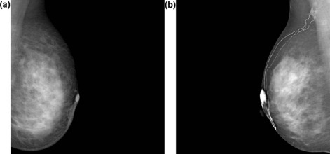
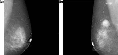
Patients who underwent mammography with Breast Imaging Reporting and Data System (BI-RADS) category 4 or 5 lesions and had abnormal mammographic findings in the upper-outer quadrant of the breast were enrolled. Next, MLG was performed. A water-soluble agent was subcutaneously injected into the upper-outer periareolar region of the bilateral breast, and mammography was then performed. Morphologic characteristics, including lymphatic vessel development, the presence of lymphatic vessel defects, and dilation, were recorded for evaluation.
Results
Fifty-one patients with BI-RADS category 4 lesions and 40 patients with BI-RADS category 5 lesions were included in the study. Sixty-one patients were found to have malignant disease, whereas 30 patients were found to have benign disease. Morphologic characteristics were recorded for evaluation. The interobserver agreement was evaluated and was classified as excellent according to kappa analysis. The PPV of MLG characteristics for malignant breast disease and lymphatic metastasis was analyzed by logistic regression, and the presentation of a lymphatic vessel defect was the most predictive characteristic of a malignancy (PPV: 0.89; P value: 0.02) in patients with BI-RADS category 4 lesions. Meanwhile, in patients with malignant breast disease, the PPVs for predicting lymphatic metastasis with lymphatic vessel defect and dilation were 0.50 ( P value: 0.02) and 0.67 ( P value: <0.01), respectively.
Conclusion
The assessment of morphologic characteristics by MLG has the potential to predict malignant breast disease and lymphatic metastasis.
Introduction
Breast cancer is the most frequently diagnosed cancer and the leading cause of cancer deaths among females, accounting for 23% of the total cancer cases and 14% of the cancer deaths worldwide . Malignant spread to axillary lymph nodes is one of the most important predictors of survival in patients with breast cancer, increasing the 10-year recurrence rate from 20%–30% to nearly 70% . Sentinel lymph node (SLN) mapping and biopsy are currently performed intraoperatively via a radiocolloid scintigraphic method combined with the use of a blue dye . Although this method has proven to be successful for SLN mapping, a technique is still required to determine the condition of the lymphatic system preoperatively. Imaging via the intradermal injection of a contrast agent in the breast under another modality is thought to be a safe method to evaluate the condition of the lymphatic system. A previous study demonstrated that computed tomographic (CT) lymphography using the non-ionic contrast medium iopamidol is useful in SLN evaluation, and patients with a positive result tend to have some typical features in lymphography that are absent in patients proven to be negative by SLN biopsy . However, the true value of these features in diagnosis has not yet been properly discussed. Theoretically, the lymphatic vessel may also be visible in mammography through the use of intradermal contrast injection under CT. Thus, the objective of this study was to retrospectively investigate the positive predictive value (PPV) of imaging characteristics in mammographic lymphography (MLG) to predict lymphatic metastasis in patients in a typical clinical setting. Meanwhile, the value of the imaging characteristics associated with MLG in predictive malignancy is also discussed. To the best of our knowledge, this report is the first attempt to study the value of lymphography under mammography.
Materials and Methods
The protocol in our study was approved by the institutional review board of our hospital, and informed consent was obtained from all patients. Patients who underwent mammography and were assigned Breast Imaging Reporting and Data System (BI-RADS) classifications of 4 or 5 from June 2009 through January 2011 were all enrolled in our study, whereas pregnant patients and patients with a history of axillary lymphadenectomy and renal insufficiency were excluded.
Contrast Agent
Get Radiology Tree app to read full this article<
MLG
Get Radiology Tree app to read full this article<
Image Analysis
Get Radiology Tree app to read full this article<
Get Radiology Tree app to read full this article<
Reference Standard
Get Radiology Tree app to read full this article<
Statistical Evaluation
Get Radiology Tree app to read full this article<
PPV=True positive/(True positive+False positive) PPV
=
True positive
/
(
True positive
+
False positive
)
Get Radiology Tree app to read full this article<
Get Radiology Tree app to read full this article<
Results
Get Radiology Tree app to read full this article<
Get Radiology Tree app to read full this article<
TABLE 1
Predictive Value of Morphologic Characteristics in Detecting Malignancy
Morphologic Characteristics BI-RADS 4 BI-RADS 5 TP FP TN FN PPV OR_P_ value TP FP TN FN PPV OR_P_ value Poor lymphatic vessel development 2 3 25 21 0.40 0.79 0.80 1 2 0 35 0.33 0 0.99 Lymphatic vessel defect 8 1 27 15 0.89 14.4 0.02 18 0 2 18 1.00 1 1.00 Lymphatic vessel dilation 3 3 25 20 0.50 1.25 0.73 12 0 2 24 1.00 1 1.00 Patients in group 51/23 N/A 40/38 N/A
BI-RADS, Breast Imaging Reporting and Data System; FN, false negative; FP, false positive; OR, odds ratio; PPV, positive predicting value; TN, true negative; TP, true positive.
Get Radiology Tree app to read full this article<
Get Radiology Tree app to read full this article<
TABLE 2
Predictive Value of Morphologic Characteristics in Detecting Lymphatic Metastasis
Morphologic Characteristics TP FP TN FN PPV OR_P_ value Poor lymphatic vessel development 2 3 39 17 0.40 1.53 0.07 Lymphatic vessel defect 13 13 29 6 0.50 4.83 0.02 Lymphatic vessel dilation 10 5 37 9 0.67 8.22 <0.01 Without 59/19 N/A
FN, false negative; FP, false positive; OR, odds ratio; PPV, positive predicting value; TN, true negative; TP, true positive.
Get Radiology Tree app to read full this article<
Discussion
Get Radiology Tree app to read full this article<
Get Radiology Tree app to read full this article<
Get Radiology Tree app to read full this article<
Get Radiology Tree app to read full this article<
Get Radiology Tree app to read full this article<
Get Radiology Tree app to read full this article<
Get Radiology Tree app to read full this article<
References
1. Jemal A., Bray F., Center M.M., et. al.: Global cancer statistics. CA Cancer J Clin 2011; 61: pp. 69-90.
2. Fitzgibbons P.L., Page D.L., Weaver D., et. al.: Prognostic factors in breast cancer. College of American Pathologists Consensus Statement 1999. Arch Pathol Lab Med 2000; 124: pp. 966-978.
3. Carney P.A., Miglioretti D.L., Yankaskas B.C., et. al.: Individual and combined effects of age, breast density, and hormone replacement therapy use on the accuracy of screening mammography. Ann Intern Med 2003; 138: pp. 168-175.
4. Minohata J., Takao S., Hirokaga K.: Sentinel lymph node biopsy using CT lymphography in breast cancer. Breast Cancer 2011; 18: pp. 129-136.
5. Yamamoto S., Maeda N., Tamesa M., et. al.: Prospective ultrasonographic prediction of sentinel lymph node metastasis by real-time virtual sonography constructed with three-dimensional computed tomography-lymphography in breast cancer patients. Breast Cancer 2012; 19: pp. 77-82.
6. McCormick B., Ottesen R.A., Hughes M.E., et. al.: Impact of guideline changes on use or omission of radiation in the elderly with early breast cancer: practice patterns at National Comprehensive Cancer Network institutions. J Am Coll Surg 2014; 219: pp. 796-802.
7. Gradishar W.J., Anderson B.O., Blair S.L., et. al.: Breast cancer version 3.2014. J Natl Compr Canc Netw 2014; 12: pp. 542-590.
8. Magrini S.M., Buglione M., Costa L.: Optimizing radiation treatment decisions for patients who receive neoadjuvant chemotherapy. J Natl Cancer Inst Monogr 2015; 51: pp. 9-10.
9. Jankowski C., Hudry D., Vaillant D., et. al.: Evaluation of axillary involvement by ultrasound-guided lymph node biopsy: a prospective study. Gynecol Obstet Fertil 2015; 43: pp. 431-436.
10. Yamashita K., Haga S.: 3D-CT mammary lymphography for sentinel node biopsy. Nihon Rinsho 2012; 70: pp. 377-380.
11. Yamamoto S., Maeda N., Yoshimura K., et. al.: Intraoperative detection of sentinel lymph nodes in breast cancer patients using ultrasonography-guided direct indocyanine green dye-marking by real-time virtual sonography constructed with three-dimensional computed tomography-lymphography. Breast 2013; 22: pp. 933-937.
12. Motomura K., Ishitobi M., Komoike Y., et. al.: SPIO-enhanced magnetic resonance imaging for the detection of metastases in sentinel nodes localized by computed tomography lymphography in patients with breast cancer. Ann Surg Oncol 2011; 18: pp. 3422-3429.
13. Lu Q., Bui D., Liu N.F., et. al.: Magnetic resonance lymphography at 3T: a promising noninvasive approach to characterise inguinal lymphatic vessel leakage. Eur J Vasc Endovasc Surg 2012; 43: pp. 106-111.
14. Borgstein P.J., Meijer S., Pijpers R.J., et. al.: Functional lymphatic anatomy for sentinel node biopsy in breast cancer: echoes from the past and the periareolar blue method. Ann Surg 2000; 232: pp. 81-89.
15. D’Orsi C.J., Bassett L.W., Berg W.A., et. al.: Breast Imaging Reporting and Data System: ACR BI-RADS-mammography.4th ed2003.American College of RadiologyReston, VA
16. Sedgwick E.L., Ebuoma L., Hamame A., et. al.: BI-RADS update for breast cancer caregivers. Breast Cancer Res Treat 2015; 150: pp. 243-254.
17. Szuba A., Shin W.S., Strauss H.W., et. al.: The third circulation: radionuclide lymphoscintigraphy in the evaluation of lymphedema. J Nucl Med 2003; 44: pp. 43-57.