
Rationale and Objectives
The aim of this study was to compare effective radiation doses between prospective and retrospective electrocardiographic gating during coronary computed tomographic angiography for coronary artery bypass grafting evaluation.
Materials and Methods
Fifty consecutive coronary computed tomographic angiographic exams for coronary artery bypass grafting evaluation, 25 prospectively gated and 25 retrospectively gated, were reviewed from January 8, 2008, to June 16, 2009. Body mass index and image quality were also compared between the two groups. To minimize the potential bias introduced by differences in torso length, the effective radiation dose from each exam was measured and normalized to a 24-cm z -axis scan length for all patients. Pooled t tests were used to compare the prospectively and retrospectively gated groups.
Results
The average effective doses delivered in the retrospective and prospective groups were 40.8 mSv (standard error [SE], 1.8 mSv) and 8.6 mSv (SE, 0.7 mSv), respectively. When normalized to the average z -axis scan length of 24 cm, the effective dose in the retrospective group, 38.4 mSv (SE, 1.3 mSv), was still >4 times greater than that in the prospective group, 9.1 mSv (SE, 0.7 mSv) ( P < .0001). There was no significant difference in body mass index or image quality between the groups.
Conclusions
Effective radiation dose in coronary computed tomographic angiography for coronary artery bypass grafting evaluation is very high because of long scan lengths. Prospective electrocardiographic gating significantly reduces effective radiation dose by an average of 76% compared to retrospectively gated scans (9.1 vs 38.4 mSv). In the coronary artery bypass grafting population, prospective electrocardiographic gating should be used whenever ventricular functional assessment is not required.
Coronary computed tomographic (CT) angiography (CCTA) has become an increasingly popular modality for the noninvasive evaluation of coronary anatomy and pathology. CCTA is an accurate method for the noninvasive evaluation of coronary vasculature, with a high negative predictive value for obstructive coronary disease . Risks from ionizing radiation are well documented, and recent innovations have been developed to reduce patient dose. Traditional retrospective electrocardiographic (ECG) gating is performed using continuous radiation and data acquisition and retrospective selection of relevant data for image reconstruction . Prospective ECG gating is a newer technique using the prediction of R-wave timing to allow for intermittent data acquisition alternating with table movement . Typically, prospective gating allows for the x-ray beam to be “on” for approximately 26% of every other R-R interval, resulting in a 77% average dose reduction .
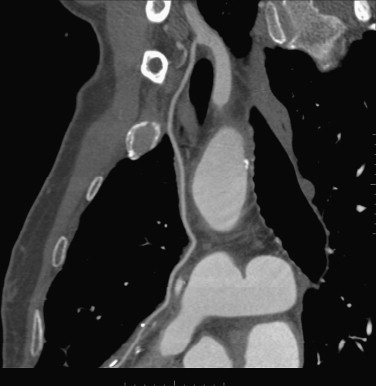
Recent studies have demonstrated the utility of CCTA in patients with prior coronary artery bypass grafting (CABG), with 98% sensitivity and 97% specificity for the detection of obstructive bypass graft disease . Despite this diagnostic performance, CCTA is not without risks. CABG patients require a longer z -axis scan length, and therefore higher radiation dose, to image the entire graft length. For example, evaluation of a left internal mammary artery graft requires images from the thoracic inlet to the diaphragm, whereas a native heart only requires images from the main pulmonary artery to the diaphragm. Our hypothesis was that CABG patients undergoing CCTA receive a significantly increased radiation dose, making traditional retrospective ECG gating potentially unwarranted.
Materials and methods
Get Radiology Tree app to read full this article<
Patients
Get Radiology Tree app to read full this article<
Table 1
Patient Characteristics
Variable Prospective ( n = 25) Retrospective ( n = 25)P Weight (lb) 174.0 ± 28.3 183.7 ± 45.4 .37 Height (in) 67.2 ± 4.2 68.2 ± 3.1 .32 Body mass index 27.1 ± 3.8 27.6 ± 6.1 .71 Heart rate (beats/min) 59.4 ± 7.7 68.7 ± 16.0 .01 ∗ Age 64.7 ± 11.9 72.3 ± 11.3 .03 ∗ Men 19 (76%) 22 (88%) .27 †
Data are expressed as mean ± standard deviation or as number (percentage). Except as indicated, comparisons were made using t test.
Get Radiology Tree app to read full this article<
Get Radiology Tree app to read full this article<
Get Radiology Tree app to read full this article<
Get Radiology Tree app to read full this article<
CT Technique
Get Radiology Tree app to read full this article<
Get Radiology Tree app to read full this article<
Get Radiology Tree app to read full this article<
Get Radiology Tree app to read full this article<
Get Radiology Tree app to read full this article<
Get Radiology Tree app to read full this article<
Radiation Dose
Get Radiology Tree app to read full this article<
Image Analysis
Get Radiology Tree app to read full this article<
Get Radiology Tree app to read full this article<
Statistical Analysis
Get Radiology Tree app to read full this article<
Get Radiology Tree app to read full this article<
Get Radiology Tree app to read full this article<
Results
Group Differences
Get Radiology Tree app to read full this article<
Radiation Dose
Get Radiology Tree app to read full this article<
Table 2
Effective Radiation Dose
Variable Prospective ( n = 25) Retrospective ( n = 25)P Dose 8.6 ± 0.7 40.8 ± 1.8 <.0001 ∗ Normalized dose 9.1 ± 0.7 38.4 ± 1.3 <.0001 ∗
Data are expressed as mean ± standard deviation. Comparisons were made using t tests.
Get Radiology Tree app to read full this article<
Get Radiology Tree app to read full this article<
Image Analysis
Get Radiology Tree app to read full this article<
Table 3
Image Quality Score
Variable Prospective ( n = 72) Retrospective ( n = 73) Total ( n = 143) Reader 1 3.49 (0.11) 3.56 (0.11) 3.52 (0.08) Reader 2 3.47 (0.12) 3.51 (0.11) 3.49 (0.08) Unweighted κ 0.71 (0.55–0.88) 0.70 (0.54–0.87) 0.71 (0.59–0.83) Weighted κ 0.81 (0.68–0.95) 0.81 (0.69–0.94) 0.81 (0.72–0.91)
Data are reported as mean (standard error) or as κ estimate (95% confidence interval). Quality ratings were 4 (excellent), with no stair-step artifacts and no motion blur; 3 (good), with artifacts affecting <25% of the vessel diameter; 2 (moderate), with artifacts affecting 25% to 50% of the vessel diameter; and 1 (poor), with artifacts affecting >50% of the vessel diameter.
Table 4
Image Quality: Effect of Readers and Type of Gating
Effect_F_ Numerator df Denominator df__P Reader 0.86 1 141 .35 Reader–type of gating 0.32 1 141 .58 Between-subjects effect 0.13 .71
Get Radiology Tree app to read full this article<
Discussion
Get Radiology Tree app to read full this article<
Get Radiology Tree app to read full this article<
Get Radiology Tree app to read full this article<
Get Radiology Tree app to read full this article<
Get Radiology Tree app to read full this article<
Get Radiology Tree app to read full this article<
Get Radiology Tree app to read full this article<
References
1. Raff G.L., Gallagher M.J., O’Neill W.W., et. al.: Diagnostic accuracy of non-invasive coronary angiography using 64-slice spiral computed tomography. J Am Coll Cardiol 2005; 46: pp. 552-557.
2. Leber A.W., Becker A., Knez A., et. al.: Accuracy of 64-slice computed tomography to classify and quantify plaque volumes in the proximal coronary system: a comparative study using intravascular ultrasound. J Am Coll Cardiol 2006; 47: pp. 672-677.
3. Hoffmann U., Moselewski F., Cury R.C., et. al.: Predictive value of 16-slice multidetector spiral computed tomography to detect significant obstructive coronary artery disease in patients at high risk for coronary artery disease. Circulation 2004; 110: pp. 2638-2643.
4. Mollet N.R., Cademartiri F., van Mieghem C.A.G., et. al.: High-resolution spiral computed tomography coronary angiography in patients referred for diagnostic conventional coronary angiography. Circulation 2005; 112: pp. 2318-2323.
5. Hoffmann M.H.K., Shi H., Schmitz B.L., et. al.: Noninvasive coronary angiography with multislice computed tomography. JAMA 2005; 293: pp. 2471-2478.
6. Leber A., Knez A., von Ziegler F., et. al.: Quantification of obstructive and nonobstructive coronary lesions by 64-slice computed tomography. J Am Coll Cardiol 2005; 46: pp. 147-154.
7. Miller J.M., Rochitte C.E., Dewey M., et. al.: Diagnostic performance of coronary angiography by 64-row CT. N Engl J Med 2008; 359: pp. 2324-2336.
8. Achenbach S., Ulzheimer S., Baum U., et. al.: Noninvasive coronary angiography by retrospectively ECG-gated multislice spiral CT. Circulation 2000; 102: pp. 2823.
9. Earls J.P., Berman E.L., Urban B.A., et. al.: Prospectively gated transverse coronary CT angiography versus retrospectively gated helical technique: improved image quality and reduced radiation dose. Radiology 2008; 246: pp. 742-753.
10. Hirai N., Horiguchi J., Fujioka C., et. al.: Prospective versus retrospective ECG-gated 64-detector coronary CT angiography: assessment of image quality, stenosis, and radiation dose. Radiology 2008; 248: pp. 424-430.
11. Knowles N.G., Patel S., Kazerooni E.A.: Cardiac CT for acute chest pain in the emergency department: advantages of prospective triggering. Int J Cardiovasc Imaging 2009; 25: pp. 255-265.
12. Gopal A., Mao S.S., Karlsberg D., et. al.: Radiation reduction with prospective ECG-triggering acquisition using 64-multidetector computed tomographic angiography. Int J Cardiovasc Imaging 2009; 25: pp. 405-416.
13. Shuman W.P., Branch K.R., May J.M., et. al.: Prospective versus retrospective ECG gating for 64-detector CT of the coronary arteries: comparison of image quality and patient radiation dose. Radiology 2008; 248: pp. 431-437.
14. Hamon M., Lepage O., Malagutti P., et. al.: Diagnostic performance of 16- and 64-section spiral CT for coronary artery bypass graft assessment: meta-analysis. Radiology 2008; 247: pp. 679-686.
15. Jakobs T.F., Becker C.R., Ohnesorge B., et. al.: Multislice helical CT of the heart with retrospective ECG gating: reduction of radiation exposure by ECG-controlled tube current modulation. Eur Radiol 2002; 12: pp. 1081-1086.
16. Bongartz G., Golding S.J., Jurik A.J., et. al.: European guidelines for multislice computed tomography: report EUR 16262 EN 2004.2004.European CommissionLuxembourg
17. Soeken K.L., Prescott P.A.: Issues of the use of kappa to estimate reliability. Med Care 1986; 24: pp. 733-741.
18. Hausleiter J., Meyer T., Hermann F., et. al.: estimated radiation dose associated with cardiac CT angiography. JAMA 2009; 301: pp. 500-507.
19. Hamon M., Biondi-Zoccai G., Malagutti P., et. al.: Diagnostic performance of multislice spiral computed tomography of coronary arteries as compared against conventional invasive coronary angiography: a meta-analysis. J Am Coll Cardiol 2006; 48: pp. 1896-1910.
20. Yamamoto M., Kimura F., Niinami H., et. al.: Noninvasive assessment of off-pump coronary artery bypass surgery by 16-channel multidetector-row computed tomography. Ann Thorac Surg 2006; 81: pp. 820-827.
21. Hurwitz L.M., Reiman R.E., Yoshizumi T.T., et. al.: Radiation dose from contemporary cardiothoracic multidetect CT protocols with an anthropomorphic female phantom: implications for cancer induction. Radiology 2007; 245: pp. 742-750.
22. Committee to Assess Health Risks From Exposure to Low Levels of Ionizing Radiation, National Research Council. Health risk from exposure to low levels of ionizing radiation: BEIR VII phase 2.2006.National Academy PressWashington, DC
23. Einstein A.J., Henzlova M.J., Rajagopalan S.: Estimating risk of cancer associated with radiation exposure from 64-slice computed tomography coronary angiography. JAMA 2007; 298: pp. 317-323.
24. 1990 recommendations of the International Commission on Radiological Protection. Ann ICRP 1990; 21-1-201
25. Kim K.P., Einstein A.J.: Berrington de González A. Coronary artery calcification screening: estimated radiation dose and cancer risk. Arch Intern Med 2009; 169: pp. 1188-1194.
26. Brenner D.J., Hall E.J.: Computed tomography—an increasing source of radiation exposure. N Engl J Med 2007; 357: pp. 2277-2284.
27. Scanlon P.J., Faxon D.P., Audet A., et. al.: ACC/AHA guidelines for coronary angiography: a report of the American College of Cardiology/American Heart Association Task Force on Practice Guidelines (Committee on Coronary Angiography) developed in collaboration with the Society for Cardiac Angiography and Interventions. J Am Coll Cardiol 1999; 33: pp. 1756-1824.
28. Takakuwa K.M., Halpern E.J.: Evaluation of a “triple rule-out” coronary CT angiography protocol: use of 64-Section CT in low-to-moderate risk emergency department patients suspected of having acute coronary syndrome. Radiology 2008; 248: pp. 438-446.
29. Takakuwa K.M., Halpern E.J., Gingold E.L., et. al.: Radiation dose in a “triple rule-out” coronary CT angiography protocol of emergency department patients using 64-MDCT: the impact of ECG-based tube current modulation on age, sex, and body mass index. AJR Am J Roentgenol 2009; 192: pp. 866-872.
30. Leschka S., Wildermuth S., Boehm T., et. al.: Noninvasive coronary angiography with 64-section CT: effect of average heart rate and heart rate variability on image quality. Radiology 2006; 241: pp. 378-385.
31. Ropers U., Ropers D., Pflederer T., et. al.: Influence of heart rate on the diagnostic accuracy of dual-source computed tomography coronary angiography. J Am Coll Cardiol 2007; 50: pp. 2393-2398.