
Rationale and Objectives
The posterior inferior cerebellar artery aneurysm (PICAA), especially distal PICAA, is easily missed by a doctor, leading to misdiagnosis and treatment delays. The objective of this article is to report the computed tomography angiography (CTA) presentations of 30 cases of PICAA proved by digital subtraction angiography (DSA) or surgical operation, and analyze the causes of misdiagnosis of PICAA by CTA.
Materials and Methods
Thirty cases of patients with PICAA that were proved by DSA or surgical operation were included in this study, all of whom underwent CTA before surgical procedure. The relationship between the locations of PICAA and the rates of missed diagnosis by CTA was analyzed. The detection rates of the PICAA by volume rendering (VR) images and original thin axial images of CTA were compared.
Results
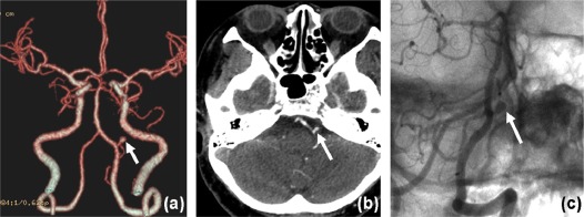
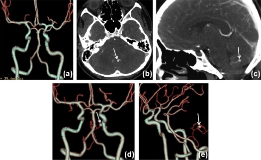
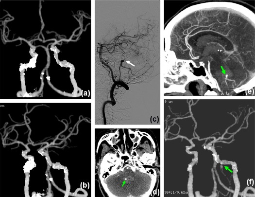
Twelve cases (12 of 30, 40%) of aneurysm lied on the proximal end of posterior inferior cerebellar artery (PICA) (border with vertebral artery) and all of them (12 of 12,100%) were clearly displayed on the VR images of CTA and correctly diagnosed by doctors. Eighteen cases (18 of 30, 60%) of aneurysm lied on the distal part of the PICA, whereas only 2 of them (2/18, 11.1%) were displayed on the VR images and correctly diagnosed before surgical procedure. After surgical operation, the respective review of the CTA images demonstrated that all aneurysms (30 of 30, 100%) can be found on the thin axial images after careful observation and are shown on VR images after adjusting the display threshold when the locations of the PICAA through thin axial images were known, including the distal PICAA.
Conclusions
Thin axial CT images are most important and reliable for the detection of distal PICAA. Overdependence on three-dimensional VR images of CTA is the main cause of misdiagnosis.
Introduction
Posterior inferior cerebellar artery aneurysm (PICAA) is relatively rare, accounts for about 0.5%–1% of intracranial aneurysm, but it is easy to re-rupture after the acute phase of first-time hemorrhage and causes death. So it is extremely important to determine the existence of PICAA and give surgical treatment as earlier as possible . Although the posterior inferior cerebellar artery (PICA) is the last and largest branch of vertebral artery, its thin lumen makes it difficult to be shown on volume rendering (VR) images of computed tomography angiography (CTA) under conventional or standard protocols . The posterior inferior cerebellar artery aneurysm (PICAA), especially distal PICAA, is easily missed by a doctor, leading to misdiagnosis and treatment delays. In our routine work, we found that most of the PICAAs were missed on CTA diagnosis and nearly all of the distal PICAAs were missed by CTA. The objective of this article is to report the CTA presentations of PICAA and analyze the causes of misdiagnosis of PICAA by CTA.
Materials and Methods
Patients
The study was conducted under the approval of the institutional review board of our hospital. Written consents were obtained from all patients before the study.
Get Radiology Tree app to read full this article<
Get Radiology Tree app to read full this article<
Examination
Get Radiology Tree app to read full this article<
Get Radiology Tree app to read full this article<
Get Radiology Tree app to read full this article<
Get Radiology Tree app to read full this article<
Analytical Methods
Get Radiology Tree app to read full this article<
Statistical Analysis
Get Radiology Tree app to read full this article<
Results
Get Radiology Tree app to read full this article<
Get Radiology Tree app to read full this article<
Discussion
Get Radiology Tree app to read full this article<
Get Radiology Tree app to read full this article<
Get Radiology Tree app to read full this article<
Get Radiology Tree app to read full this article<
Limitations
Get Radiology Tree app to read full this article<
Conclusions
Get Radiology Tree app to read full this article<
Get Radiology Tree app to read full this article<
References
1. Sejkorová A., Cihlář F., Hejčl A., et. al.: Microsurgery and endovascular treatment of posterior inferior cerebellar artery aneurysms. Neurosurg Rev 2016; 39: pp. 159-168.
2. Madaelil T.P., Wallace A.N., Chatterjee A.N., et. al.: Endovascular parent vessel sacrifice in ruptured dissecting vertebral and posterior inferior cerebellar artery aneurysms: clinical outcomes and review of the literature. J Neurointerv Surg 2016; 8: pp. 796-801.
3. Williamson R.W., Wilson D.A., Abla A.A., et. al.: Clinical characteristics and long-term outcomes in patients with ruptured posterior inferior cerebellar artery aneurysms: a comparative analysis. J Neurosurg 2015; 123: pp. 441-445.
4. Lehto H., Kivisaari R., Niemelä M., et. al.: Seventy aneurysms of the posterior inferior cerebellar artery: anatomical features and value of computed tomography angiography in microneurosurgery. World Neurosurg 2014; 82: pp. 1106-1112.
5. Kovač J.D., Stanković A., Stanković D., et. al.: Intracranial arterial variations: a comprehensive evaluation using CT angiography. Med Sci Monit 2014; 20: pp. 420-427.
6. Kim M.S.: Developmental anomalies of the distal vertebral artery and posterior inferior cerebellar artery: diagnosis by CT angiography and literature review. Surg Radilo Anat 2016; 38: pp. 997-1006.
7. Tang J., Wei L., Li L., et. al.: Endovascular treatment of distal posterior inferior cerebellar artery aneurysms. Neurosci (Riyadh) 2016; 21: pp. 236-240.
8. Huynh-Le P., Matsushima T., Miyazono M., et. al.: Three-dimensional CT angiography for the surgical management of the vertebral artery-posterior inferior cerebellar artery aneurysms. Acta Neurochir (Wien) 2004; 146: pp. 329-335.
9. Singh R.K., Behari S., Kumar V., et. al.: Posterior inferior cerebellar artery aneurysm: anatomical variations and surgical strategies. Asian J Neurosurg 2012; 7: pp. 2-11.
10. Jovanović V.T., Rakić M., Djurović B.M., et. al.: Distal posterior cerebellar artery aneurysm: case report. Acta Chir Iugosl 2008; 55: pp. 93-96.
11. Wu Q., Wang H.D., Zhang Q.R., et. al.: Parent artery occlusion with Onyx for distal aneurysms of posterior inferior cerebellar artery: a single-centre experience in a series of 15 patients. Neurol India 2013; 61: pp. 265-269.