
Rationale and Objectives
The purpose of our study was to compare test bolus techniques using undiluted or diluted contrast material for their ability to predict aortic enhancement on coronary computed tomographic angiography (c-CTA) images.
Materials and Methods
We divided 200 consecutive patients who underwent c-CTA on a 64-MDCT scanner into two groups. In group A ( n = 100), we used a test bolus of undiluted contrast material and in group B ( n = 100), the contrast material was diluted. The injection volume was body weight × 0.2 (contrast material 100%) in group A and body weight × 0.7 (contrast material 30%, saline 70%) in group B. We then compared the CT number in the ascending aorta on c-CTA images obtained with undiluted and diluted contrast media to the CT number on c-CTA images.
Results
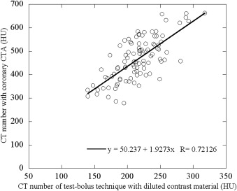
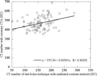
The mean CT number in the ascending aorta was significantly higher in group B than group A (217.1 vs. 157.4 HU, P < .001). There was a significant difference in the correlation between the CT number of the ascending aorta on c-CTA images and on images acquired with the test bolus using undiluted or diluted test bolus ( P < .001). In group B, the correlation had a strong positive linear relationship ( r = 0.72, P < .001), whereas in group A the positive linear relationship was weak ( r = 0.36).
Conclusions
The test bolus with diluted contrast material was useful for predicting aortic enhancement before c-CTA scanning.
Introduction
Coronary computed tomographic angiography (c-CTA) is an accurate noninvasive modality to assess coronary artery disease. To be diagnostically useful the c-CTA images must be of the highest quality. One index of their quality is the contrast-to-noise ratio (CNR) of the coronary artery determined by the contrast enhancement of the coronary artery and the background noise. Although the image noise can be regulated with the tube current at CT scanning, patient variability renders the regulation of coronary artery enhancement difficult. However, with the aid of the time-density curve of the test bolus, arterial enhancement in individual patients can be predicted before c-CTA , and this may make it possible to obtain the optimal CNR for c-CTA by selecting the appropriate tube current second setting. With the test bolus technique, not only the aortic time-density curve, but also aortic peak enhancement can be obtained before c-CTA. The bolus-tracking technique, on the other hand, is a real-time method and it cannot be used to predict enhancement before c-CTA. In general, the total contrast material volume used for the test bolus is low (approximately 10 mL), and large interpatient variability in arterial enhancement renders its prediction for CTA difficult . We hypothesized that the injection of a larger volume of diluted contrast material would yield a better correlation between arterial enhancement obtained with the test bolus and c-CTA than the injection of the conventional volume of undiluted contrast material.
The purpose of our study was to compare the value of test bolus techniques that delivered diluted or undiluted contrast material for predicting aortic enhancement on c-CTA images.
Materials and methods
Get Radiology Tree app to read full this article<
Patient Selection
Get Radiology Tree app to read full this article<
Table 1
Patient Characteristics for Test Bolus Technique with Undiluted and Diluted Contrast Materials
Undiluted Contrast Material Diluted Contrast Material_P_ Value Number of patients ( n ) 100 100 Sex (male/female) 48/52 44/56 .57 Age (y) 67.5 ± 10.9 (38–92) 70.0 ± 11.3 (40–89) .31 Body height (cm) 158.5 ± 8.6 (137–178) 156.5 ± 8.5 (140–182) .1 Body weight (kg) 57.0 ± 7.7 (39–69) 55.0 ± 7.1 (39–69) .16 Body surface area (m 2 ) 1.6 ± 0.1 1.58 ± 0.1 .19 Body mass index (kg/m 2 ) 22.3 ± 2.4 22.5 ± 2.6 .81 Heart rate (bpm) 59.0 ± 7.2 (44–85) 60.5 ± 7.2 (44–80) .51 Ejection fraction (%) 66.0 ± 8.9 (35–85) 68.0 ± 6.2 (51–88) .39
Get Radiology Tree app to read full this article<
CT and Contrast Injection Protocols
Get Radiology Tree app to read full this article<
Get Radiology Tree app to read full this article<
Table 2
Contrast Injection Protocol for Test Bolus and Coronary Computed Tomographic Angiography with Undiluted and Diluted Contrast Materials
Parameter Undiluted Contrast Material Diluted Contrast Material Test bolus Iodine dose (mgI/kg) 70.0 70.0 Injection volume (mL) Body weight (BW) (kg) × 0.2 (contrast material: 100%) BW (kg) × 0.7 (contrast material: 30%, saline: 70%) Injection rate (mL/s) BW (kg) × 0.07 BW (kg) × 0.07 Injection duration (s) 3.0 10.0 Saline chaser (mL) 20.0 20.0 Coronary computed tomographic angiography Iodine dose (mgI/kg) 245.0 Injection volume (mL) BW (kg) × 0.7 (contrast material: 100%) Injection rate (mL/s) BW (kg) × 0.07 Injection duration (s) 10.0 Saline chaser (mL) 20.0
Get Radiology Tree app to read full this article<
Get Radiology Tree app to read full this article<
c-CTA Scanning
Get Radiology Tree app to read full this article<
Data Analysis
Get Radiology Tree app to read full this article<
Statistical Analysis
Get Radiology Tree app to read full this article<
Results
Test Bolus Technique
Get Radiology Tree app to read full this article<
Table 3
Contrast Administration Parameters
Parameter Undiluted Contrast Material Diluted Contrast Material_P_ Value Arrival time (sec) 19.0 ± 2.9 20.0 ± 2.5 .27 Injection rate (mL/sec) 3.9 ± 0.5 3.9 ± 3.1 .63 Injection volume (mL) (Test-bolus) Contrast material 11.0 ± 1.6 11.0 ± 1.7 .77 Saline solution – 27.0 ± 6.8 Total volume 11.0 ± 1.5 39.0 ± 5.1 Injection volume (mL) (Coronary CTA) Contrast material 40.0 ± 5.2 39.0 ± 5.1 .29
Get Radiology Tree app to read full this article<
Coronary CT Angiography
Get Radiology Tree app to read full this article<
Get Radiology Tree app to read full this article<
Get Radiology Tree app to read full this article<
Discussion
Get Radiology Tree app to read full this article<
Get Radiology Tree app to read full this article<
Get Radiology Tree app to read full this article<
Get Radiology Tree app to read full this article<
Get Radiology Tree app to read full this article<
Get Radiology Tree app to read full this article<
Conclusions
Get Radiology Tree app to read full this article<
References
1. Cademartiri F., Mollet N.R., Lemos P.A., et. al.: Higher intracoronary attenuation improves diagnostic accuracy in MDCT coronary angiography. AJR Am J Roentgenol 2006; 187: pp. W430-W433.
2. Cademartiri F., Maffei E., Palumbo A.A., et. al.: Influence of intra-coronary enhancement on diagnostic accuracy with 64-slice CT coronary angiography. Eur Radiol 2008; 18: pp. 576-583.
3. Fleischmann D., Hittmair K.: Mathematical analysis of arterial enhancement and optimization of bolus geometry for CT angiography using the discrete Fourier transform. J Comput Assist Tomogr 1999; 23: pp. 474-484.
4. Hittmair K., Fleischmann D.: Accuracy of predicting and controlling time-dependent aortic enhancement from a test bolus injection. J Comput Assist Tomogr 2001; 25: pp. 287-294.
5. Fleischmann D.: CT angiography: injection and acquisition technique. Radiol Clin North Am 2010; 48: pp. 237-247. vii
6. Nakaura T., Awai K., Yanaga Y., et. al.: Low-dose contrast protocol using the test bolus technique for 64-detector computed tomography coronary angiography. Jpn J Radiol 2011; 29: pp. 457-465.
7. Kidoh M., Nakaura T., Awai K., et. al.: Compact-bolus dynamic CT protocol with a test bolus technique in 64-MDCT coronary angiography: comparison of fixed injection rate and duration protocol. Jpn J Radiol 2013; 31: pp. 115-122.
8. Nakaura T., Awai K., Yauaga Y., et. al.: Contrast injection protocols for coronary computed tomography angiography using a 64-detector scanner: comparison between patient weight-adjusted- and fixed iodine-dose protocols. Invest Radiol 2008; 43: pp. 512-519.
9. Meng X.-L., Rosenthal R., Rubin D.B.: Comparing correlated correlation coefficients. Psychol Bull 1992; 111: pp. 172-175.
10. Awai K., Hatcho A., Nakayama Y., et. al.: Simulation of aortic peak enhancement on MDCT using a contrast material flow phantom: feasibility study. AJR Am J Roentgenol 2006; 186: pp. 379-385.
11. Awai K., Hori S.: Effect of contrast injection protocol with dose tailored to patient weight and fixed injection duration on aortic and hepatic enhancement at multidetector-row helical CT. Eur Radiol 2003; 13: pp. 2155-2160.
12. Kerl J.M., Lehnert T., Schell B., et. al.: Intravenous contrast material administration at high-pitch dual-source CT pulmonary angiography: test bolus versus bolus-tracking technique. Eur J Radiol 2012; 81: pp. 2887-2891.
13. Svensson A., Ripsweden J., Rück A., et. al.: Heart rate variability and heat sensation during CT coronary angiography: low-osmolar versus iso-osmolar contrast media. Acta Radiol 2010; 51: pp. 722-726.
14. Vrachliotis T.G., Bis K.G., Haidary A., et. al.: Atypical chest pain: coronary, aortic, and pulmonary vasculature enhancement at biphasic single-injection 64-section CT angiography. Radiology 2007; 243: pp. 368-376.
15. Funama Y., Sugaya Y., Miyazaki O., et. al.: Automatic exposure control at MDCT based on the contrast-to-noise ratio: theoretical background and phantom study. Phys Med 2013; 29: pp. 39-47.
16. Bae K.T., Seeck B.A., Hildebolt C.F., et. al.: Contrast enhancement in cardiovascular MDCT: effect of body weight, height, body surface area, body mass index, and obesity. AJR Am J Roentgenol 2008; 190: pp. 777-784.
17. Yamamuro M., Tadamura E., Kanao S., et. al.: Coronary angiography by 64-detector row computed tomography using low dose of contrast material with saline chaser: influence of total injection volume on vessel attenuation. J Comput Assist Tomogr 2007; 31: pp. 272-280.
18. Layritz C., Muschiol G., Flohr T., et. al.: Automated attenuation-based selection of tube voltage and tube current for coronary CT angiography: reduction of radiation exposure versus a BMI-based strategy with an expert investigator. J Cardiovasc Comput Tomogr 2013; 7: pp. 303-310.