
Rationale and Objectives
To assess the prognostic value of a diverticular disease severity score (DDSS) based on computed tomography colonography (CTC) after acute diverticulitis (AD).
Materials and Methods
Of 252 patients who had an AD episode, we finally selected 46 patients who underwent both conventional CT at the acute event and CTC after 9 ± 7 weeks. Of these 46 patients, 17 underwent elective surgery after CTC. Disease severity was assessed with a 0–4 modified Hinchey CT-based score and a 1–4 CTC-based DDSS. A phone survey was performed 27 months later (range 4–52) for the 29 patients not surgically treated.
Results
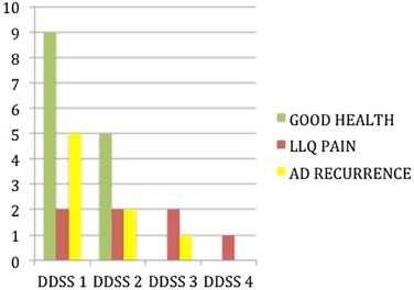
Significant correlation was found between CTC-based DDSS and clinical follow-up ( P = 0.022) or elective surgery ( P = 0.007), but not between clinical follow-up and CT-based score, extraluminal gas, C-reactive protein serum level, age, gender, or first versus recurrent AD episode. CTC demonstrated relevant additional findings in five of 46 (11%) patients: two AD complications (enterocolic and enterotubal fistulae), two colon cancers, and one extracolonic (lung) cancer.
Conclusion s
The CTC-based DDSS showed a prognostic value and correlated with the risk of undergoing surgery, and clinically relevant additional findings were found in more than 10% of patients. CTC could be the preferred test in patients recovering after AD.
Introduction
he appropriate follow-up of patients recovering from an acute episode of colonic diverticulitis is controversial. In particular, some authors are not in favor of further testing because an increased risk of colon cancer was not observed in these patients. On the contrary, others deem follow-up useful because it allows an opportunity to monitor the severity of the diverticular disease, and to confirm the absence of any other mimicking disease (e.g. colorectal cancer), which may not have been diagnosed in the acute stage. Recently, computed tomography colonography (CTC) has been proposed as a useful test in the evaluation of patients recovering from acute diverticulitis (AD) . Advocates point out the advantages of CTC compared to alternative tests routinely performed in the past, namely double-contrast barium enema and conventional colonoscopy. In particular, Flor et al. have proposed a diverticular disease severity score (DDSS) based on CTC findings performed after the acute episode. While some authors aimed at testing the validity and prognostic value of conventional CT examination performed at the time of an acute inflammatory episode , there are no similar evaluation studies for CTC in patients recovering after AD.
Hence, the purpose of our work was to assess the prognostic value of CTC in patients recovering after AD by using the DDSS and to evaluate the clinical relevance of any additional information gained from CTC in this setting.
Materials and Methods
Population
Get Radiology Tree app to read full this article<
Get Radiology Tree app to read full this article<
Imaging Protocol
Get Radiology Tree app to read full this article<
CT Colonography
Get Radiology Tree app to read full this article<
Get Radiology Tree app to read full this article<
Get Radiology Tree app to read full this article<
Image Analysis
Get Radiology Tree app to read full this article<
Table 1
Hinchey Modified Diverticular Disease Classification Adapted from Wasvary et al. Based on Conventional CT
0 Mild clinical diverticulitis Mild-Moderate 1a Confined pericolic inflammation—phlegmon 1b Confined pericolic abscess Severe 2 Pelvic, distant intra-abdominal, or retroperitoneal abscess 3 Generalized purulent peritonitis 4 Fecal peritonitis
CT, computed tomography.
Table 2
Diverticular Disease Severity Score Based on CTC
Maximum Wall Thickening (mm) Minimum Lumen Diameter (mm) 1 <3 ≥15 Mild-Moderate 2 3–8 ≥5 3 ≥8 ≥5 Severe 4 ≥8 <5
CTC, computed tomography colonography.
Get Radiology Tree app to read full this article<
Clinical Follow-up
Get Radiology Tree app to read full this article<
Statistical Analysis
Get Radiology Tree app to read full this article<
Get Radiology Tree app to read full this article<
Get Radiology Tree app to read full this article<
Get Radiology Tree app to read full this article<
Results
Get Radiology Tree app to read full this article<
Get Radiology Tree app to read full this article<
Clinical Findings and Laboratory Testing
Get Radiology Tree app to read full this article<
Get Radiology Tree app to read full this article<
Severity of Diverticular Disease Based on Conventional CT at Time of Acute Event
Get Radiology Tree app to read full this article<
Severity of Diverticular Disease Based on CTC (DDSS)
Get Radiology Tree app to read full this article<
Additional Value Added by CTC
Get Radiology Tree app to read full this article<
Get Radiology Tree app to read full this article<
Get Radiology Tree app to read full this article<
Get Radiology Tree app to read full this article<
Get Radiology Tree app to read full this article<
Get Radiology Tree app to read full this article<
Get Radiology Tree app to read full this article<
Get Radiology Tree app to read full this article<
Elective Surgery for Diverticular Disease
Get Radiology Tree app to read full this article<
Get Radiology Tree app to read full this article<
Survey on Clinical Follow-up
Get Radiology Tree app to read full this article<
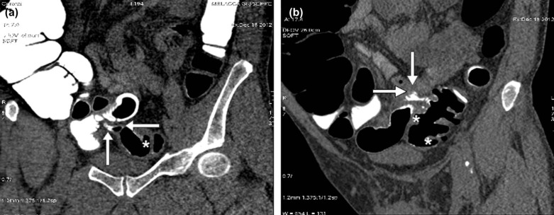
![Figure 2, Images in a patient in good health at follow-up. (a) Axial two-dimensional (2D) supine conventional computed tomography (CT) shows acute diverticulitis in the presence of sigmoid colon wall thickening, inflamed diverticula ( arrows ), mesenteric fat stranding, and thickening of mesenteric fasciae (modified Hinchey Ib). (b) Axial 2D supine computed tomography colonography (CTC) image showing sigmoid colon diverticula without wall thickening and lumen stenosis (diverticular disease severity score [DDSS] 1).](https://storage.googleapis.com/dl.dentistrykey.com/clinical/PrognosticValueoftheDiverticularDiseaseSeverityScoreBasedonCTColonography/1_1s20S1076633215003700.jpg)
![Figure 3, Images in a patient with diverticulitis recurrence at follow-up. (a) Axial two-dimensional (2D) supine conventional computed tomography (CT) shows acute diverticulitis in the presence of sigmoid colon wall thickening, inflamed diverticula ( arrows ), mesenteric fat stranding, and thickening of mesenteric fasciae (modified Hinchey Ib). (b) Axial 2D supine computed tomography colonography (CTC) image showing sigmoid colon diverticula in the presence of severe wall thickening associated with severe focal lumen stenosis (diverticular disease severity score [DDSS] 4).](https://storage.googleapis.com/dl.dentistrykey.com/clinical/PrognosticValueoftheDiverticularDiseaseSeverityScoreBasedonCTColonography/2_1s20S1076633215003700.jpg)
Get Radiology Tree app to read full this article<
Get Radiology Tree app to read full this article<
Get Radiology Tree app to read full this article<
Discussion
Get Radiology Tree app to read full this article<
Get Radiology Tree app to read full this article<
Get Radiology Tree app to read full this article<
Get Radiology Tree app to read full this article<
Get Radiology Tree app to read full this article<
Get Radiology Tree app to read full this article<
Get Radiology Tree app to read full this article<
Get Radiology Tree app to read full this article<
Get Radiology Tree app to read full this article<
Get Radiology Tree app to read full this article<
Get Radiology Tree app to read full this article<
Get Radiology Tree app to read full this article<
Get Radiology Tree app to read full this article<
Get Radiology Tree app to read full this article<
Get Radiology Tree app to read full this article<
References
1. Sai V.F., Velayos F., Neuhaus J., et. al.: Colonoscopy after CT diagnosis of diverticulitis to exclude colon cancer: a systematic literature review. Radiology 2012; 263: pp. 383-390.
2. Westwood D.A., Eglinton T.W., Frizelle F.A.: Routine colonoscopy following acute uncomplicated diverticulitis. Br J Surg 2011; 98: pp. 1630-1634.
3. Feingold D., Steele S.R., Lee S., et. al.: Practice parameters for the treatment of sigmoid diverticulitis. Dis Colon Rectum 2014; 57: pp. 284-294.
4. Rafferty J., Shellito P., Hyman N.H., et. al.: Practice parameters for sigmoid diverticulitis. Dis Colon Rectum 2006; 49: pp. 939-944.
5. Andersen J.C., Bundgaard L., Elbrønd H., et. al.: Danish national guidelines for treatment of diverticular disease. Dan Med J 2012; 59: pp. C4453.
6. Elmi A., Hedgire S.S., Pargaonkar V., et. al.: Is early colonoscopy beneficial in patients with CT-diagnosed diverticulitis?. AJR Am J Roentgenol 2013; 200: pp. 1269-1274.
7. Agarwal A.K., Karanjawala B.E., Maykel J.A., et. al.: Routine colonic endoscopic evaluation following resolution of acute diverticulitis: is it necessary?. World J Gastroenterol 2014; 20: pp. 12509-12516.
8. Hjern F., Jonas E., Holmstrom B., et. al.: CT colonography versus colonoscopy in the follow-up of patients after diverticulitis. A prospective, comparative study. Clin Radiol 2007; 62: pp. 645-650.
9. Flor N., Rigamonti P., Pisani Ceretti A., et. al.: Diverticular disease severity score based on CT colonography. Eur Radiol 2013; 23: pp. 2723-2729.
10. Chabok A., Smedh K., Nilsson S., et. al.: CT-colonography in the follow-up of acute diverticulitis: patient acceptance and diagnostic accuracy. Scand J Gastroenterol 2013; 48: pp. 979-986.
11. Ambrosetti P.: Acute diverticulitis of the left colon: value of the initial CT and timing of elective colectomy. J Gastrointest Surg 2008; 12: pp. 1318-1320.
12. Kaiser A.M., Jiang J.K., Lake J.P., et. al.: The management of complicated diverticulitis and the role of computed tomography. Am J Gastroenterol 2005; 100: pp. 910-917.
13. Buchach C.M., Kim D.H., Pickhardt P.J.: Performing an additional decubitus series at CT colonography. Abd Imaging 2001; 36: pp. 538-544.
14. Wasvary H., Turfah F., Kadro O., et. al.: Same hospitalization resection for acute diverticulitis. Am Surg 1999; 65: pp. 632-635.
15. Zalis M.E., Barish M.A., Choi J.R., et. al.: CT colonography reporting and data system: a consensus proposal. Radiology 2005; 236: pp. 3-9.
16. Spada C., Stoker J., Alarcon O., et. al.: Clinical indications for computed tomography colonography. European Society of Gastrointestinal Endoscopy (ESGE) and European Society of Gastrointestinal and Abdominal Radiology (ESGAR) guideline. Eur Radiol 2014; 46: pp. 897-915.
17. Nguyen G.C., Sam J., Anand N.: Epidemiological trends and geographic variation in hospital admissions for diverticulitis in the United States. World J Gastroenterology 2011; 17: pp. 1600-1605.
18. Surgical treatment of diverticulitis. Patient care committee of the Society for Surgery of the Alimentary Tract (SSAT). J Gastrointest Surg 1999; 3: pp. 212-213.
19. Margolin D.A.: Timing of elective surgery for diverticular disease. Clin Colon Recal Surg 2009; 22: pp. 169-172.
20. Pickhardt P.J., Hassan C., Halligan S., et. al.: Colorectal cancer: CT colonography for detection—systematic review and meta-analysis. Radiology 2001; 259: pp. 393-405.
21. Flor N., Rigamonti P., Di Leo G., et. al.: Technical quality of CT colonography in relation with diverticular disease. Eur J Radiol 2012; 81: pp. e250-e254.
22. Sanford M., Pickhardt P.J.: Diagnostic performance of primary 3-dimensional computed tomography colonography in the setting of colonic diverticular disease. Clin Gastroenterol Hepatol 2006; 4: pp. 1039-1047.
23. Flor N., Sardanelli F., Pickhardt P.J.: Diagnostic accuracy of CT colonography for the detection of polyps in the diverticular disease. Scand J Gastroenterol 2014; 49: pp. 383-384.
24. Pickhardt P.J.: Missed lesions at CT colonography: lessons learned. Abdom Imaging 2013; 38: pp. 82-97.
25. Coppola F., Regge D., Flor N., et. al.: Flat lesions missed at conventional colonoscopy (CC) and visualized by CT colonography (CTC): a pictorial essay. Abdom Imaging 2014; 39: pp. 25-32.
26. Dafnis G., Granath F., Pahlman L., et. al.: Patient factors influencing the completion rate in colonoscopy. Dig Liver Dis 2005; 37: pp. 113-118.
27. Hanson M.E., Pickhardt P.J., Kim D.H., et. al.: Anatomic factors predictive of incomplete colonoscopy based on findings at CT colonography. AJR 2007; 189: pp. 774-779.
28. Ambrosetti P., Grossholz M., Becker C., et. al.: Computed tomography in acute left colonic diverticulitis. Br J Surg 1997; 84: pp. 532-534.
29. Retert H., Nldge G., Encke J., et. al.: The value of CT for the diagnosis of acute diverticulitis. Radiologe 2003; 43: pp. 51-58.
30. Buckley O., Geoghegan T., O’Riordain D.S., et. al.: Computed tomography in the imaging of colonic diverticulitis. Clin Radiol 2004; 59: pp. 977-983.