
Rationale and Objectives
Phosphorous magnetic resonance spectroscopy ( 31 P MRS) has been used to evaluate and predict treatment response in squamous cell carcinoma of the head and neck (HNSCC). Several studies have also shown the potential of proton MRS ( 1 H MRS) in assessing response in HNSCC. In view of the inherent limitations associated with performing 31 P MRS in clinical settings, the current study was performed to explore whether 1 H MRS could provide similar or complementary metabolic information in HNSCC.
Materials and Methods
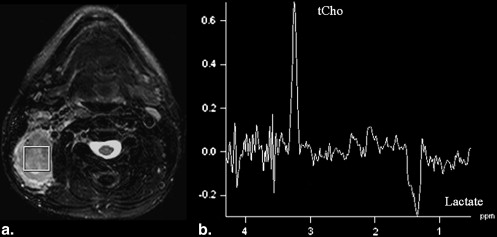
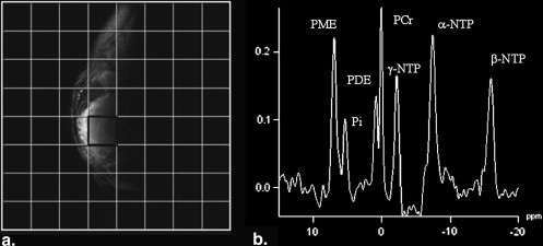
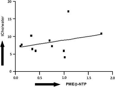
Fifteen patients with HNSCC underwent pretreatment magnetic resonance imaging. Both 1 H MRS and 31 P MRS were performed on viable solid parts of the metastatic lymph nodes of these patients. Peak areas of total choline (tCho) and unsuppressed water as observed on 1 H MRS and phosphomonoester (PME) and β-nucleotide triphosphate (β-NTP) on 31 P MRS were computed. Pearson’s correlation coefficient was used to correlate the tCho/water and PME/β-NTP ratios.
Results
In all patients, the metastatic nodes appeared hyperintense on T2-weighted images and hypointense on T1-weighted images with variable signal intensity. A prominent resonance of tCho on 1 H MRS and a resonance of PME on 31 P MRS from the metastatic nodes of all patients were observed. A moderate correlation of 0.31 was observed between tCho/water and PME/β-NTP ( P > .05).
Conclusions
The biochemical pathways involved in 1 H MRS of tCho may be different from the phospholipid metabolites seen on 31 P MRS of head and neck cancers, and thus the two MRS techniques may be complementary to each other.
Squamous cell carcinoma of the head and neck (HNSCC) is a malignant tumor arising most frequently in the nonkeratinized epithelial tissue of the upper aerodigestive tract. This disease accounts for about 5% of all cancers and about 90% of all malignant tumors of the head and neck region . These tumors are characterized by a multiphasic and multifactorial etiopathogenesis . Despite recent progress in diagnosis and advances in local and systemic therapeutic approaches, HNSCC remains a clinically challenging disease . It is expected that developments in treatment modalities, along with consistent, reliable, and reproducible prognostic biomarkers that monitor and predict therapeutic response, may improve the clinical outcome of patients with HNSCC.
Phosphorous magnetic resonance (MR) spectroscopy ( 31 P MRS) provides a window for assessing tissue bioenergetics and metabolism of membrane phospholipids . Specifically, the 31 P MRS spectrum demonstrates signals from phosphomonoesters (PMEs; phospholipid precursors) and phosphodiesters (PDEs; phospholipid catabolites), inorganic phosphate (Pi), phosphocreatine (PCr), and nucleotide triphosphates (NTPs) related to energy metabolism. Using 31 P MRS, it has been reported that head and neck tumors exhibit elevated PME/β-NTP ratios , and this ratio has been used as a response indicator for chemotherapy and/or radiation therapy in head and neck tumors .
Get Radiology Tree app to read full this article<
Get Radiology Tree app to read full this article<
Materials and methods
Subjects
Get Radiology Tree app to read full this article<
Data Acquisition
MR Imaging
Get Radiology Tree app to read full this article<
1 H MRS
Get Radiology Tree app to read full this article<
31 P MRS
Get Radiology Tree app to read full this article<
Data Analysis
Get Radiology Tree app to read full this article<
Get Radiology Tree app to read full this article<
SI=ρ[1−exp(−TR/T1)][exp(−TE/T2)] SI
=
ρ
[
1
−
exp
(
−
TR
/
T
1
)
]
[
exp
(
−
TE
/
T2
)
]
where SI is the signal intensity of the unsuppressed water signal, ρ is proton density, T1 is the spin-lattice relaxation time, and T2 is the spin-spin relaxation time measured from each patient. At a TR of 1500 ms and a TE of 135 ms, the value of ρ was determined from equation 1 using the T1 and T2 values from each metastatic node. Quantitative T2 and T1 measurements in each case were performed by acquiring a series of T2-weighted images (at four different TEs: 13, 53, 80, and 120 ms [TR, 2000 ms]) and T1-weighted images [at five different inversion times: 60, 200, 400, 800, and 1600 ms [TR, 1880 ms; TE, 4.38 ms]), respectively. The tCho/ρ ratio was computed from all patients.
Get Radiology Tree app to read full this article<
Get Radiology Tree app to read full this article<
Get Radiology Tree app to read full this article<
Results
Get Radiology Tree app to read full this article<
Get Radiology Tree app to read full this article<
Get Radiology Tree app to read full this article<
Get Radiology Tree app to read full this article<
Discussion
Get Radiology Tree app to read full this article<
Get Radiology Tree app to read full this article<
Get Radiology Tree app to read full this article<
Get Radiology Tree app to read full this article<
Get Radiology Tree app to read full this article<
Get Radiology Tree app to read full this article<
Acknowledgments
Get Radiology Tree app to read full this article<
Get Radiology Tree app to read full this article<
References
1. Funk G.F., Karnell L.H., Robinson R.A., Zhen W.K., Trask D.K., Hoffman H.T.: Presentation, treatment, and outcome of oral cavity cancer: a National Cancer data base report. Head Neck 2002; 24: pp. 165-180.
2. Scully C., Field J.K., Tanzawa H.: Genetic aberrations in oral or head and neck squamous cell carcinoma (SCCHN): 1. Carcinogen metabolism, DNA repair and cell cycle control. Oral Oncol 2000; 36: pp. 256-263.
3. Zorat P.L., Paccagnella A., Cavaniglia G., et. al.: Randomized phase III trial of neoadjuvant chemotherapy in head and neck cancer: 10-year follow-up. J Natl Cancer Inst 2004; 96: pp. 1714-1717.
4. Podo F.: Tumour phospholipid metabolism. NMR Biomed 1999; 12: pp. 413-439.
5. McKenna W.G., Lenkinski R.E., Hendrix R.A., Vogele K.E., Bloch P.: The use of magnetic resonance imaging and spectroscopy in the assessment of patients with head and neck and other superficial human malignancies. Cancer 1989; 64: pp. 2069-2075.
6. Maldonado X., Alonso J., Giralt J., et. al.: 31Phosphorus magnetic resonance spectroscopy in the assessment of head and neck tumors. Int J Radiat Oncol Biol Phys 1998; 40: pp. 309-312.
7. Shukla-Dave A., Poptani H., Loevner L.A., et. al.: Prediction of treatment response of head and neck cancers with P-31 MR spectroscopy from pretreatment relative phosphomonoester levels. Acad Radiol 2002; 9: pp. 688-694.
8. Arias-Mendoza F., Smith M.R., Brown T.R.: Predicting treatment response in non-Hodgkin’s lymphoma from the pretreatment tumor content of phosphoethanolamine plus phosphocholine. Acad Radiol 2004; 11: pp. 368-376.
9. Arias-Mendoza F., Zakian K., Schwartz A., et. al.: Methodological standardization for a multi-institutional in vivo trial of localized 31P MR spectroscopy in human cancer research. In vitro and normal volunteer studies. NMR Biomed 2004; 17: pp. 382-391.
10. Gadian D.G., Isaacs E.B., Cross J.H., et. al.: Lateralization of brain function in childhood revealed by magnetic resonance spectroscopy. Neurology 1996; 46: pp. 974-977.
11. Bourne R.M., Stanwell P., Stretch J.R., et. al.: In vivo and ex vivo proton MR spectroscopy of primary and secondary melanoma. Eur J Radiol 2005; 53: pp. 506-513.
12. Hollingworth W., Medina L.S., Lenkinski R.E., et. al.: A systematic literature review of magnetic resonance spectroscopy for the characterization of brain tumors. AJNR Am J Neuroradiol 2006; 27: pp. 1404-1411.
13. Miller B.L.: A review of chemical issues in 1H NMR spectroscopy: N-acetyl-L-aspartate, creatine and choline. NMR Biomed 1991; 4: pp. 47-52.
14. Mukherji S.K., Schiro S., Castillo M., Kwock L., Muller K.E., Blackstock W.: Proton MR spectroscopy of squamous cell carcinoma of the extracranial head and neck: in vitro and in vivo studies. AJNR Am J Neuroradiol 1997; 18: pp. 1057-1072.
15. Mukherji S.K., Gerstle R.J.: In vitro 1H MR spectroscopy of squamous cell carcinoma of the extracranial head and neck. Neuroimaging Clin N Am 1998; 8: pp. 835-847.
16. Bisdas S., Baghi M., Huebner F., et. al.: In vivo proton MR spectroscopy of primary tumours, nodal and recurrent disease of the extracranial head and neck. Eur Radiol 2007; 17: pp. 251-257.
17. Kuesel A.C., Graschew G., Hull W.E., Lorenz W., Thielmann H.W.: 31P NMR studies of cultured human tumor cells. Influence of pH on phospholipid metabolite levels and the detection of cytidine 5′-diphosphate choline. NMR Biomed 1990; 3: pp. 78-89.
18. Star-Lack J.M., Adalsteinsson E., Adam M.F., et. al.: In vivo 1H MR spectroscopy of human head and neck lymph node metastasis and comparison with oxygen tension measurements. AJNR Am J Neuroradiol 2000; 21: pp. 183-193.
19. Gerstle R.J., Aylward S.R., Kromhout-Schiro S., Mukherji S.K.: The role of neural networks in improving the accuracy of MR spectroscopy for the diagnosis of head and neck squamous cell carcinoma. AJNR Am J Neuroradiol 2000; 21: pp. 1133-1138.
20. Maheshwari S.R., Mukherji S.K., Neelon B., et. al.: The choline/creatine ratio in five benign neoplasms: comparison with squamous cell carcinoma by use of in vitro MR spectroscopy. AJNR Am J Neuroradiol 2000; 21: pp. 1930-1935.
21. Bezabeh T., Odlum O., Nason R., et. al.: Prediction of treatment response in head and neck cancer by magnetic resonance spectroscopy. AJNR Am J Neuroradiol 2005; 26: pp. 2108-2113.
22. Christiansen P., Toft P.B., Gideon P., Danielsen E.R., Ring P., Henriksen O.: MR-visible water content in human brain: a proton MRS study. Magn Reson Imaging 1994; 12: pp. 1237-1244.
23. Poptani H., Gupta R.K., Roy R., Pandey R., Jain V.K., Chhabra D.K.: Characterization of intracranial mass lesions with in vivo proton MR spectroscopy. AJNR Am J Neuroradiol 1995; 16: pp. 1593-1603.
24. Mishra A.M., Gupta R.K., Jaggi R.S., et. al.: Role of diffusion-weighted imaging and in vivo proton magnetic resonance spectroscopy in the differential diagnosis of ring-enhancing intracranial cystic mass lesions. J Comput Assist Tomogr 2004; 28: pp. 540-547.
25. Vogl T., Peer F., Schedel H., et. al.: 31P-spectroscopy of head and neck tumors–surface coil technique. Magn Reson Imaging 1989; 7: pp. 425-435.
26. Hendrix R.A., Lenkinski R.E., Vogele K., Bloch P., McKenna W.G.: 31P localized magnetic resonance spectroscopy of head and neck tumors—preliminary findings. Otolaryngol Head Neck Surg 1990; 103: pp. 775-783.
27. Karczmar G.S., Meyerhoff D.J., Boska M.D., et. al.: P-31 spectroscopy study of response of superficial human tumors to therapy. Radiology 1991; 179: pp. 149-153.
28. Ruiz-Cabello J., Cohen J.S.: Phospholipid metabolites as indicators of cancer cell function. NMR Biomed 1992; 5: pp. 226-233.
29. Luyten P.R., Bruntink G., Sloff F.M., et. al.: Broadband proton decoupling in human 31P NMR spectroscopy. NMR Biomed 1989; 1: pp. 177-183.
30. Li C.W., Negendank W.G., Murphy-Boesch J., Padavic-Shaller K., Brown T.R.: Molar quantitation of hepatic metabolites in vivo in proton-decoupled, nuclear Overhauser effect enhanced 31P NMR spectra localized by three-dimensional chemical shift imaging. NMR Biomed 1996; 9: pp. 141-155.
31. Mukherji S.K., Schiro S., Castillo M., et. al.: Proton MR spectroscopy of squamous cell carcinoma of the upper aerodigestive tract: in vitro characteristics. AJNR Am J Neuroradiol 1996; 17: pp. 1485-1490.
32. El-Sayed S., Bezabeh T., Odlum O., et. al.: An ex vivo study exploring the diagnostic potential of 1H magnetic resonance spectroscopy in squamous cell carcinoma of the head and neck region. Head Neck 2002; 24: pp. 766-772.
33. Huang W., Roche P., Shindo M., Madoff D., Geronimo C., Button T.: Evaluation of head and neck tumor response to therapy using in vivo 1H MR spectroscopy: correlation with pathology. Proc Int Soc Magn Reson 2000; 8: pp. 552.
34. McKnight T.R.: Proton magnetic resonance spectroscopic evaluation of brain tumor metabolism. Semin Oncol 2004; 31: pp. 605-617.
35. Barker P.B., Breiter S.N., Soher B.J., et. al.: Quantitative proton spectroscopy of canine brain: in vivo and in vitro correlations. Magn Reson Med 1994; 32: pp. 157-163.
36. Albers M.J., Krieger M.D., Gonzalez-Gomez I., et. al.: Proton-decoupled 31P MRS in untreated pediatric brain tumors. Magn Reson Med 2005; 53: pp. 22-29.
37. Usenius J.P., Vainio P., Hernesniemi J., Kauppinen R.A.: Choline-containing compounds in human astrocytomas studied by 1H NMR spectroscopy in vivo and in vitro. J Neurochem 1994; 63: pp. 1538-1543.
38. Smith T.A., Glaholm J., Leach M.O., Machin L., McCready V.R.: The effect of intra-tumour heterogeneity on the distribution of phosphorus-containing metabolites within human breast tumours: an in vitro study using 31P NMR spectroscopy. NMR Biomed 1991; 4: pp. 262-267.
39. Aboagye E.O., Bhujwalla Z.M.: Malignant transformation alters membrane choline phospholipid metabolism of human mammary epithelial cells. Cancer Res 1999; 59: pp. 80-84.
40. Cai H., Erhardt P., Troppmair J., et. al.: Hydrolysis of phosphatidylcholine couples Ras to activation of Raf protein kinase during mitogenic signal transduction. Mol Cell Biol 1993; 13: pp. 7645-7651.
41. Daly P.F., Lyon R.C., Faustino P.J., Cohen J.S.: Phospholipid metabolism in cancer cells monitored by 31P NMR spectroscopy. J Biol Chem 1987; 262: pp. 14875-14878.
42. Miceli M.V., Kan L.S., Newsome D.A.: Phosphorus-31 nuclear magnetic resonance spectroscopy of human retinoblastoma cells: correlation with metabolic indices. Biochim Biophys Acta 1988; 970: pp. 262-269.
43. Bruhn H., Frahm J., Gyngell M.L., et. al.: Noninvasive differentiation of tumors with use of localized H-1 MR spectroscopy in vivo: initial experience in patients with cerebral tumors. Radiology 1989; 172: pp. 541-548.
44. Kinoshita Y., Yokota A., Koga Y.: Phosphorylethanolamine content of human brain tumors. Neurol Med Chir (Tokyo) 1994; 34: pp. 803-806.
45. Clejan S.: HPLC analytical methods for the separation of molecular species of fatty acids in diacylglycerol and cellular phospholipids. Methods Mol Biol 1998; 105: pp. 255-274.
46. Kalra R., Wade K.E., Hands L., et. al.: Phosphomonoester is associated with proliferation in human breast cancer: a 31P MRS study. Br J Cancer 1993; 67: pp. 1145-1153.
47. Maris J.M., Evans A.E., McLaughlin A.C., et. al.: 31P nuclear magnetic resonance spectroscopic investigation of human neuroblastoma in situ. N Engl J Med 1985; 312: pp. 1500-1505.
48. Kinoshita Y., Yokota A.: Absolute concentrations of metabolites in human brain tumors using in vitro proton magnetic resonance spectroscopy. NMR Biomed 1997; 10: pp. 2-12.
49. Dixon R.M., Tian M.: Phospholipid synthesis in the lymphomatous mouse liver studied by 31P nuclear magnetic resonance spectroscopy in vitro and by administration of 14C-radiolabelled compounds in vivo. Biochim Biophys Acta 1993; 1181: pp. 111-121.
50. Cullis P.R., de Kruijff B., Hope M.J., Nayar R., Schmid S.L.: Phospholipids and membrane transport. Can J Biochem 1980; 58: pp. 1091-1100.
51. Kiss Z., Crilly K.S., Anderson W.H.: Carcinogens stimulate phosphorylation of ethanolamine derived from increased hydrolysis of phosphatidylethanolamine in C3H/101/2 fibroblasts. FEBS Lett 1993; 336: pp. 115-118.
52. Gillham H., Brindle K.M.: 31P NMR measurements of the effects of unsaturated fatty acids on cellular phospholipid metabolism. Magn Reson Med 1996; 35: pp. 481-488.
53. Smith T.A., Glaholm J., Leach M.O., et. al.: A comparison of in vivo and in vitro 31P NMR spectra from human breast tumours: variations in phospholipid metabolism. Br J Cancer 1991; 63: pp. 514-516.