
Rationale and Objectives
Chest radiographs can be demanding, making this an area of focus during most first-year resident chest rotations. This often comes at a cost of cross-sectional imaging, and new residents are often not initially comfortable with reading chest computed tomographic angiograms (CTAs) for pulmonary embolisms (PEs). We created a teaching file of CTAs to improve the detection of PEs.
Materials and Methods
For initial testing, we used videos of 25 cases, which played for 90 seconds (to allow multiple passes) to residents with and without call experience. The presence and location of PEs and the readers’ confidence scores were recorded. After initial testing, first-year residents without call experience were given 20 separate known positive CTA videos to scroll through on their own. The goal of this was to allow for individual review and development of individual search strategies. A second testing was done with all levels of residents with the same initial 25 cases, re-randomized to evaluate for improvement.
Results
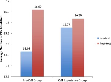
Initially, first-year residents without call experience identified an average of 14.7 of 18 examinations positive for PEs (versus 15.8 for more senior residents; P < .04). After reviewing the 20 known positive cases, the first-year residents improved, averaging 16.6 (versus 14.7 earlier; P < .01).
Conclusions
We created a fast, simple way to expose novice residents to CTA examinations and increase their accuracy in identifying PEs. After using a teaching file, the ability to recognize PEs improved significantly, and scores were no longer significantly different from those of residents with call experience.
The task of the radiology resident on call is no longer limited to reading a tall stack of plain films. The transition to digital imaging and the development of serial imaging have changed the duties of the novice on-call radiology resident dramatically. As the job description of “taking call” has changed, residency programs have been challenged to modify their training methods to ensure that residents are prepared for their call duties.
With the recent change in format of the radiology boards, there is an increased emphasis on going through the material that was traditionally covered before oral examination (4 years) in a shorter period, before the new core examination (3 years). There are several ways programs attempt to accomplish this, ranging from fewer rotations in specific areas, to shorter rotations. Regardless of how this new challenge is handled, the fact is that there is a shorter period of time to cover information; therefore, it is essential to make teaching as effective as possible to best prepare residents for success on boards and in the workplace.
Get Radiology Tree app to read full this article<
Get Radiology Tree app to read full this article<
Get Radiology Tree app to read full this article<
Get Radiology Tree app to read full this article<
Get Radiology Tree app to read full this article<
Get Radiology Tree app to read full this article<
Get Radiology Tree app to read full this article<
Get Radiology Tree app to read full this article<
Methods and materials
Get Radiology Tree app to read full this article<
Get Radiology Tree app to read full this article<
Get Radiology Tree app to read full this article<
Get Radiology Tree app to read full this article<
Get Radiology Tree app to read full this article<
Get Radiology Tree app to read full this article<
Results
Get Radiology Tree app to read full this article<
Get Radiology Tree app to read full this article<
Get Radiology Tree app to read full this article<
Get Radiology Tree app to read full this article<
Get Radiology Tree app to read full this article<
Discussion
Get Radiology Tree app to read full this article<
Get Radiology Tree app to read full this article<
Get Radiology Tree app to read full this article<
Get Radiology Tree app to read full this article<
Get Radiology Tree app to read full this article<
Get Radiology Tree app to read full this article<
Get Radiology Tree app to read full this article<
Get Radiology Tree app to read full this article<
Get Radiology Tree app to read full this article<
Get Radiology Tree app to read full this article<
Get Radiology Tree app to read full this article<
Get Radiology Tree app to read full this article<
Get Radiology Tree app to read full this article<
Get Radiology Tree app to read full this article<
Get Radiology Tree app to read full this article<
References
1. Rojas C., Hamza J., Chung J.: The new era of radiology teaching files. AJR Am J Roentgenol 2012; 198: pp. 773-776.
2. Tran T., Roach N., O’Kane P., et. al.: Computers in radiology: creating a digital radiographic teaching file and database using a PC and common software. AJR Am J Roentgenol 2000; 175: pp. 325-327.
3. Talanow R.: Radiology Teacher: a free, Internet-base radiology teaching file server. J Am Coll Radiol 2009; 6: pp. 871-875.
4. LoRusso A., Bassignani J., Harvey J.: Enhanced teaching of screening mammography using an electronic format. Acad Radiol 2006; 13: pp. 782-788.
5. Kamauu A., Whipple J., DuVall S., et. al.: IHE teaching file and clinical trial export integration profile: functional examples. RadioGraphics 2008; 28: pp. 933-945.
6. Scarsbrook J., Graham R., Perriss R.: Radiology education: a glimpse into the future. Clin Radiol 2006; 61: pp. 640-648.
7. Towbin A., Paterson B., Chang P.: Computer-based simulator for radiology: an educational tool. RadioGraphics 2008; 28: pp. 309-316.
8. Towbin A., Paterson B., Chang P.: A computer-based radiology simulator as a learning tool to help prepare first-year residents for being on call. Acad Radiol 2007; 14: pp. 1271-1283.