
Objectives
This study aimed to intra-individually and inter-individually compare image quality, radiation dose, and diagnostic accuracy of dual-source dual-energy computed tomography pulmonary angiography (CTPA) protocols in patients with suspected pulmonary embolism (PE).
Methods
Thirty-three patients with suspected PE underwent initial and follow-up dual-energy CTPA at 80/Sn140 kVp (group A) or 100/Sn140 kVp (group B), which were assigned based on tube voltages. Subjective and objective CTPA image quality and lung perfusion map image quality were evaluated. Diagnostic accuracies of CTPA and perfusion maps were assessed by two radiologists independently. Effective dose (ED) was calculated and compared.
Results
Mean computed tomography (CT) values of pulmonary arteries were higher in group A than group B ( P = .006). There was no difference in signal-to-noise ratio and contrast-to-noise ratio between the two groups (both P > .05). Interobserver agreement for evaluating subjective image quality of CTPA and color-coded perfusion images was either good (κ = 0.784) or excellent (κ = 0.887). Perfusion defect scores and diagnostic accuracy of CTPA showed no difference between both groups (both P > .05). Effective dose of group A was reduced by 45.8% compared to group B ( P < .001).
Conclusions
Second-generation dual-source dual-energy CTPA with 80/Sn140 kVp allows for sufficient image quality and diagnostic accuracy for detecting PE while substantially reducing radiation dose.
Introduction
Acute pulmonary embolism (PE) ranks as the third most common cardiovascular condition, after coronary artery disease and stroke . Due to the nonspecific clinical symptoms, imaging plays an important role in detection and follow-up of acute PE . Currently, computed tomography pulmonary angiography (CTPA) is considered the reference standard in diagnosis of acute PE . Dual-energy CTPA is capable of providing anatomic, perfusion, ventilation, and pulmonary vascular tree information for the whole lung, which makes it an important tool for comprehensive evaluation of PE .
With the application of dual-energy computed tomography (CT) in the lungs, diagnostic accuracy for acute and chronic PE, as well as other vascular disorders such as lung malignancies and parenchymal diseases, has been improved . Compared to routine single-energy CTPA, dual-energy CTPA showed advantages in detecting small peripheral PE . With concerns regarding radiation risk, the clinical use of CTPA has increasingly come into focus. Thieme et al. indicated that according to the European guidelines on quality criteria for CT, the radiation dose with first-generation dual-source dual-energy CTPA (80/140 kVp) is lower than the reference value of 650 mGy⋅cm. With the development of second-generation dual-source CT, multiple scanning parameters of dual-source dual-energy CTPA, such as 80/Sn140 kVp and 100/Sn140 kVp, can be selected. Recently, Bauer et al. demonstrated that second-generation dual-energy CT with 80/Sn140 kVp allowed for significant dose reduction with the same image quality compared to 120 kVp CTPA. However, this study did not include an intra-individual comparison and did not analyze lung perfusion images. Both Thieme et al. and Bauer et al. provide important information for the diagnosis and prognosis evaluation of PE . Thus, further studies seem warranted to expand on these aspects. The purpose of this study was to intra-individually and inter-individually compare image quality, radiation dose, and diagnostic accuracy of dual-energy CTPA with the two different scanning parameters, and determine the optimized scanning protocols for dual-energy CTPA in clinical routine.
Materials and Methods
Patients
Get Radiology Tree app to read full this article<
CT Scanning Protocol
Get Radiology Tree app to read full this article<
Get Radiology Tree app to read full this article<
Get Radiology Tree app to read full this article<
Dual-Energy CTPA Image Quality Evaluation
Objective Image Quality Evaluation
Get Radiology Tree app to read full this article<
Subjective Image Quality Evaluation
Get Radiology Tree app to read full this article<
Dual-Energy Lung Perfusion Image Evaluation
Get Radiology Tree app to read full this article<
Get Radiology Tree app to read full this article<
Get Radiology Tree app to read full this article<
Detection of Pulmonary Emboli
Get Radiology Tree app to read full this article<
Radiation Dose Estimation
Get Radiology Tree app to read full this article<
Statistical Analysis
Get Radiology Tree app to read full this article<
Results
Subjects
Get Radiology Tree app to read full this article<
Dual-Energy CTPA Image Quality Evaluation
Get Radiology Tree app to read full this article<
Table 1
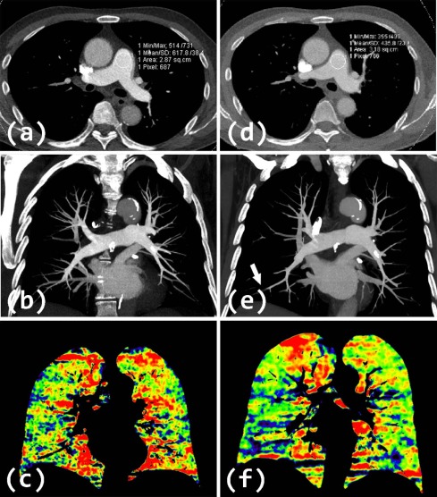
Objective Image Quality Measurements in the Two CTPA Groups
Location Group A Group B_P_ Value MPA CT number (HU) \* 386.3 ± 94.1 318.2 ± 94.8 .003 SNR 41.4 ± 12.4 46.3 ± 16.5 .145 CNR 36.0 ± 11.7 38.6 ± 15.7 .419 RPA CT number (HU) \* 357.0 ± 86.3 302.5 ± 92.7 .008 SNR 38.2 ± 11.0 44.1 ± 16.2 .064 CNR 32.8 ± 10.4 36.3 ± 15.4 .235 LPA CT number (HU) \* 366.9 ± 91.9 307.7 ± 95.3 .007 SNR 39.3 ± 11.7 44.8 ± 16.2 .102 CNR 33.8 ± 11.1 37.1 ± 15.4 .315 RULA CT number (HU) \* 373.8 ± 96.9 302.4 ± 108.2 .003 SNR 40.1 ± 12.3 44.0 ± 17.2 .257 CNR 34.6 ± 11.6 36.2 ± 16.5 .628 RILA CT number (HU) 340.6 ± 96.6 301.5 ± 110.4 .075 SNR \* 36.2 ± 10.7 43.8 ± 17.2 .031 CNR 30.8 ± 10.4 36.0 ± 16.5 .114 LULA CT number (HU) \* 366.7 ± 102.2 299.1 ± 106.2 .006 SNR 39.3 ± 12.7 43.4 ± 15.9 .251 CNR 33.8 ± 12.0 35.6 ± 15.3 .608 LILA CT number (HU) \* 356.1 ± 95.9 303.8 ± 107.4 .007 SNR \* 38.0 ± 11.2 44.2 ± 16.5 .047 CNR 32.5 ± 10.7 36.4 ± 15.8 .184 Mean CT number (HU) \* 363.9 ± 90.7 305.0 ± 100.4 .006 SNR 38.9 ± 11.3 44.4 ± 16.3 .099 CNR 33.5 ± 10.7 36.6 ± 15.5 .317 Muscle (HU) 50.7 ± 9.7 53.6 ± 7.5 .125 Noise (HU) \* 9.6 ± 1.4 7.1 ± 1.3 <.001
CNR, contrast-to-noise ratio; CT, computed tomography; CTPA, computed tomography pulmonary angiography; HU, Hounsfield units; LILA, left inferior lobe pulmonary artery; LPA, left main pulmonary artery; LULA, left upper lobe pulmonary artery; MPA, main pulmonary artery; RILA, right inferior lobe pulmonary artery; RPA, right main pulmonary artery; RULA, right upper lobe pulmonary artery; SNR, signal-to-noise ratio.
Get Radiology Tree app to read full this article<
Get Radiology Tree app to read full this article<
Get Radiology Tree app to read full this article<
Table 2
Subjective Image Quality Evaluation in the Two Dual-Energy CTPA Groups
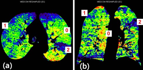
Readers Group A Group B_P_ Value CTPA images Reader 1 1.09 ± 0.05 1.06 ± 0.04 .655 Reader 2 1.06 ± 0.04 1.03 ± 0.03 .564 Kappa value 0.784 0.653 – Perfusion images Reader 1 2.64 ± 0.49 2.79 ± 0.42 .096 Reader 2 2.67 ± 0.48 2.79 ± 0.42 .157 Kappa value 0.933 0.819 –
CTPA, computed tomography pulmonary angiography.
Get Radiology Tree app to read full this article<
Dual-Energy Lung Perfusion Image Quality Evaluation
Get Radiology Tree app to read full this article<
Get Radiology Tree app to read full this article<
Detection of Pulmonary Emboli
Get Radiology Tree app to read full this article<
Table 3
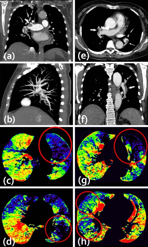
Pulmonary Emboli Detected by Two Readers
True Positive False Negative False Positive True Negative Group A Group B Group A Group B Group A Group B Group A Group B Patients Reader 1 17 19 1 0 2 1 13 13 Reader 2 17 17 1 2 1 0 14 14 Central PE Reader 1 21 22 1 0 4 2 271 273 Reader 2 21 20 2 2 2 1 272 274 Peripheral PE Reader 1 42 20 8 5 2 1 608 634 Reader 2 39 18 11 7 4 1 606 634
PE, pulmonary embolism.
The data for the central arteries refer to the number of affected main and lobar pulmonary arteries. The data for the peripheral arteries refer to the number of affected pulmonary segmental and subsegmental arteries.
Table 4
Diagnostic Accuracy Comparison of the Two CTPA Protocols
Sensitivity Specificity PPV NPV Accuracy_P_ Value Group A Group B Group A Group B Group A Group B Group A Group B Group A Group B Patient Reader 1 94.4
(74.2–99.0) 100
(83.2–100) 86.7
(62.1–96.3) 92.9
(68.5–98.7) 89.5
(76.4–99.1) 95.0
(76.4–99.1) 92.9
(68.5–98.7) 100
(77.2–100) 90.9
(76.4–96.9) 97.0
(84.7–99.5) .30 Reader 2 94.4
(74.2–99.0) 89.5
(68.6–97.1) 93.3
(70.2–98.8) 100
(78.5–100) 94.4
(74.2–99.0) 100
(81.6–100) 93.3
(70.2–98.8) 87.5
(64.0–96.5) 93.9
(80.4–98.3) 93.9
(80.4–98.3) 1 Central PE Reader 1 95.5
(78.2–99.2) 100
(85.1–100) 98.5
(96.3–99.4) 99.3
(97.4–99.8) 84.0
(65.4–93.6) 91.7
(74.2–97.7) 99.6
(98.0–99.9) 100
(98.6–100) 98.3
(96.1–99.3) 99.3
(97.6–99.8) .25 Reader 2 91.3
(73.2–97.6) 90.9
(72.2–97.5) 99.3
(97.4–99.8) 99.6
(98.0–99.9) 91.3
(73.2–97.6) 95.2
(77.3–99.2) 99.3
(97.4–99.8) 99.3
(97.4–99.8) 98.7
(96.6–99.5) 99.0
(97.1–99.7) .70 Peripheral PE Reader 1 84.0
(71.5–91.7) 80.0
(60.9–91.1) 99.7
(98.8–99.9) 99.8
(99.1–100) 95.5
(84.9–98.7) 95.2
(77.3–99.2) 98.7
(97.5–99.3) 99.2
(98.2–99.7) 98.5
(97.2–99.2) 99.1
(98.0–99.6) .31 Reader 2 78.0
(64.8–87.3) 72.0
(52.4–85.7) 99.3
(98.3–99.7) 99.8
(99.1–100) 90.7
(78.4–96.3) 94.7
(75.4–99.1) 98.2
(96.8–99.0) 98.9
(97.8–99.5) 97.7
(96.3–98.6) 98.8
(97.6–99.4) .14
NPV, negative predictive value; PE, pulmonary embolism; PPV, positive predictive value.
Data in parentheses indicate 95% confidence interval.
Get Radiology Tree app to read full this article<
Radiation Dose Comparison
Get Radiology Tree app to read full this article<
Table 5
Radiation Dose Estimation and Comparison in This Study
Parameters Group A Group B_P_ Value CTDIvol (mGy) 3.0 ± 0.7 5.6 ± 1.5 <.001 DLP (mGy⋅cm) 93.3 ± 25.6 173.5 ± 44.9 <.001 ED (mSv) 1.3 ± 0.4 2.4 ± 0.6 <.001
CTDIvol, volume CT dose index; DLP, dose-length product; ED, effective dose.
Get Radiology Tree app to read full this article<
Discussion
Get Radiology Tree app to read full this article<
Get Radiology Tree app to read full this article<
Get Radiology Tree app to read full this article<
Get Radiology Tree app to read full this article<
Get Radiology Tree app to read full this article<
Get Radiology Tree app to read full this article<
Get Radiology Tree app to read full this article<
Acknowledgment
Get Radiology Tree app to read full this article<
References
1. Doğan H., de Roos A., Geleijins J., et. al.: The role of computed tomography in the diagnosis of acute and chronic pulmonary embolism. Diagn Interv Radiol 2015; 21: pp. 307-316.
2. Meier A., Wurnig M., Desbiolles L., et. al.: Advanced virtual monoenergetic images: improving the contrast of dual-energy CT pulmonary angiography. Clin Radiol 2015; 70: pp. 1244-1251.
3. Zhang L.J., Zhang Z., Li S.J., et. al.: Pulmonary embolism and renal vein thrombosis in patients with nephrotic syndrome: prospective evaluation of prevalence and risk factors with CT. Radiology 2014; 273: pp. 897-906.
4. Mayo J., Thakur Y.: Pulmonary CT angiography as first-line imaging for PE: image quality and radiation dose considerations. AJR Am J Roentgenol 2014; 200: pp. 522-528.
5. Zhang L.J., Zhao Y.E., Wu S.Y., et. al.: Pulmonary embolism detection with dual-energy CT: experimental study of dual-source CT in rabbits. Radiology 2009; 252: pp. 61-70.
6. Bettmann M.A., Baginski S.G., White R.D., et. al.: ACR Appropriateness Criteria® acute chest pain–suspected pulmonary embolism. J Thorac Imaging 2012; 27: pp. W28-W31.
7. Bauer R.W., Kramer S., Renker M., et. al.: Dose and image quality at CT pulmonary angiography-comparison of first and second generation dual-energy CT and 64-slice CT. Eur Radiol 2011; 21: pp. 2139-2147.
8. Zhang L.J., Chai X., Wu S.Y., et. al.: Detection of pulmonary embolism by dual energy CT: correlation with perfusion scintigraphy and histopathological findings in rabbits. Eur Radiol 2009; 19: pp. 2844-2854.
9. Lu G.M., Zhao Y., Zhang L.J., et. al.: Dual-energy CT of the lung. AJR Am J Roentgenol 2012; 199: pp. S40-S53.
10. Wildberger J.E., Schoepf U.J., Mahnken A.H., et. al.: Approaches to CT perfusion imaging in pulmonary embolism. Semin Roentgenol 2005; 40: pp. 64-73.
11. Apfaltrer P., Bachmann V., Meyer M., et. al.: Prognostic value of perfusion defect volume at dual energy CTA in patients with pulmonary embolism: correlation with CTA obstruction scores, CT parameters of right ventricular dysfunction and adverse clinical outcome. Eur J Radiol 2012; 81: pp. 3592-3597.
12. Lu G.M., Wu S.Y., Yeh B.M., et. al.: Dual-energy computed tomography in pulmonary embolism. Br J Radiol 2010; 83: pp. 707-718.
13. Thieme S.F., Johnson T.R., Reiser M.F., et. al.: Dual-energy lung perfusion computed tomography: a novel pulmonary functional imaging method. Semin Ultrasound CT MR 2010; 31: pp. 301-308.
14. Albrecht M.H., Trommer J., Wichmann J.L., et. al.: Comprehensive comparison of virtual monoenergetic and linearly blended reconstruction techniques in third-generation dual-source dual-energy computed tomography angiography of the thorax and abdomen. Invest Radiol 2016; 51: pp. 582-590.
15. Li Q., Yu H., Zhang L., et. al.: Combining low tube voltage and iterative reconstruction for contrast-enhanced CT imaging of the chest-initial clinical experience. Clin Radiol 2013; 68: pp. e249-e253.
16. Hou D.J., Tso D.K., Davison C., et. al.: Clinical utility of ultra high pitch dual source thoracic CT imaging of acute pulmonary embolism in the emergency department: are we one step closer towards a non-gated triple rule out?. Eur J Radiol 2013; 82: pp. 1793-1798.
17. Bauer R.W., Frellesen C., Renker M., et. al.: Dual energy CT pulmonary blood volume assessment in acute pulmonary embolism—correlation with D-dimer level, right heart strain and clinical outcome. Eur Radiol 2011; 21: pp. 1914-1921.
18. Chae E.J., Seo J.B., Jang Y.M., et. al.: Dual-energy CT for assessment of the severity of acute pulmonary embolism: pulmonary perfusion defect score compared with CT angiographic obstruction score and right ventricular/left ventricular diameter ratio. AJR Am J Roentgenol 2010; 194: pp. 604-610.
19. Lu G.M., Luo S., Meinel F.G., et. al.: High-pitch computed tomography pulmonary angiography with iterative reconstruction at 80 kVp and 20 mL contrast agent volume. Eur Radiol 2014; 24: pp. 3260-3268.
20. Szucs-Farkas Z., Schaller C., Bensler S., et. al.: Detection of pulmonary emboli with CT angiography at reduced radiation exposure and contrast material volume: comparison of 80 kVp and 120 kVp protocols in a matched cohort. Invest Radiol 2009; 44: pp. 793-799.
21. Co S.J., Mayo J., Liang T., et. al.: Iterative reconstructed ultra high pitch CT pulmonary angiography with cardiac bowtie-shaped filter in the acute setting: effect on dose and image quality. Eur J Radiol 2013; 82: pp. 1571-1576.
22. Cohen J.: A coefficient of agreement for nominal scales. Educ Psychol Meas 1960; 20: pp. 37-46.
23. Sodickson A.: Strategies for reducing radiation exposure in multi-detector row CT. Radiol Clin North Am 2012; 50: pp. 1-14.
24. Schueller-Weidekamm C., Schaefer-Prokop C.M., Weber M., et. al.: CT angiography of pulmonary arteries to detect pulmonary embolism: improvement of vascular enhancement with low kilovoltage settings. Radiology 2006; 241: pp. 899-907.
25. Gunn M.L., Kohr J.R.: State of the art: technologies for computed tomography dose reduction. Emerg Radiol 2010; 17: pp. 209-218.
26. Szucs-Farkas Z., Christe A., Megyeri B., et. al.: Diagnostic accuracy of computed tomography pulmonary angiography with reduced radiation and contrast material dose: a prospective randomized clinical trial. Invest Radiol 2014; 49: pp. 201-208.
27. Viteri-Ramírez G., García-Lallana A., Simón-Yarza I., et. al.: Low radiation and low-contrast dose pulmonary CT angiography: comparison of 80 kVp/60 ml and 100 kVp/80 ml protocols. Clin Radiol 2012; 67: pp. 833-839.
28. Faggioni L., Neri E., Sbragia P., et. al.: 80-kV Pulmonary CT angiography with 40 mL of iodinated contrast material in lean patients: comparison of vascular enhancement with iodixanol (320 mg I/mL) and iomeprol (400 mg I/mL). AJR Am J Roentgenol 2012; 199: pp. 1220-1225.
29. Wang G., Gao J., Zhao S., et. al.: Achieving consistent image quality and overall radiation dose reduction for coronary CT angiography with body mass index-dependent tube voltage and tube current selection. Clin Radiol 2014; 69: pp. 945-951.
30. Kaza R.K., Platt J.F., Goodsitt M.M., et. al.: Emerging techniques for dose optimization in abdominal CT. Radiographics 2014; 34: pp. 4-17.
31. Waaijer A., Prokop M., Velthuis B.K., et. al.: Circle of Willis at CT angiography: dose reduction and image quality–reducing tube voltage and increasing tube current settings. Radiology 2007; 242: pp. 832-839.
32. Qi L., Meinel F.G., Zhou C.S., et. al.: Image quality and radiation dose of lower extremity CT angiography using 70 kVp, high pitch acquisition and sinogram-affirmed iterative reconstruction. PLoS ONE 2014; 9: pp. e99112.
33. Hwang H.J., Seo J.B., Lee H.J., et. al.: Low-dose chest computed tomography with sinogram-affirmed iterative reconstruction, iterative reconstruction in image space, and filtered back projection: studies on image quality. J Comput Assist Tomogr 2013; 37: pp. 610-617.
34. Li X., Ni Q.Q., Schoepf U.J., et. al.: 70-kVp High-pitch computed tomography pulmonary angiography with 40 mL contrast agent: initial experience. Acad Radiol 2015; 22: pp. 1562-1570.
35. De Zordo T., von Lutterotti K., Dejaco C., et. al.: Comparison of image quality and radiation dose of different pulmonary CTA protocols on a 128-slice CT: high-pitch dual source CT, dual energy CT and conventional spiral CT. Eur Radiol 2012; 22: pp. 279-286.