
Rationale and Objectives
Three-dimensional image reconstruction by volume rendering and rapid prototyping has made it possible to visualize anatomic structures in three dimensions for interventional planning and academic research.
Methods
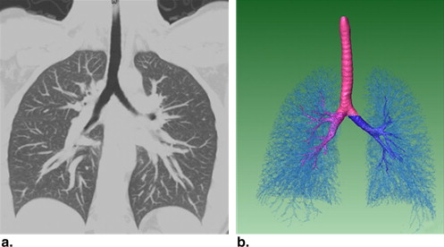
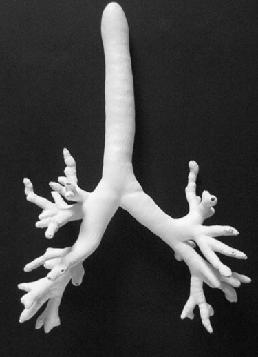
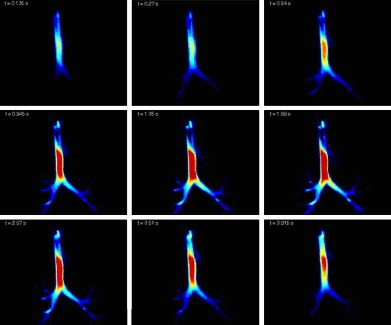
Volumetric chest computed tomography was performed on a healthy volunteer. Computed tomographic images of the larger bronchial branches were segmented by an extended three-dimensional region-growing algorithm, converted into a stereolithography file, and used for computer-aided design on a laser sintering machine. The injection of gases for respiratory flow modeling and measurements using magnetic resonance imaging were done on a hollow cast.
Results
Manufacturing the rapid prototype took about 40 minutes and included the airway tree from trackea to segmental bronchi (fifth generation). The branching of the airways are clearly visible in the 3 He images, and the radial imaging has the potential to elucidate the airway dimensions.
Conclusion
The results for flow patterns in the human bronchial tree using the rapid-prototype model with hyperpolarized helium-3 magnetic resonance imaging show the value of this model for flow phantom studies.
Radiologic imaging provides a noninvasive modality to visualize human organ structures. With the revolutionary technological developments of the past decade, radiology has grown beyond the visualization of two-dimensional structures. Multislice helical computed tomography can handle up to 320 slices within <1 second, recording volumetric data at submillimeter spatial resolution, which has improved quality in diagnostic radiology. Three-dimensional image reconstruction by volume rendering has made it possible to visualize anatomic structures in three dimensions for interventional preoperative planning and procedures. The recently introduced method of rapid prototyping is one modality with a promising future in academic and industrial research.
Rapid prototyping can be used to produce models of living organs from high-resolution in vivo images representing the actual structure in three dimensions. Humans are considered to be the most evolved and complicated organisms, yet we are still uncertain about many human physiologic processes, because in vitro models are used to mimic in vivo processes.
Get Radiology Tree app to read full this article<
Materials and methods
Patient and Imaging
Get Radiology Tree app to read full this article<
Rapid Prototyping
Get Radiology Tree app to read full this article<
Helium-3 MRI
Get Radiology Tree app to read full this article<
Get Radiology Tree app to read full this article<
Results
Get Radiology Tree app to read full this article<
Get Radiology Tree app to read full this article<
Get Radiology Tree app to read full this article<
Get Radiology Tree app to read full this article<
Get Radiology Tree app to read full this article<
Discussion
Get Radiology Tree app to read full this article<
Get Radiology Tree app to read full this article<
Get Radiology Tree app to read full this article<
Get Radiology Tree app to read full this article<
Acknowledgment
Get Radiology Tree app to read full this article<
References
1. Kim M.S., Hansgen A.R., Wink O., et. al.: Rapid prototyping: a new tool in understanding and treating structural heart disease. Circulation 2008; 117: pp. 2388-2394.
2. De Zanche N., Chhina N., Teh K., Randell C., Pruessmann K.P., Wild J.M.: Asymmetric quadrature split birdcage coil for hyperpolarized (3)He lung MRI at 1.5T. Magn Reson Med 2008; 60: pp. 431-438.
3. Wild J.M., Paley M.N.J., Kasuboski L., Swift A., Fichele S., Woodhouse N., van Beek E.J.R.: Dynamic radial projection MRI of inhaled hyperpolarised 3He. Magn Reson Med 2003; 49: pp. 991-997.
4. Lewis T.A., Tzeng Y.S., McKinstry E.L., et. al.: Quantification of airway diameters and 3D airway tree rendering from dynamic hyperpolarized 3He magnetic resonance imaging. Magn Reson Med 2005; 53: pp. 474-478.
5. Tschirren J., McLennan G., Palagyi K., Hoffman E.A., Sonka M.: Matching and anatomical labelling of human airway tree. IEEE Trans Med Imaging 2005; 24: pp. 1540-1547.
6. Tzeng Y.S., Hoffman E.A., Cook-Granroth J., et. al.: Comparison of airway diameter measurements from an anthropomorphic airway tree phantom using hyperpolarized 3He MRI and high-resolution computed tomography. Magn Reson Med 2007; 58: pp. 636-642.
7. de Rochefort L., Vial L., Fodil R., et. al.: In vitro validation of computational fluid dynamic simulation in human proximal airways with hyperpolarized 3He magnetic resonance phase-contrast velocimetry. J Appl Physiol 2007; 102: pp. 2012-2023.
8. Brown N.J., Salome C.M., Berend N., Thorpe C.W., King G.G.: Airway distensibility in adults with asthma and healthy adults, measured by forced oscillation technique. Am J Respir Crit Care Med 2007; 176: pp. 129-137.