
Rationale and Objectives
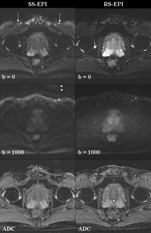
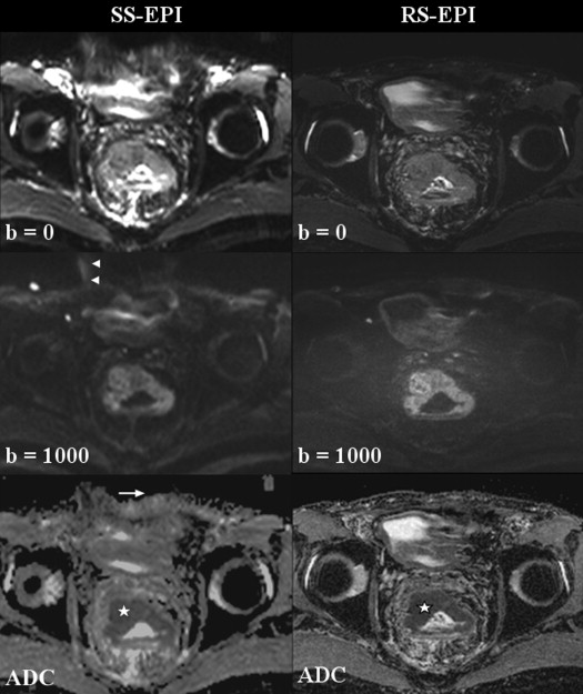
Diffusion-weighted imaging (DWI) of the pelvis at 3T is prone to artifacts that diminish the image quality. Readout-segmented echo-planar imaging (RS-EPI) is a new DWI technique that can reduce the artifacts associated with standard single-shot echo-planar imaging (SS-EPI) DWI. The purpose of this study was to evaluate the feasibility and image quality of RS-EPI in pelvic DWI compared to SS-EPI on a 3T imaging system.
Materials and Methods
Thirty patients underwent pelvic DWI on a 3T scanner with SS-EPI and RS-EPI techniques. Two blinded readers independently assessed each set of images for geometric distortion, image blurring, ghosting artifacts, lesion conspicuity, and overall image quality on a 7-point scale. Qualitative image scores were compared using paired Wilcoxon signed rank test. Interreader correlation was assessed by Spearman rank correlation.
Results
Geometric distortion, imaging blurring, ghosting artifacts, lesion conspicuity, and overall image quality were rated significantly better by both readers for RS-EPI technique ( P < .01 for all parameters). There was moderate–high correlation between the readers ( r = 0.649–0.752) for all parameters apart from lesion conspicuity ( r = 0.351). Both readers preferred the RS-EPI set of DWI images in most of the cases (reader 1: 0.87, 95% CI 0.74–0.99; reader 2: 0.77, 95% CI 0.61–0.93). Mean difference and limits of agreement between apparent diffusion coefficient (ADC) values obtained from the two methods were 0.01 (−0.08, 0.10) × 10 −3 mm 2 /s.
Conclusions
RS-EPI DWI images showed improved image quality compared to SS-EPI technique at 3T. RS-EPI is a feasible technique in the pelvis for producing high-resolution DWI.
Diffusion-weighted imaging (DWI) has emerged in the past decade as an important functional imaging technique in extracranial oncologic imaging. In the pelvis, DWI has been applied to imaging of rectal, prostate, endometrial, and cervical cancers . DWI provides an excellent contrast mechanism that is useful for tumor detection and delineating disease extent.
Currently the most widely used sequence for clinical DWI in the pelvis is single-shot echo-planar imaging (SS-EPI), whereby all the lines in k-space are filled by multiple gradient reversals in a single acquisition after a single radiofrequency pulse. However, the SS-EPI technique suffers from significant artifacts that reduce the image quality of DWI. It is vulnerable to T2*-induced blurring and geometric distortions due to magnetic field inhomogeneities that cause accumulation of phase errors . In the pelvis, the presence of gas-containing viscera, such as the rectum, small and large bowel, and vagina, frequently exacerbates these artifacts. Distortion and blurring artifacts worsen with higher field strength and higher resolution, limiting the achievable resolution with SS-EPI before these effects become prohibitive . The functional image quality and resolution achieved with DWI thus presently lags considerably behind the high-resolution T2-weighted sequences used for morphologic assessment and anatomic correlation in the pelvis.
Get Radiology Tree app to read full this article<
Get Radiology Tree app to read full this article<
Materials and methods
Get Radiology Tree app to read full this article<
MR Imaging
Get Radiology Tree app to read full this article<
Table 1
Sequence Parameters for Single-shot and Readout-segmented Echo-planar Imaging
Sequence Parameter Single-shot Echo-planar Imaging Readout-segmented Echo-planar Imaging Diffusion directions Three-direction trace Three-direction trace Diffusion encoding scheme Monopolar, Stejskal-Tanner Monopolar, Stejskal-Tanner b-value (s/mm 2 ) 0, 100, 1000 0, 1000 Fat suppression Inversion recovery, gradient reversal Inversion recovery, gradient reversal TR (millisecond) 5300–6900 7800–8200 TE (millisecond) 62–79 (minimum) 63–80 (minimum) Field of view (mm) ∗ 260 240 Matrix ∗ 160 × 120 192 × 164 Number of sections 30–34 30–34 Section thickness (mm) 4 4 Number of readout segments 1 7 Number of signals acquired 6 1 Parallel imaging GRAPPA GRAPPA Acceleration factor 2 2 Acquisition time (minute:second) 3:40 4:17
GRAPPA, generalized autocalibrating partially parallel acquisition; TE, echo time; TR, repetition time.
Get Radiology Tree app to read full this article<
Get Radiology Tree app to read full this article<
Get Radiology Tree app to read full this article<
Image Assessment
Get Radiology Tree app to read full this article<
Get Radiology Tree app to read full this article<
Get Radiology Tree app to read full this article<
Statistical Analysis
Get Radiology Tree app to read full this article<
Results
Get Radiology Tree app to read full this article<
Qualitative Image Assessment
Get Radiology Tree app to read full this article<
Get Radiology Tree app to read full this article<
Get Radiology Tree app to read full this article<
Get Radiology Tree app to read full this article<
Table 2
Comparison of Qualitative Scores of DWIs Using SS-EPI Technique and RS-EPI Technique
Parameter Reader 1 (Mean Score ± SD)P † Reader 2 (Mean Score ± SD)P † Spearman Correlation ( r__s ) ‡ SS-EPI RS-EPI SS-EPI RS-EPI Geometric distortion 3.77 ± 0.57 5.93 ± 0.25 <.01 ∗ 3.90 ± 0.55 5.13 ± 0.68 <.001 ∗ 0.752 ( P < .001) ∗ Image blurring 3.93 ± 0.37 6.00 ± 0.26 <.001 ∗ 4.00 ± 0.26 4.93 ± 0.83 <.001 ∗ 0.649 ( P < .001) ∗ Ghosting artifacts 3.87 ± 0.78 5.60 ± 0.62 <.001 ∗ 3.93 ± 0.64 5.13 ± 0.73 <.001 ∗ 0.711 ( P < .001) ∗ Lesion conspicuity 4.58 ± 1.26 5.74 ± 1.24 .002 ∗ 4.05 ± 0.40 5.05 ± 0.91 .004 ∗ 0.351 ( P = 1) Overall image quality 3.97 ± 0.41 5.83 ± 0.38 <.001 ∗ 4.10 ± 0.31 5.10 ± 0.61 <.001 ∗ 0.737 ( P < .001) ∗
DWI, diffusion-weighted imaging; RS-EPI, readout-segmented echo-planar imaging; SD, standard deviation; SS-EPI, single-shot echo-planar imaging.
Get Radiology Tree app to read full this article<
Get Radiology Tree app to read full this article<
Get Radiology Tree app to read full this article<
Get Radiology Tree app to read full this article<
ADC and SNR Analysis
Get Radiology Tree app to read full this article<
![Figure 3, Bland–Altman plot shows good agreement of ADC measurements between SS-EPI and RS-EPI techniques. The mean ADC difference between both techniques and the limits of agreement (±1.96 times the standard deviation [SD]) are displayed. ADC, apparent diffusion coefficient; RS-EPI, readout-segmented echo-planar imaging; SS-EPI, single-shot echo-planar imaging.](https://storage.googleapis.com/dl.dentistrykey.com/clinical/ReadoutsegmentedEchoplanarImagingforDiffusionweightedImaginginthePelvisat3TAFeasibilityStudy/2_1s20S1076633214000087.jpg)
Get Radiology Tree app to read full this article<
Get Radiology Tree app to read full this article<
Discussion
Get Radiology Tree app to read full this article<
Get Radiology Tree app to read full this article<
Get Radiology Tree app to read full this article<
Get Radiology Tree app to read full this article<
Get Radiology Tree app to read full this article<
Get Radiology Tree app to read full this article<
Get Radiology Tree app to read full this article<
Get Radiology Tree app to read full this article<
Get Radiology Tree app to read full this article<
Get Radiology Tree app to read full this article<
Get Radiology Tree app to read full this article<
References
1. Lambregts D.M., Cappendijk V.C., Maas M., et. al.: Value of MRI and diffusion-weighted MRI for the diagnosis of locally recurrent rectal cancer. Eur Radiol 2011; 21: pp. 1250-1258.
2. Park M.J., Kim S.H., Lee S.J., et. al.: Locally advanced rectal cancer: added value of diffusion-weighted MR imaging for predicting tumor clearance of the mesorectal fascia after neoadjuvant chemotherapy and radiation therapy. Radiology 2011; 260: pp. 771-780.
3. Haider M.A., van der Kwast T.H., Tanguay J., et. al.: Combined T2-weighted and diffusion-weighted MRI for localization of prostate cancer. AJR Am J Roentgenol 2007; 189: pp. 323-328.
4. Barentsz J.O., Richenberg J., Clements R., et. al.: ESUR prostate MR guidelines 2012. Eur Radiol 2012; 22: pp. 746-757.
5. Tamai K., Koyama T., Saga T., et. al.: Diffusion-weighted MR imaging of uterine endometrial cancer. J Magn Reson Imaging 2007; 26: pp. 682-687.
6. Kuang F., Ren J., Zhong Q., et. al.: The value of apparent diffusion coefficient in the assessment of cervical cancer. Eur Radiol 2013; 23: pp. 1050-1058.
7. Tsao J.: Ultrafast imaging: principles, pitfalls, solutions, and applications. J Magn Reson Imaging 2010; 32: pp. 252-266.
8. Le Bihan D., Poupon C., Amadon A., et. al.: Artifacts and pitfalls in diffusion MRI. J Magn Reson Imaging 2006; 24: pp. 478-488.
9. Porter D.A., Heidemann R.M.: High resolution diffusion-weighted imaging using readout-segmented echo-planar imaging, parallel imaging and a two-dimensional navigator-based reacquisition. Magn Reson Med 2009; 62: pp. 468-475.
10. Holdsworth S.J., Yeom K., Skare S., et. al.: Clinical application of readout-segmented- echo-planar imaging for diffusion-weighted imaging in pediatric brain. AJNR Am J Neuroradiol 2011; 32: pp. 1274-1279.
11. Yeom K.W., Holdsworth S.J., Van A.T., et. al.: Comparison of readout-segmented echo-planar imaging (EPI) and single-shot EPI in clinical application of diffusion-weighted imaging of the pediatric brain. AJR Am J Roentgenol 2013; 200: pp. W437-W443.
12. Morelli J., Porter D., Ai F., et. al.: Clinical evaluation of single-shot and readout-segmented diffusion-weighted imaging in stroke patients at 3 T. Acta Radiol 2013; 54: pp. 299-306.
13. Heverhagen J.T.: Noise measurement and estimation in MR imaging experiments. Radiology 2007; 245: pp. 638-639.
14. Kaufman L., Kramer D.M., Crooks L.E., et. al.: Measuring signal-to-noise ratios in MR imaging. Radiology 1989; 173: pp. 265-267.
15. Bogner W., Pinker-Domenig K., Bickel H., et. al.: Readout-segmented echo-planar imaging improves the diagnostic performance of diffusion-weighted MR breast examinations at 3.0 T. Radiology 2012; 263: pp. 64-76.
16. Rumpel H., Chong Y., Porter D.A., et. al.: Benign versus metastatic vertebral compression fractures: combined diffusion-weighted MRI and MR spectroscopy aids differentiation. Eur Radiol 2013; 23: pp. 541-550.
17. Liu Y., Bai R., Sun H., et. al.: Diffusion-weighted imaging in predicting and monitoring the response of uterine cervical cancer to combined chemoradiation. Clin Radiol 2009; 64: pp. 1067-1074.
18. Lim H.K., Kim J.K., Kim K.A., et. al.: Prostate cancer: apparent diffusion coefficient map with T2-weighted images for detection–a multireader study. Radiology 2009; 250: pp. 145-151.
19. Hunsche S., Moseley M.E., Stoeter P., et. al.: Diffusion-tensor MR imaging at 1.5 and 3.0 T: initial observations. Radiology 2001; 221: pp. 550-556.
20. Kuhl C.K., Textor J., Gieseke J., et. al.: Acute and subacute ischemic stroke at high-field-strength (3.0-T) diffusion-weighted MR imaging: intraindividual comparative study. Radiology 2005; 234: pp. 509-516.
21. Mazaheri Y., Vargas H.A., Nyman G., et. al.: Image artifacts on prostate diffusion-weighted magnetic resonance imaging: trade-offs at 1.5 Tesla and 3.0 Tesla. Acad Radiol 2013 Aug; 20: pp. 1041-1047.
22. Rosenkrantz A.B., Oei M., Babb J.S., et. al.: Diffusion-weighted imaging of the abdomen at 3.0 Tesla: image quality and apparent diffusion coefficient reproducibility compared with 1.5 Tesla. J Magn Reson Imaging 2010; 33: pp. 128-135.
23. Sasaki M., Yamada K., Watanabe Y., et. al.: Variability in absolute apparent diffusion coefficient values across different platforms may be substantial: a multivendor, multi-institutional comparison study. Radiology 2008; 249: pp. 624-630.
24. Girometti R., Furlan A., Esposito G., et. al.: Relevance of b-values in evaluating liver fibrosis: a study in healthy and cirrhotic subjects using two single-shot spin-echo echo-planar diffusion-weighted sequences. J Magn Reson Imaging 2008; 28: pp. 411-419.
25. Kwee T.C., Takahara T., Koh D.M., et. al.: Comparison and reproducibility of ADC measurements in breathhold, respiratory triggered, and free-breathing diffusion-weighted MR imaging of the liver. J Magn Reson Imaging 2008; 28: pp. 1141-1148.
26. Padhani A.R., Liu G., Koh D.M., et. al.: Diffusion-weighted magnetic resonance imaging as a cancer biomarker: consensus and recommendations. Neoplasia 2009; 11: pp. 102-125.
27. Holdsworth S.J., Skare S., Newbould R.D., et. al.: Readout-segmented EPI for rapid high resolution diffusion imaging at 3 T. Eur J Radiol 2008; 65: pp. 36-46.
28. Robson P.M., Grant A.K., Madhuranthakam A.J., et. al.: Comprehensive quantification of signal-to-noise ratio and g-factor for image-based and k-space-based parallel imaging reconstructions. Magn Reson Med 2008; 60: pp. 895-907.