
Rationale and Objectives
The recent increasing utilization of imaging has increased the population exposure to ionizing radiation. With increasing knowledge of the potential harm of radiation exposure, efforts should be made to minimize patient radiation whenever possible, especially in young children. The purpose of this study was to use the exposure index (EI) standard to assess the potential for reducing radiation dose to babies by removing a soft comfort pad, often placed underneath the baby. The pad is located between the baby and the image detector plate. As such it absorbs x-rays that have already passed through the baby but have not yet reached the imaging detector plate.
Materials and Methods
Using a thoracic infant phantom and fixed exposure factors, we measured the percentage of the radiation exiting a neonatal chest phantom that was absorbed/attenuated by the comfort pad, before it hit the detector to create the image. We studied comfort pads of 4 different thicknesses, ranging from 0.5″ to 8″.
Results
Radiation beam attenuation, ranging from 12% to 72.1%, was found with all comfort pads, with increased x-ray beam attenuation occurring with increasing pad thickness.
Conclusions
The study shows that comfort pads cause a high attenuation of the radiation beam, after it exits the chest phantom. As such, removal of the pads prior to radiographic exposure of babies is a method of potentially reducing patient radiation exposure in the newborn nursery.
Campaigns such as the “Image Gently Campaign” have led to increasing recent discussion of radiation safety, with special considerations given to pediatric patients . As children live longer than adults, it is thought that they have an increased risk of developing long-term complications from radiation, such as cancer . The newborn intensive care nursery is a most important location for radiation exposure monitoring as it contains our youngest patients, many of whom may require serial imaging at high frequency.
After passing through the patient the x-ray beam hits an image detector plate, which converts the energy in the x-ray beam into a digital image. Measuring the radiation dose at the detector plate is an indirect method of measuring the radiation that has been used. Older methods of determining the radiation dose at the image detector plate varied by manufacturer of the image equipment . To avoid the confusion of this variability, a new universal standard has been agreed upon and the new term for describing the radiation dose, measured at the level of the detector plate, is called the exposure index . This exposure index essentially provides the ability to monitor patient’s radiation exposure. The exposure index increases in a linear manner as the mAs is increased. Doubling the mAs will double the exposure index . The response to change in kVp is not linear.
Get Radiology Tree app to read full this article<
Get Radiology Tree app to read full this article<
Methods
Get Radiology Tree app to read full this article<
Get Radiology Tree app to read full this article<
Get Radiology Tree app to read full this article<
Get Radiology Tree app to read full this article<
Get Radiology Tree app to read full this article<
Percent exposure index reduction=exposure index without pad−exposure index with padexposure index without pad Percent exposure index reduction
=
exposure index without pad
−
exposure index with pad
exposure index without pad
Get Radiology Tree app to read full this article<
Get Radiology Tree app to read full this article<
Results
Get Radiology Tree app to read full this article<
Table 1
Exposure Index Reduction Relationship to Pad Thickness at Varying kVp
Kvp Pad Thickness in Inches Exposure Index without Pad Exposure Index with Pad Percent Reduction in Exposure Index with Pad in Place 56
p 0.0475 0.5″ 203 190 6.4 1.0′ 228 183 19.7 3.0″ 235 116 50.6 8.0″ 235 60 74.5 66
p 0.0277 0.5″ 499 439 12 1.0′ 503 367 27 3.0″ 484 280 42.4 8.0″ 484 135 72.1 76
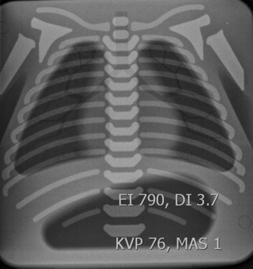
p 0.0415 0.5″ 790 714 9.6 1.0′ 793 661 16.6 3.0″ 790 444 43.7 8.0″ 790 245 69
p values <0.05 confirm a significant difference in the exposure indices without and with the pad for all pad thicknesses at each kVp.
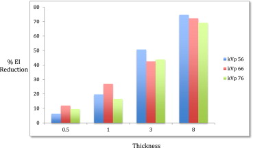
Table 2
Standard Deviation of Percent Exposure Index Reduction for Each Pad Thickness Group at Constant mAs is Maximum 5.3%
0.5′′ Thick 1′′ Thick 3′′ Thick 8′′ Thick kVp 56 6.4 19.7 50.6 74.5 kVp 66 12 27 42.4 72.1 kVp 76 9.6 16.6 43.7 69 SD 2.8 5.3 4.4 2.8
Get Radiology Tree app to read full this article<
Discussion
Get Radiology Tree app to read full this article<
Get Radiology Tree app to read full this article<
Get Radiology Tree app to read full this article<
Get Radiology Tree app to read full this article<
Get Radiology Tree app to read full this article<
Get Radiology Tree app to read full this article<
Get Radiology Tree app to read full this article<
Conclusions
Get Radiology Tree app to read full this article<
Get Radiology Tree app to read full this article<
References
1. Goske M.J., Applegate K.E., Boylan J., et. al.: The Image Gently Campaign: working together to change practice. AJR Am J Roentgenol 2008; 190: pp. 273-274.
2. Uffmann M., Schaefer-Prokop C.: Digital radiography: the balance between image quality and required radiation dose. Eur J Radiol 2009; 72: pp. 202-208.
3. Don S.: Radiosensitivity in children: potential for overexposure in CR and DR and magnitude of doses in ordinary radiographic examinations. Pediatr Radiol 2004; 34: pp. S167-S172. discussion S234–S231
4. Siebert J.A.: Tradeoffs between image quality and dose. Pediatr Radiol 2004; 34: pp. S183-S195.
5. Schaetzing R.: Management of pediatric radiation dose using Agfa computed radiography. Pediatr Radiol 2004; 34: pp. S234-S241.
6. Cohen M.D., Cooper M.L., Piersall K., et. al.: Quality assurance: using the exposure index and the deviation index to monitor radiation exposure for portable chest radiographs in neonates. Pediatr Radiol 2011; 41: pp. 592-601.
7. Shepard S.J., Wang J., Flynn M., et. al.: An exposure indicator for digital radiography: AAPM Task Group 116 (executive summary). Med Phys 2009; 36: pp. 2898-2914.
8. International Electrotechnical Commission (2008) Medical electrical equipment –exposure index of digital x-ray imaging systems –part 1: definitions and requirements for general radiography. IEC 62494 -1:5.
9. Sundance Enterprises Inc. White Planes, New York. Web reference. http://sundancesolutions.com/neonatal/ .