
Rationale and Objectives
Cardiac computed tomographic (CT) scans for the assessment of coronary calcium scores include approximately 70% of the lung volume and may be useful for the quantitative assessment of emphysema. The reproducibility of lung density measures from cardiac computed tomography and their validity compared to lung density measures from full-lung scans is unknown.
Materials and Methods
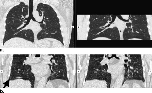
The Multi-Ethnic Study of Atherosclerosis (MESA) performed paired cardiac CT scans for 6814 participants at baseline and at follow-up. The MESA-Lung Study assessed lung density measures in the lung fields of these cardiac scans, counting voxels below −910 HU as moderate-to-severe emphysema-like lung regions. We evaluated: 1) the reproducibility of lung density measures among 120 randomly selected participants; 2) the comparability of measures acquired on electron beam CT (EBCT) and multidetector CT (MDCT) scanners among 10 participants; and 3) the validity of these measures compared to full-lung scans among 42 participants. Limits of agreement were determined using Bland-Altman approaches.
Results
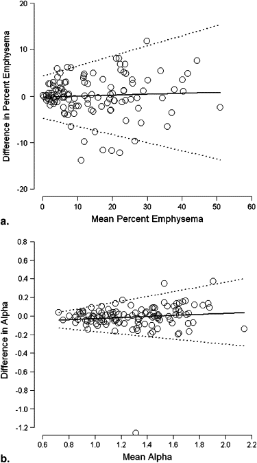
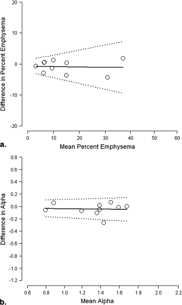
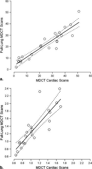
Percent emphysema measures from paired cardiac scans were highly correlated ( r = 0.92–0.95) with mean difference of −0.05% (95% limits of agreement: −8.3, 8.4%). Measures from EBCT and MDCT scanners were comparable (mean difference −0.9%; 95% limits of agreement: −5.1, 3.3%). Percent emphysema measures from MDCT cardiac and MDCT full-lung scans were highly correlated ( r = 0.93) and demonstrated reasonable agreement (mean difference 2.2%; 95% limits of agreement: −9.2, 13.8%).
Conclusions
Although full-lung imaging is preferred for the quantification of emphysema, the lung imaging from paired cardiac computed tomography provided a reproducible and valid quantitative assessment of emphysema in a population-based sample.
Chronic obstructive pulmonary disease (COPD) is the fourth leading cause of death in the United States. Morbidity and mortality from COPD continue to rise—in contrast to declines in mortality from cardiovascular disease, cancer, and stroke . COPD is defined by airflow obstruction on post-bronchodilator spirometry testing; however, millions of Americans have symptomatic COPD that is undiagnosed because of difficulties with access, testing, and interpretation of spirometry . Emphysema is defined by a loss of lung tissue in the absence of fibrosis and overlaps considerably with COPD. Emphysema can be quantitated on full-lung computed tomographic (CT) scans by various measures of CT lung density , which provide supplemental information to spirometry .
Cardiac CT scans for the assessment of coronary calcium scores can be obtained rapidly with low radiation exposure . Coronary calcium scores strongly predict near-term risk of coronary heart disease independent of established risk factors . Cardiac CT scans might provide an opportunity for the assessment of emphysema because they image about 70% of the lung volume (approximately the carina to T11). Cardiac CT scans differ, however, from full-lung CT scans that are performed for lung density measurements not just by the volume of the lung imaged but also by coaching of the patient to full inspiration, cardiac gating, and the image acquisition protocol itself. It is therefore uncertain if lung density measures from cardiac scans are reproducible and valid measures of full-lung emphysema.
Get Radiology Tree app to read full this article<
Materials and methods
Study Sample
Get Radiology Tree app to read full this article<
Get Radiology Tree app to read full this article<
Get Radiology Tree app to read full this article<
Get Radiology Tree app to read full this article<
CT Scanning Protocols
MESA Cardiac CT Protocol
Get Radiology Tree app to read full this article<
Table 1
Computed Tomography Protocols of Coronary Calcium and Full-lung Scans in the Multi-Ethnic Study of Atherosclerosis
Scanner Scan Mode mA/mAs ∗ kV Rotation Time (s) Scan Aperture (s) Slice Thickness (mm) Slice Spacing (mm) Kernel Cardiac scans (ECG-gated) † GE Light Speed QXi Axial 200 mA 120 0.800 0.52 2.5 2.5 Standard GE Light Speed Plus Axial 320 mA 120 0.500 0.33 2.5 2.5 Standard Siemens Volume Zoom Axial 139 mA/50 mAs 120 0.361 0.36 2.5 2.5 Standard Imatron C-150 Axial 630 mA 120 0.100 0.10 2.5 2.5 Sharp Siemens Sensation 64 (comparability study) Axial 50 mAs 120 0.220 0.22 3 3 B30f GE Light Speed Pro 16 (validation study) Axial 320 mA 120 0.500 0.33 2.5 2.5 Standard Full lung scans (non-gated) GE Light Speed Pro 16 Spiral 75 mAs 120 0.500 0.5 0.75 1.8 Standard/lung Siemens Sensation 64 Spiral 50 mAs 120 0.500 0.5 0.75 1.8 B30/B50
Get Radiology Tree app to read full this article<
Get Radiology Tree app to read full this article<
Get Radiology Tree app to read full this article<
Full-lung Chest CT Protocol
Get Radiology Tree app to read full this article<
Lung Density Measures
Get Radiology Tree app to read full this article<
Get Radiology Tree app to read full this article<
Get Radiology Tree app to read full this article<
Get Radiology Tree app to read full this article<
Additional Measures
Get Radiology Tree app to read full this article<
Statistical Analysis
Get Radiology Tree app to read full this article<
Get Radiology Tree app to read full this article<
Results
Get Radiology Tree app to read full this article<
Table 2
Characteristics of Patients Included in the Reproducibility Study of Cardiac CT Scans, Comparability Study of EBCT versus MDCT Cardiac CT Scans, and Validation Study of Cardiac versus Full-lung CT Scans
Reproducibility Study ( n = 119) Comparability Study ( n = 10) Validation Study ( n = 42) Age, y, mean ± SD 58.9 ± 9.1 63.8 ± 9.9 62.3 ± 10.7 Male, % 42.0 60 57.1 Race/ethnicity, % Caucasian 31.1 60 14.3 African-American 37.8 20 76.2 Hispanic 21.9 20 9.5 Chinese 9.2 0 0 Smoking status, % Never 37.3 20 51.2 Former 37.3 60 39.0 Current 25.4 20 9.8 Pack-years among ever smokers, mean ± SD 21.6 ± 23.2 15.7 ± 17.3 25.9 ± 32.6 Height, cm, mean ± SD 167 ± 10.2 169 ± 10.1 169 ± 9.6 Weight >220 lbs, % 19.3 10 23.8 BMI, kg/m 2 , mean ± SD 29.5 ± 5.8 27.2 ± 3.7 30.1 ± 5.7 FVC %predicted, mean ± SD 96.9 ± 15.5 95.1 ± 15.6 88.7 ± 15.8 FEV1 %predicted, mean ± SD 94.4 ± 15.1 89.6 ± 18.2 85.9 ± 18.3 FEV1/FVC %, mean ± SD 75.3 ± 6.6 71.0 ± 8.3 73.8 ± 9.6 Airflow obstruction, ∗ % 5.4 30 12.5 CT % emphysema -910 HU , median (interquartile range) † 13.6 (5.88, 25.2) 10.9 (6.94, 18.5) 18.4 (8.23, 28.9) CT alpha -910 HU , median (interquartile range) † 1.25 (1.01, 1.50) 1.45 (1.26, 1.61) 1.27 (0.96, 1.58) Apical-basilar difference in %emphysema -910 HU , median (interquartile range) † 0.08 (−4.04, 3.26) −3.27 (−6.58, −1.00) 0.98 (−1.10, 6.00) Apical-basilar difference in alpha -910 HU , median (interquartile range) † 0.14 (−0.02, 0.29) 0.11 (0.07, 0.13) 0.04 (−0.14, 0.24)
BMI, body mass index; CT, computed tomographic; EBCT, electron-beam CT; FEV1, volume expired during the first 1 second of a forced expiratory volume maneuver starting at total lung capacity; FVC, forced vital capacity; HU, Hounsfield units; MDCT, multidetector CT; SD, standard deviation.
Get Radiology Tree app to read full this article<
Get Radiology Tree app to read full this article<
Get Radiology Tree app to read full this article<
Get Radiology Tree app to read full this article<
Get Radiology Tree app to read full this article<
Reproducibility of Lung Density Measures from Cardiac CT Scans
Get Radiology Tree app to read full this article<
Table 3
Reproducibility of Lung Density Measures from Pairs of Cardiac Computed Tomographic (CT) Scans at Baseline and at Follow-up
Paired Cardiac CT Scans at Baseline ( n = 119 participants) Paired Cardiac CT Scans at Follow-up ( n = 118 participants) Spearman’s Correlation Coefficient Mean Difference (95%CI) Intraclass Correlation Coefficient ∗ 95% Limits of Agreement Spearman’s Correlation Coefficient Mean Difference (95% CI) Intraclass Correlation Coefficient ∗ 95% Limits of Agreement %Emphysema -910 HU 0.92 0.05 (−0.71, 0.81) 0.93 −8.28, 8.38 0.93 0.41 (−0.66, 1.48) 0.89 −11.2, 12.0 Alpha -910 HU 0.91 −0.012 (−0.04, 0.01) 0.88 −0.31, 0.28 0.93 −0.03 (−0.05, −0.008) 0.91 −0.28, −0.22 Apical-basilar difference in %emphysema -910 HU 0.82 0.05 (−0.63, 0.74) 0.84 −7.40, 7.50 0.76 0.85 (−0.15, 1.84) 0.70 −9.98, 11.7 Apical-basilar difference in alpha -910 HU 0.71 −0.006 (−0.05, 0.04) 0.68 −0.46, 0.45 0.68 −0.003 (−0.05, 0.04) 0.64 −0.50, 0.50
Get Radiology Tree app to read full this article<
Get Radiology Tree app to read full this article<
Get Radiology Tree app to read full this article<
Get Radiology Tree app to read full this article<
Get Radiology Tree app to read full this article<
Table 4
Reproducibility of Lung Density Measures from Pairs of Cardiac CT scans, Stratified by Scanner Type
Paired Cardiac CT Scans at Baseline Paired Cardiac CT Scans at Follow-up EBCT (n = 59) Multidetector Sub-second CT MDCT ∗ (n = 60) EBCT (n = 58) Multidetector Sub-second CT MDCT ∗ (n = 60) Spearman’s Correlation Coefficient Intraclass Correlation Coefficient † Spearman’s Correlation Coefficient Intraclass Correlation Coefficient † Spearman’s Correlation Coefficient Intraclass Correlation Coefficient † Spearman’s Correlation Coefficient Intraclass Correlation Coefficient † %Emphysema -910 HU 0.87 0.93 0.94 0.93 0.93 0.91 0.91 0.88 Alpha -910 HU 0.83 0.80 0.96 0.96 0.92 0.92 0.93 0.90 Apical-basilar difference in %emphysema -910 HU 0.80 0.78 0.84 0.87 0.80 0.75 0.73 0.68 Apical-basilar difference in alpha -910 HU 0.71 0.53 0.68 0.78 0.64 0.49 0.73 0.72
CT, computed tomographic; EBCT, electron beam CT; HU, Hounsfield units; MDCT, multidetector CT.
Get Radiology Tree app to read full this article<
Get Radiology Tree app to read full this article<
Get Radiology Tree app to read full this article<
Get Radiology Tree app to read full this article<
Comparability of Lung Density Measures from EBCT versus MDCT Cardiac CT Scans
Get Radiology Tree app to read full this article<
Table 5
Comparability of Lung Density Measures from Cardiac CT Scans on EBCT vs. MDCT Scanners
n = 10 Median Value from EBT Cardiac Scan (Interquartile Range) Median Value from MDCT ∗ Cardiac Scan (Interquartile Range) Spearman’s Correlation Coefficient Mean Difference † (95% CI) 95% Limits of Agreement %Emphysema -910 HU 10.8 (7.35, 16.9) 10.9 (6.94, 18.5) 0.94 −0.87 (−2.20, 0.46) −5.07, 3.34 Alpha -910 HU 1.38 (1.19, 1.59) 1.45 (1.26, 1.61) 0.83 −0.04 (−0.10, 0.02) −0.23, 0.14 Apical-basilar difference in %emphysema -910 HU 0.12 (−4.35, 1.63) −3.27 (−6.58, −1.00) 0.75 2.04 (0.05, 4.03) −4.25, 8.33 Apical-basilar difference in alpha -910 HU 0.13 (0.09, 0.35) 0.11 (0.07, 0.13) 0.22 0.09 (−0.03, 0.22) −0.30, 0.49
CT, computed tomography; EBCT, electron beam CT; HU, Hounsfield units; MDCT, multidetector CT.
Get Radiology Tree app to read full this article<
Get Radiology Tree app to read full this article<
Get Radiology Tree app to read full this article<
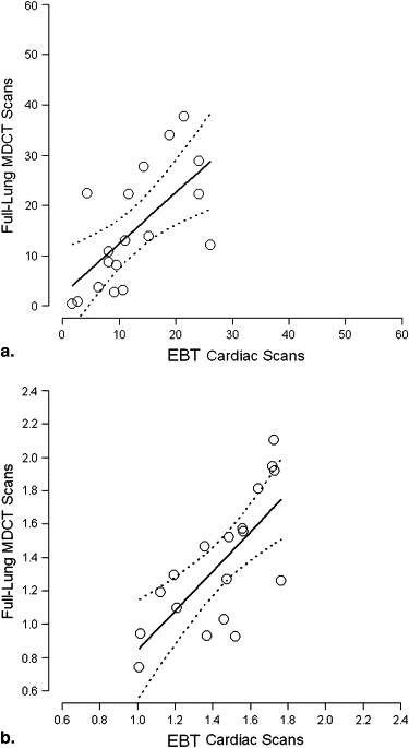
Validation of Lung Density Measures from Cardiac versus Full-lung CT Scans
Get Radiology Tree app to read full this article<
Table 6
Validation of Lung Density Measures from Cardiac CT Scan on MDCT Scanner vs. Full-lung CT Scan on MDCT Scanner
n =24 Median Value from MDCT ∗ Cardiac Scan (Interquartile Range) Median Value from MDCT ∗ Full-lung Scan (Interquartile Range) Spearman’s Correlation Coefficient Mean Difference (95% CI) 95% Limits of Agreement %emphysema -910 HU 22.6 (5.88, 34.9) 20.4 (9.59, 29.5) 0.93 2.25 (−0.10, 4.60) −9.25, 13.8 Alpha -910 HU 1.01 (0.82, 1.43) 1.24 (0.96, 1.65) 0.88 −0.25 (−0.35, −0.15) −0.75, 0.25 Apical-basilar difference in %Emphysema -910 HU 3.02 (−0.19, 14.1) 4.74 (−0.66, 11.2) 0.76 −0.57 (−3.83, 2.40) −15.1, 13.9 Apical-basilar difference in alpha -910 HU −0.01 (−0.13, 0.11) −0.03 (−0.18, 0.18) 0.65 0.01 (−0.09, 0.12) −0.51, 0.54
CT, computed tomographic; EBCT, electron beam CT; HU, Hounsfield units; MDCT, multidetector CT.
Get Radiology Tree app to read full this article<
Get Radiology Tree app to read full this article<
Get Radiology Tree app to read full this article<
Get Radiology Tree app to read full this article<
Table 7
Validation of Lung Density Measures from Cardiac CT Scan on EBCT Scanner versus Full-lung CT Scan on MDCT Scanner
n =18 Median Value from EBCT Cardiac Scan (Interquartile Range) Median Value from MDCT ∗ Full-lung Scan (Interquartile Range) Spearman’s Correlation Coefficient Mean Difference (95% CI) 95% Limits of Agreement %Emphysema -910 HU 10.6 (8.00, 18.9) 12.7 (3.68, 22.6) 0.70 −2.99 (−7.12, 1.15) −20.5, 14.6 Alpha -910 HU 1.49 (1.28, 1.65) 1.29 (1.03, 1.58) 0.69 0.08 (−0.05, 0.21) −0.46, 0.62 Apical-basilar difference in %emphysema -910 HU 3.81 (−2.12, 5.74) −0.38 (−4.11, 2.19) 0.67 2.23 (−0.26, 4.92) −8.68, 13.3 Apical-basilar difference in alpha -910 HU 0.12 (−0.08, 0.32) 0.06 (−0.03, 0.24) 0.49 0.00 (−0.12, 0.12) −0.51, 0.52
CT, computed tomography; EBCT, electron beam CT; HU, Hounsfield units; MDCT, multidetector CT.
Get Radiology Tree app to read full this article<
Get Radiology Tree app to read full this article<
Get Radiology Tree app to read full this article<
Discussion
Get Radiology Tree app to read full this article<
Get Radiology Tree app to read full this article<
Get Radiology Tree app to read full this article<
Get Radiology Tree app to read full this article<
Get Radiology Tree app to read full this article<
Get Radiology Tree app to read full this article<
Get Radiology Tree app to read full this article<
Get Radiology Tree app to read full this article<
Get Radiology Tree app to read full this article<
Acknowledgment
Get Radiology Tree app to read full this article<
Get Radiology Tree app to read full this article<
Get Radiology Tree app to read full this article<
References
1. Petty T.L., Weinmann G.G.: Building a national strategy for the prevention and management of and research in chronic obstructive pulmonary disease. National Heart, Lung, and Blood Institute Workshop Summary. JAMA 1997; 277: pp. 246-253.
2. Mannino D.M., Homa D.M., Akinbami L.J., et. al.: Chronic obstructive pulmonary disease surveillance—United States, 1971–2000. In: Surveillance Summaries, August 2, 2002. MMWR 2002; 51: pp. 1-8.
3. Ferguson G.T., Enright P.L., Buist A.S., et. al.: Office spirometry for lung health assessment in adults: a consensus statement from the National Lung Health Education Program. Chest 2000; 117: pp. 1146-1161.
4. Enright P.L., Kaminsky D.A.: Strategies for screening for chronic obstructive pulmonary disease. Respir Care 2003; 48: pp. 1194-1201.
5. Coxson H.O., Mayo J.R., Behzad H., et. al.: Measurement of lung expansion with computed tomography and comparison with quantitative histology. J Appl Physiol 1995; 79: pp. 1525-1530.
6. Gevenois P.A., de Maertelaer V., De Vuyst P., et. al.: Comparison of computed density and macroscopic morphometry in pulmonary emphysema. Am J Resp Crit Care Med 1995; 152: pp. 653-657.
7. Gevenois P.A., Scillia P., de Maertelaer V., et. al.: The effects of age, sex, lung size, and hyperinflation on CT lung densitometry. AJR Am J Roentgenol 1996; 167: pp. 1169-1173.
8. Kauczor H.U., Hast J., Heussel C.P., et. al.: CT attenuation of paired HRCT scans obtained at full inspiratory/expiratory position: comparison with pulmonary function tests. Eur Radiol 2002; 12: pp. 2757-2763.
9. Nakano Y., Sakai H., Muro S., et. al.: Comparison of low attenuation areas on computed tomographic scans between inner and outer segments of the lung in patients with chronic obstructive pulmonary disease: incidence and contribution to lung function. Thorax 1999; 54: pp. 384-389.
10. Nishimura K., Murata K., Yamagishi M., et. al.: Comparison of different computed tomography scanning methods for quantifying emphysema. J Thorac Imaging 1998; 13: pp. 193-198.
11. Parr D.G., Stoel B.C., Stolk J., et. al.: Validation of computed tomographic lung densitometry for monitoring emphysema in alpha1-antitrypsin deficiency. Thorax 2006; 61: pp. 485-490.
12. Shaker S.B., Dirksen A., Laursen L.C., et. al.: Short-term reproducibility of computed tomography-based lung density measurements in alpha-1 antitrypsin deficiency and smokers with emphysema. Acta Radiol 2004; 45: pp. 424-430.
13. Stolk J., Dirksen A., van der Lugt A.A., et. al.: Repeatability of lung density measurements with low-dose computed tomography in subjects with alpha-1-antitrypsin deficiency-associated emphysema. Invest Radiol 2001; 36: pp. 648-651.
14. Yuan R., Mayo J.R., Hogg J.C., et. al.: The effects of radiation dose and CT manufacturer on measurements of lung densitometry. Chest 2007; 132: pp. 617-623.
15. Hoffman E.A., Simon B.A., McLennan G.: A structural and functional assessment of the lung vai multidetector-row computed tomography: phenotyping chronic obstructive pulmonary disease. Proc Am Thorac Soc 2006; 3: 519–334
16. Carr J.J., Nelson J.C., Wong N.D., et. al.: Calcified coronary artery plaque measurement with cardiac CT in population-based studies: standardized protocol of Multi-Ethnic Study of Atherosclerosis (MESA) and Coronary Artery Risk Development in Young Adults (CARDIA) study. Radiology 2005; 234: pp. 35-43.
17. Detrano R., Guerci A., Carr J.J., et. al.: Coronary calcium as a predictor of coronary events in four racial or ethnic groups. N Engl J Med 2008; 358: pp. 1336-1345.
18. Bild D.E., Bluemke D.A., Burke G.L., et. al.: Multi-Ethnic Study of Atherosclerosis: objectives and design. Am J Epidemiol 2002; 156: pp. 871-881.
19. Aberle D.R., Gamsu G., Henschke C.I., et. al.: A consensus statement of the Society of Thoracic Radiology: screening for lung cancer with helical computed tomography. J Thorac Imaging 2001; 16: pp. 65-68.
20. Guo J., Reinhardt J.M., Kitaoka H., et. al.: Integrated system for CT-based assessment of Parenchymal lung disease. IEEE Int Symp Biomed Imaging 2002; pp. 871-874.
21. Rationale and design of The National Emphysema Treatment Trial: a prospective randomized trial of lung volume reduction surgery. The National Emphysema Treatment Trial Research Group. Chest 1999; 116: pp. 1750-1761.
22. Fishman A., Martinez F., Naunheim K., et. al.: A randomized trial comparing lung-volume-reduction surgery with medical therapy for severe emphysema. N Engl J Med 2003; 348: pp. 2059-2073.
23. Tschirren J., McLennan G., Palagyi K., et. al.: Matching and anatomical labeling of human airway tree. IEEE Trans Medi Imaging 2005; 24: pp. 1540-1547.
24. Hu S., Hoffman E.A., Reinhardt J.M.: Automatic lung segmentation for accurate quantitation of volumetric X-ray CT images. IEEE Trans Medi Imaging 2001; 20: pp. 490-498.
25. Zhang L., Hoffman E.A., Reinhardt J.M.: Atlas-driven lung lobe segmentation in volumetric X-ray CT images. IEEE Trans Med Imaging 2006; 25: pp. 1-16.
26. Mishima M., Hirai T., Itoh H., et. al.: Complexity of terminal airspace geometry assessed by lung computed tomography in normal subjects and patients with chronic obstructive pulmonary disease. Proc Natl Acad Sci U S A 1999; 96: pp. 8829-8834.
27. Miller M.R., Hankinson J., Brusasco V., et. al.: Standardisation of spirometry. Eur Respir J 2005; 26: pp. 319-338.
28. Stukovsky K., Hankinson J.H., Jiang R., et. al.: Participant and technician predictors of spirometry quality factors in the Multi-Ethnic Study of Atherosclerosis (MESA)-Lung Study (abstract). Eur Respir J 2006; pp. 451-452s.
29. Hankinson J.L., Kawut S.M., Shahar E., et. al.: Performance of spirometry reference values in a multiethnic population. The Mesa-Lung Study. (abstract). Am J Respir Crit Care Med 2007; 175: pp. A605.
30. Bland J.M., Altman D.G.: Statistical method for assessing agreement between two methods of clinical measurement. Lancet 1986; i: pp. 307-310.
31. Bland J.M., Altman D.G.: Measuring agreement in method comparison studies. Stat Methods Med Res 1999; 8: pp. 135-160.
32. Gietema H.A., Schilham A.M., van Ginneken B., et. al.: Monitoring of smoking-induced emphysema with CT in a lung cancer screening setting: detection of real increase in extent of emphysema. Radiology 2007; 244: pp. 890-897.
33. Wei J.H., Hoffman E.A., Ritman E.L., et. al.: Cardiogenic motion of right lung parenchyma in anesthetized intact dogs. J Appl Physiol 1985; 58: pp. 384-391.
34. Reddy S., Barr R.G., Berbaum K., et. al.: Quantitative evaluation of emphysema presence and distribution via coronary calcium CT compared with full lung study (abstract). Presented at the international conference of the Radiological Society of North America 2005;
35. Haraguchi M., Shimura S., Hida W., et. al.: Pulmonary function and regional distribution of emphysema as determined by high-resolution computed tomography. Respiration 1998; 65: pp. 125-129.