
Objective
To report our clinical experience with a 256-slice multidetector computed tomography (MDCT) with a 270-ms gantry rotation system in performing CT coronary angiograms (CTCA) using both prospectively gated step and shoot (PGSS) and retrospectively gated helical (RGH) techniques.
Materials and Methods
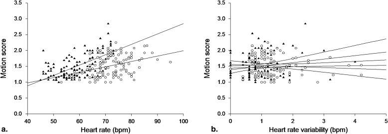
We studied 252 patients who received CTCA; 126 patients having mean heart rate (HR) of 72.1 were imaged with RGH CTCA and 126 patients having mean HR of 58.7 were imaged with PGSS CTCA. For patients with a prescan HR ≤70 beats/min, a PGSS acquisitions trigger was used, whereas patients whose prescan HR was >70 beats/min were imaged using an RGH acquisition. The blood vessel accessibility of both PGSS and RGH techniques was evaluated by grading the image quality score from 1 (no motion artifacts) to 4 (severe motion artifacts preventing diagnosis) for each coronary artery segment. Radiation doses of the techniques were also compared.
Results
In both groups, more than 50% of segments received the best imaging score. The overall image quality scores for RGH and PGSS groups were 1.522 ± 0.317 and 1.500 ± 0.374, respectively. There was no significant difference in right coronary artery, left anterior descending artery, and left circumflex artery image quality between the two groups. Only 0.1% of segments were nonevaluative with the PGSS technique and all segments were evaluative with RGH. PGSS was associated with a 62% reduction in effective radiation dose as compared to RGH (PGSS, 5.1 mSv; RGH, 13.2 mSv).
Conclusions
There is no significant difference in image quality between PGSS and RGH in this study. Although providing similar image quality as RGH, PGSS was associated with a 62% reduction in effective radiation dose. Further study to confirm the diagnostic accuracy as compared to coronary artery angiography is warranted.
Computed tomography coronary angiography (CTCA) using 64-slice multidetector computed tomography (MDCT) and dual-source CT (DSCT) is highly accurate for the detection of coronary artery disease (CAD). Although the noninvasive nature of CT is attractive, the radiation dose of CTCA compared to catheterization remains a significant hurdle because doses for conventional coronary angiography range from 5 to 12 mSv , whereas those of retrospectively gated helical (RGH) CTCA range from 12 to 28 mSv .
With RGH, image data are acquired continuously throughout the cardiac cycle and the reconstruction phase with the least amount of motion artifact is retrospectively chosen, usually during ventricular diastole . On the other hand, prospectively gated step and shoot (PGSS) is a CT acquisition technique in which x-rays are turned on only during the cardiac phase of interest. Being prospective in nature, PGSS requires precise synchronization of the x-ray with the cardiac cycle, and its successful implementation also depends on the matching between the selected phase of the cardiac cycle and the phase with least motion. The continuous application of radiation throughout the cardiac cycle with RGH results in a higher radiation dose as compared to PGSS .
Get Radiology Tree app to read full this article<
Get Radiology Tree app to read full this article<
Materials and methods
Study Population
Get Radiology Tree app to read full this article<
The 256-slice MDCT Protocol
Get Radiology Tree app to read full this article<
Get Radiology Tree app to read full this article<
Get Radiology Tree app to read full this article<
Get Radiology Tree app to read full this article<
Table 1
Patient Demographic and RGH and PGSS Technical Data
RGH ( n = 126) PGSS ( n = 126)P Value Age (y) 56.5 ± 9.8 58.0 ± 8.0 .190 Male gender 77 (61.1) 69 (78.4) .007 ∗ Body mass index (kg/m 2 ) 25.6 ± 4.0 25.5 ± 3.3 .930 MHR (beats/min) 72.1 ± 6.9 58.7 ± 7.3 <.001 ∗ HRV (beats/min) 1.3 ± 0.7 1.0 ± 0.8 <.001 ∗ Received beta-blockers 35 (27.8) 5 (5.7) <.001 ∗ Tube voltage (kV) 120 120 N/A Tube current (mA) 565.7 ± 91.7 780.3 ± 97.8 <.001 ∗ Scan time (seconds) 4.9 ± 0.3 3.9 ± 1.4 <.001 ∗ Number of shots Continuous exposure 2–3 N/A CTDI vol (mGy) 60.0 ± 10.5 22.9 ± 3.1 <.001 ∗ Scan length (mm) 129.7 ± 13.8 131.7 ± 15.6 .300 Effective dose (mSv) 13.2 ± 2.7 5.1 ± 0.9 <.001 ∗
CTDI vol , volume computed tomography dose index; HRV, heart rate variability; MHR, mean heart rate; PGSS, prospectively gated step and shoot; RGH, retrospectively gated helical.
Data are presented as mean ± standard deviation for continuous variables and number (percentage) for categorical variables.
Get Radiology Tree app to read full this article<
Get Radiology Tree app to read full this article<
CT Data Analysis
Get Radiology Tree app to read full this article<
Radiation Dose
Get Radiology Tree app to read full this article<
Statistical Analysis
Get Radiology Tree app to read full this article<
Results
Get Radiology Tree app to read full this article<
Get Radiology Tree app to read full this article<
Get Radiology Tree app to read full this article<
Get Radiology Tree app to read full this article<
Table 2
RGH and PGSS Image Quality Scores for All Segments
RGH PGSS Segment ∗ Score 1 (%) Score 2 (%) Score 3 (%) Score 4 (%) Score 1 (%) Score 2 (%) Score 3 (%) Score 4 (%) 1 57.1 (72/126) 42.1 (53/126) 0.8 (1/126) 0.0 (0/126) 58.4 (73/125) 39.2 (49/125) 2.4 (3/125) 0.0 (0/125) 2 21.4 (27/126) 59.5 (75/126) 19.0 (24/126) 0.0 (0/126) 26.4 (33/125) 54.4 (68/125) 18.4 (23/125) 0.8 (1/125) 3 56.3 (67/119) 42.9 (51/119) 0.8 (1/119) 0.0 (0/119) 53.8 (64/119) 45.4 (54/119) 0.8 (1/119) 0.0 (0/119) 4 42.7 (50/117) 54.7 (64/117) 2.6 (3/117) 0.0 (0/117) 52.9 (63/119) 46.2 (55/119) 0.8 (1/119) 0.0 (0/119) 5 87.2 (109/125) 12.8 (16/125) 0.0 (0/125) 0.0 (0/125) 80.0 (100/125) 19.2 (24/125) 0.8 (1/125) 0.0 (0/125) 6 78.6 (99/126) 21.4 (27/126) 0.0 (0/126) 0.0 (0/126) 75.2 (94/125) 22.4 (28/125) 2.4 (3/125) 0.0 (0/125) 7 53.6 (67/125) 46.4 (58/125) 0.0 (0/125) 0.0 (0/125) 54.4 (68/125) 44.8 (56/125) 0.8 (1/125) 0.0 (0/125) 8 32.8 (41/125) 64.0 (80/125) 3.2 (4/125) 0.0 (0/125) 31.2 (39/125) 64.0 (80/125) 4.8 (6/125) 0.0 (0/125) 9 48.4 (60/124) 50.0 (62/124) 1.6 (2/124) 0.0 (0/124) 62.4 (78/125) 36.8 (46/125) 0.8 (1/125) 0.0 (0/125) 10 41.5 (27/65) 55.4 (36/65) 3.1 (2/65) 0.0 (0/65) 58.1 (54/93) 39.8 (37/93) 2.2 (2/93) 0.0 (0/93) 11 64.0 (80/125) 36.0 (45/125) 0.0 (0/125) 0.0 (0/125) 67.7 (84/124) 29.8 (37/124) 2.4 (3/124) 0.0 (0/124) 12 33.6 (39/116) 64.7 (75/116) 1.7 (2/116) 0.0 (0/116) 41.4 (48/116) 55.2 (64/116) 3.4 (4/116) 0.0 (0/116) 13 40.2 (45/112) 58.0 (65/112) 1.8 (2/112) 0.0 (0/112) 43.1 (47/109) 53.2 (58/109) 3.7 (4/109) 0.0 (0/109) 14 39.7 (27/68) 57.4 (39/68) 2.9 (2/68) 0.0 (0/68) 32.9 (23/70) 61.4 (43/70) 5.7 (4/70) 0.0 (0/70) 15 62.5 (5/8) 37.5 (3/8) 0.0 (0/8) 0.0 (0/8) 60.0 (3/5) 40.0 (2/5) 0.0 (0/5) 0.0 (0/5) 16 47.6 (20/42) 52.4 (22/42) 0.0 (0/42) 0.0 (0/42) 75.0 (42/56) 23.2 (13/56) 1.8 (1/56) 0.0 (0/56) All segments 50.6 (835/1649) 46.8 (771/1649) 2.6 (43/1649) 0.0 (0/1649) 54.2 (913/1686) 42.3 (714/1686) 3.4 (58/1686) 0.1 (1/1686)
PGSS, prospectively gated step and shoot; RGH, retrospectively gated helical.
Data in parentheses are raw data used to calculate the percentage. Image quality score obtained at best R-R interval: score 1, no motion artifacts; score 2, mild motion artifacts; score 3, moderate motion artifacts; score 4, severe motion artifacts.
Get Radiology Tree app to read full this article<
Table 3
Comparison of Image Quality for RGH and PGSS Techniques
RGH PGSS Difference Between Techniques ∗ Coronary Vessel Group A1
(All HRs, n = 126) Group A2
(HR ≤75, n = 84) Group B
(HR ≤75, n = 126) Group A1 vs. B Group A2 vs. B Overall image quality 1.522 ± 0.317 1.472 ± 0.313 1.500 ± 0.374 0.023 (0.23) -0.028 (0.96) RCA 1.623 ± 0.442 1.571 ± 0.431 1.580 ± 0.465 0.043 (0.39) -0.009 (0.95) LAD 1.429 ± 0.331 1.388 ± 0.328 1.424 ± 0.377 0.005 (0.45) -0.036 (0.86) LCX 1.556 ± 0.430 1.467 ± 0.446 1.547 ± 0.469 0.009 (0.68) -0.080 (0.26)
HR, heart rate; LAD, left anterior descending coronary artery; LCX, left circumflex coronary artery; PGSS, prospectively gated step and shoot; RCA, right coronary artery; RGH, retrospectively gated helical.
Group A1 includes all patients scanned by RGH; group A2 are patients with HR <75 beats/min, scanned by RGH; group B includes patients with HR <75 beats/min, scanned by PGSS. Group data are presented as mean ± standard deviation.
Get Radiology Tree app to read full this article<
Get Radiology Tree app to read full this article<
Discussion
Get Radiology Tree app to read full this article<
Get Radiology Tree app to read full this article<
Get Radiology Tree app to read full this article<
Get Radiology Tree app to read full this article<
Get Radiology Tree app to read full this article<
Get Radiology Tree app to read full this article<
Get Radiology Tree app to read full this article<
References
1. Kocinaj D., Cioppa A., Ambrosini G., et. al.: Radiation dose exposure during cardiac and peripheral arteries catheterization. Int J Cardiol 2006; 113: pp. 283-284.
2. Klass O., Jeltsch M., Feuerlein S., et. al.: Prospectively gated axial CT coronary angiography: preliminary experiences with a novel low-dose technique. Eur Radiol 2009; 19: pp. 829-836.
3. Mori S., Nishizawa K., Kondo C., et. al.: Effective doses in subjects undergoing computed tomography cardiac imaging with the 256-multislice CT scanner. Eur J Radiol 2008; 65: pp. 442-448.
4. Gerber T.C., Kuzo R.S., Karstaedt N., et. al.: Current results and new developments of coronary angiography with use of contrast-enhanced computed tomography of the heart. Mayo Clin Proc 2002; 77: pp. 55-71.
5. Morin R.L., Gerber T.C., McCollough C.H.: Radiation dose in computed tomography of the heart. Circulation 2003; 107: pp. 917-922.
6. Earls J.P., Berman E.L., Urban B.A., et. al.: Prospectively gated transverse coronary CT angiography versus retrospectively gated helical technique: improved image quality and reduced radiation dose. Radiology 2008; 246: pp. 742-753.
7. Husmann L., Valenta I., Gaemperli O., et. al.: Feasibility of low-dose coronary CT angiography: first experience with prospective ECG-gating. Eur Heart J 2008; 29: pp. 191-197.
8. Shuman W.P., Branch K.R., May J.M., et. al.: Prospective versus retrospective ECG gating for 64-detector CT of the coronary arteries: comparison of image quality and patient radiation dose. Radiology 2008; 248: pp. 431-437.
9. Gutstein A., Wolak A., Lee C., et. al.: Predicting success of prospective and retrospective gating with dual-source coronary computed tomography angiography: development of selection criteria and initial experience. J Cardiovasc Comput Tomogr 2008; 2: pp. 81-90.
10. Weigold W.G., Olszewski M.E., Walker M.J.: Low-dose prospectively gated 256-slice coronary computed tomographic angiography. Int J Cardiovasc Imaging 2009; 25: pp. 217-230.
11. Alkadhi H., Stolzmann P., Desbiolles L., et. al.: Low-dose, 128-slice, dual-source CT coronary angiography: accuracy and radiation dose of the high-pitch and the step-and-shoot mode. Heart 2010; 96: pp. 933-938.
12. Hirai N., Horiguchi J., Fujioka C., et. al.: Prospective versus retrospective ECG-gated 64-detector coronary CT angiography: assessment of image quality, stenosis, and radiation dose. Radiology 2008; 248: pp. 424-430.
13. Xu L., Yang L., Zhang Z., et. al.: Low-dose adaptive sequential scan for dual-source CT coronary angiography in patients with high heart rate: Comparison with retrospective ECG gating. Eur J Radiol 2009 Jul 10; [Epub ahead of print]
14. Austen W.G., Edwards J.E., Frye R.L., et. al.: A reporting system on patients evaluated for coronary artery disease. Report of the Ad Hoc Committee for Grading of Coronary Artery Disease, Council on Cardiovascular Surgery, American Heart Association. Circulation 1975; 51: pp. 5-40.
15. Leschka S., Wildermuth S., Boehm T., et. al.: Noninvasive coronary angiography with 64-section CT: Effect of average heart rate and heart rate variability on image quality. Radiology 2006; 241: pp. 378-385.
16. Flohr T.G., McCollough C.H., Bruder H., et. al.: First performance evaluation of a dual-source CT (DSCT) system. Eur Radiol 2006; 16: pp. 256-268.
17. Efstathopoulos E.P., Kelekis N.L., Pantos I., et. al.: Reduction of the estimated radiation dose and associated patient risk with prospective ECG-gated 256-slice CT coronary angiography. Phys Med Biol 2009; 54: pp. 5209-5222.
18. Weustink A.C., Mollet N.R., Pugliese F., et. al.: Optimal electrocardiographic pulsing windows and heart rate: effect on image quality and radiation exposure at dual-source coronary CT angiography. Radiology 2008; 248: pp. 792-798.
19. DeFrance T., Dubois E., Gebow D., et. al.: Helical prospective ECG-gating in cardiac computed tomography: radiation dose and image quality. Int J Cardiovasc Imaging 2010; 2: pp. 99-107.
20. Dewey M.: Prospective helical acquisition for coronary CT angiography. Int J Cardiovasc Imaging 2010; 26: pp. 109-110.
21. Lehmkuhl L., Gosch D., Nagel H.D., et. al.: Quantification of radiation dose savings in cardiac computed tomography using prospectively triggered mode and ECG pulsing: a phantom study. Eur Radiol 2010; 20: pp. 2116-2125.
22. Mahnken A.H., Bruners P., Schmidt B., et. al.: Left ventricular function can reliably be assessed from dual-source CT using ECG-gated tube current modulation. Investig Radiol 2009; 44: pp. 384-389.
23. Scheffel H., Alkadhi H., Leschka S., et. al.: Low-dose CT coronary angiography in the step-and-shoot mode: diagnostic performance. Heart 2008; 94: pp. 1132-1137.
24. Stolzmann P., Leschka S., Scheffel H., et. al.: Dual source CT in step-and-shoot mode: noninvasive coronary angiography with low radiation dose. Radiology 2008; 249: pp. 71-80.
25. Hausleiter J., Meyer T., Hadamitzky M., et. al.: Radiation dose estimates from cardiac multislice computed tomography in daily practice: impact of different scanning protocols on effective dose estimates. Circulation 2006; 113: pp. 1305-1310.
26. Matt D., Scheffel H., Leschka S., et. al.: Dual-source CT coronary angiography: image quality, mean heart rate, and heart rate variability. AJR Am J Roentgenol 2007; 189: pp. 567-573.
27. Herzog B.A., Husmann L., Burkhard N., et. al.: Accuracy of low-dose computed tomography coronary angiography using prospective electrocardiogram-triggering: first clinical experience. Eur Heart J 2008; 29: pp. 3037-3042.
28. Dewey M., Zimmermann E., Deissenrieder F., et. al.: Noninvasive coronary angiography by 320-row computed tomography with lower radiation exposure and maintained diagnostic accuracy: comparison of results with cardiac catheterization in a head-to-head pilot investigation. Circulation 2009; 120: pp. 867-875.
29. Chazen J.L., Prince M.R., Yip R., et. al.: Post-CABG coronary CT angiography radiation dose and graft image quality in retrospective versus prospective ECG gating. Acad Radiol 2010; 17: pp. 1122-1127.