
Rationale and Objectives
The aim of this study was to evaluate and compare the diagnostic performance of elastography, B-mode ultrasound (US), and a combination of elastography and B-mode US for the differentiation of small breast masses.
Materials and Methods
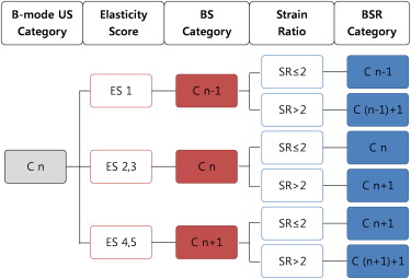
A total of 315 breast masses < 1 cm (267 benign, 48 malignant) in 278 patients were examined with B-mode US and elastography. Histopathologic results were used as a reference standard. Two radiologists retrospectively evaluated the B-mode images according to the American College of Radiology Breast Imaging Reporting and Data System and elastographic images according to the elasticity scoring classification system proposed by Itoh et al and the strain ratio. B-mode US and elastography were combined according to the cutoff value. The diagnostic performance of B-mode US, elastography, and the combination of the two modalities was compared using receiver-operating characteristic curve analysis.
Results
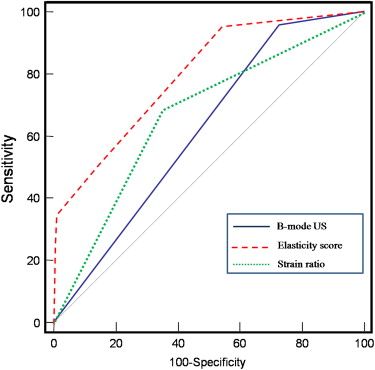
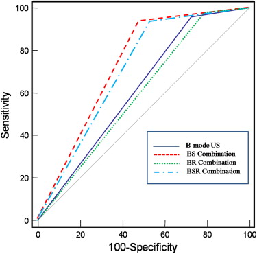
The mean elasticity score for malignant masses (3.02 ± 1.33) was significantly higher than that for benign masses (1.72 ± 0.78) ( P < .001). Areas under the receiver-operating characteristic curves were 0.616 for B-mode US, 0.784 for elasticity score, 0.668 for strain ratio, 0.727 for the combination of B-mode US and elasticity score, and 0.701 for the combination of B-mode US, elasticity score, and strain ratio. The sensitivity, specificity, positive predictive value, and negative predictive value were 93.8%, 51.7%, 25.9%, and 97.9%, respectively, when elasticity score and B-mode US were combined as follows: downgrade of B-mode US assessment category in cases with elasticity scores of 1, no change in cases with scores of 2 or 3, and upgrade in cases with scores of 4 or 5.
Conclusion
Elasticity score alone showed the best diagnostic performance, but a combination of B-mode US and elasticity score may have predictive value for the differentiation of benign and malignant lesions <1 cm.
Elastography is a method of visualizing the elasticity characteristics of a lesion. Several clinical studies have reported that elastography has the potential to differentiate benign from malignant solid breast masses and have shown similar or inferior diagnostic performance compared with conventional B-mode ultrasound (US), with sensitivity of 65% to 100% and specificity of 32% to 98.5% . The interpretation criteria in elastography consist of the qualitative parameter of elasticity score and the quantitative parameter of strain ratio . Most studies have compared the diagnostic performance of the elasticity score and the strain ratio with B-mode US, and studies of the diagnostic performance of the combination of B-mode US and elastography are rare . In such studies, on the basis of the subjective determinations of the investigators, final assessments were performed by combining the two modalities, and the results were reported not to be better than using B-mode US alone.
If the size of a mass is small, it is difficult to analyze according to the Breast Imaging Reporting and Data System, and the characterization of the lesion may not be accurate. Therefore, we evaluated the diagnostic performance of elastography for differentiation between benign and malignant small breast masses. We also compared the diagnostic performance of elastography alone, B-mode US alone, and the combination of the two modalities.
Materials and methods
Patients
Get Radiology Tree app to read full this article<
Get Radiology Tree app to read full this article<
Pathologic Analysis
Get Radiology Tree app to read full this article<
Get Radiology Tree app to read full this article<
Image Acquisition and Analysis
Get Radiology Tree app to read full this article<
Get Radiology Tree app to read full this article<
Get Radiology Tree app to read full this article<
Statistical Analysis
Get Radiology Tree app to read full this article<
Results
Get Radiology Tree app to read full this article<
Table 1
Comparison of the Diagnostic Performance of B-mode US Alone, Elastography Alone, and the Combination of the Two Modalities
B-mode US Elastography Combination of B-mode US and Elastography BI-RADS Category Elasticity Score Strain Ratio BS Method BR Method BSR Method Sensitivity 95.8% 93.8% 68.8% 93.8% 97.9% 93.8% Specificity 27.3% 45.7% 64.8% 51.7% 21.4% 46.4% PPV 19.2% 23.7% 26% 25.9% 18.3% 23.9% NPV 97.3% 97.6% 92% 97.9% 98.3% 97.6% Az (95% CI) 0.616 (0.560–0.670 0.784 (0.735–0.828) 0.668 (0.613–0.720) 0.727 (0.674–0.776) 0.596 (0.540–0.651) 0.701 (0.647–0.751)
Az, area under the receiver-operating characteristic curve; BI-RADS, Breast Imaging Reporting and Data System; BR, combination of B-mode ultrasound and strain ratio; BS, combination of B-mode ultrasound and elasticity score; BSR, combination of B-mode ultrasound, elasticity score, and strain ratio; CI, confidence interval; NPV, negative predictive value; PPV, positive predictive value; US, ultrasound.
Get Radiology Tree app to read full this article<
Conventional B-mode US
Get Radiology Tree app to read full this article<
Get Radiology Tree app to read full this article<
Get Radiology Tree app to read full this article<
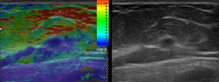
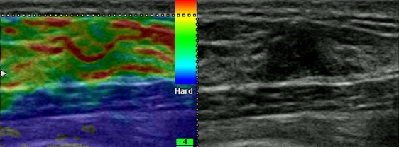
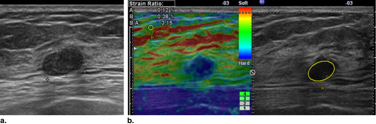
Elastography: Elasticity Score and Strain Ratio
Get Radiology Tree app to read full this article<
Get Radiology Tree app to read full this article<
Get Radiology Tree app to read full this article<
Get Radiology Tree app to read full this article<
Get Radiology Tree app to read full this article<
Get Radiology Tree app to read full this article<
Get Radiology Tree app to read full this article<
Combination of B-mode US and Elastography
Get Radiology Tree app to read full this article<
Get Radiology Tree app to read full this article<
Get Radiology Tree app to read full this article<
Get Radiology Tree app to read full this article<
Get Radiology Tree app to read full this article<
Get Radiology Tree app to read full this article<
Get Radiology Tree app to read full this article<
Get Radiology Tree app to read full this article<
Get Radiology Tree app to read full this article<
Get Radiology Tree app to read full this article<
Get Radiology Tree app to read full this article<
Get Radiology Tree app to read full this article<
Get Radiology Tree app to read full this article<
Get Radiology Tree app to read full this article<
Get Radiology Tree app to read full this article<
Discussion
Get Radiology Tree app to read full this article<
Get Radiology Tree app to read full this article<
Get Radiology Tree app to read full this article<
Get Radiology Tree app to read full this article<
Get Radiology Tree app to read full this article<
Conclusions
Get Radiology Tree app to read full this article<
References
1. Garra B.S., Cespedes E.I., Ophir J., et. al.: Elastography of breast lesions: initial clinical results. Radiology 1997; 202: pp. 79-86.
2. Itoh A., Ueno E., Tohno E., et. al.: Breast disease: clinical application of US elastography for diagnosis. Radiology 2006; 239: pp. 341-350.
3. Hall T.J., Zhu Y., Spalding C.S.: In vivo real-time freehand palpation imaging. Ultrasound Med Biol 2003; 29: pp. 427-435.
4. Thomas A., Fischer T., Frey H., et. al.: Real-time elastography—an advanced method of ultrasound: first results in 108 patients with breast lesions. Ultrasound Obstet Gynecol 2006; 28: pp. 335-340.
5. Tan S.M., Teh H.S., Mancer J.F., et. al.: Improving B mode ultrasound evaluation of breast lesions with real-time ultrasound elastography—a clinical approach. Breast 2008; 17: pp. 252-257.
6. Thomas A., Kummel S., Fritzsche F., et. al.: Real-time sonoelastography performed in addition to B-mode ultrasound and mammography: improved differentiation of breast lesions?. Acad Radiol 2006; 13: pp. 1496-1504.
7. Zhu Q.L., Jiang Y.X., Liu J.B., et. al.: Real-time ultrasound elastography: its potential role in assessment of breast lesions. Ultrasound Med Biol 2008; 34: pp. 1232-1238.
8. Zhi H., Ou B., Luo B.M., et. al.: Comparison of ultrasound elastography, mammography, and sonography in the diagnosis of solid breast lesions. J Ultrasound Med 2007; 26: pp. 807-815.
9. Cho N., Moon W.K., Park J.S., et. al.: Nonpalpable breast masses: evaluation by US elastography. Korean J Radiol 2008; 9: pp. 111-118.
10. Schaefer FK, Heer I, Schaefer PJ, et al. Breast ultrasound elastography-Results of 193 breast lesions in a prospective study with histopathologic correlation. Eur J Radiol. In press.
11. Scaperrotta G., Ferranti C., Costa C., et. al.: Role of sonoelastography in non-palpable breast lesions. Eur Radiol 2008; 18: pp. 2381-2389.
12. Cho N., Moon W.K., Kim H.Y., et. al.: Sonoelastographic strain index for differentiation of benign and malignant nonpalpable breast masses. J Ultrasound Med 2010; 29: pp. 1-7.
13. Sohn Y.M., Kim M.J., Kim E.K., et. al.: Sonographic elastography combined with conventional sonography: how much is it helpful for diagnostic performance?. J Ultrasound Med 2009; 28: pp. 413-420.
14. Thomas A., Degenhardt F., Farrokh A., et. al.: Significant differentiation of focal breast lesions calculation of strain ratio in breast sonoelastography. Acad Radiol 2010; 17: pp. 558-563.
15. Giuseppetti G.M., Martegani A., Di Cioccio B., et. al.: Elastosonography in the diagnosis of the nodular breast lesions: preliminary report. Radiol Med 2005; 110: pp. 69-76.
16. Raza S., Odulate A., Ong E.M., et. al.: Using real-time tissue elastography for breast lesion evaluation: our initial experience. J Ultrasound Med 2010; 29: pp. 551-563.
17. Regner D.M., Hesley G.K., Hangiandreou N.J., et. al.: Breast lesions: evaluation with US strain imaging—clinical experience of multiple observers. Radiology 2006; 238: pp. 425-437.