
Rationale and Objectives
To investigate whether imaging features on multiphasic multidetector computed tomography (MDCT) can help discriminate sarcomatoid renal cell carcinoma (RCC) and collecting duct carcinoma (CDC) from other solid renal masses.
Materials and Methods
With institutional review board approval for this HIPAA-compliant study, we derived a cohort of 7 sarcomatoid RCCs, 4 CDCs, 165 clear cell RCCs, 56 papillary RCCs, 22 chromophobe RCCs, 49 oncocytomas, and 16 lipid-poor angiomyolipomas with preoperative multiphasic MDCT with up to four phases (unenhanced, corticomedullary, nephrographic, and excretory). Each lesion was reviewed for contour, spread pattern, pattern of enhancement, neovascularity, and calcification.
Results
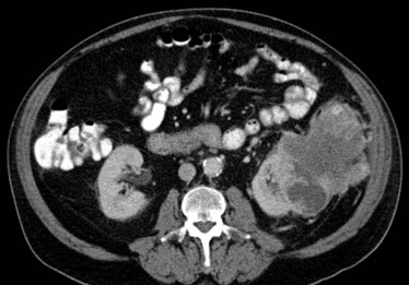
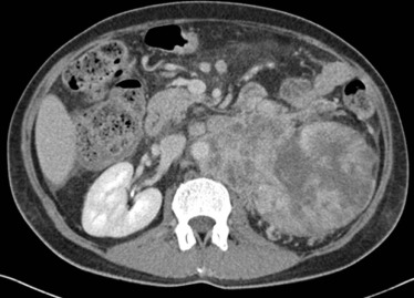
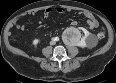
Sarcomatoid RCCs and CDCs were more likely than other solid renal masses to have an irregular contour (64% vs 2%, P < 0.001) and an infiltrative spread pattern, defined as infiltration into adjacent renal parenchyma, collecting system, or neighboring structures (82% vs 7%, P < 0.001). When used to discriminate sarcomatoid RCC and CDC from other solid renal masses, an infiltrative spread pattern had a specificity of 93% (287/308) and sensitivity of 82% (9/11), and an irregular contour had a specificity of 98% (303/308) and sensitivity of 64% (7/11).
Conclusions
Solid renal lesions with an irregular contour or an infiltrative spread pattern are suspicious for sarcomatoid RCC or CDC.
Introduction
Within the Heidelberg classification system, the most common subtype of renal cell carcinoma (RCC) is clear cell, which accounts for 70%–80% of cases of RCC, followed by papillary and chromophobe RCC, which account for 14%–17% and 4%–8% of RCCs, respectively . Of these common RCC subtypes, clear cell RCC has the worst prognosis with a 5-year survival rate of 44%–69%, accounting for 94% of metastatic RCC . On the other hand, papillary RCC has a 5-year survival rate of 82%–92% and accounts for 4% of metastatic RCC, and chromophobe RCC has a 5-year survival of 78%–87% and accounts for 2% of metastatic RCC . In contrast to these common subtypes of RCC, sarcomatoid RCC and collecting duct carcinoma are rare, aggressive variants of RCC with even more dismal prognoses with median survivals of 4–9 months and 11 months, respectively . Up to 83% of patients with collecting duct carcinoma have distant metastases at the time of diagnosis; 45%–84% of patients with sarcomatoid RCC have distant metastases at the time of diagnosis . In contrast to collecting duct carcinomas, sarcomatoid RCCs are not a distinct RCC subtype. They represent a group of RCCs with high-grade features and extensive chromosomal rearrangements that result in dedifferentiation and a spindle-like histologic morphology and can arise from any of the RCC subtypes .
Recent studies have demonstrated that enhancement on multiphasic multidetector computed tomography (MDCT) can help differentiate between clear cell RCC, papillary RCC, and chromophobe RCC, as well as oncocytoma and lipid-poor angiomyolipoma (benign mimics of RCC) . However, while there are several published case series concerning sarcomatoid RCC and collecting duct carcinoma , there are currently no robust means of differentiating sarcomatoid RCC and collecting duct carcinoma from common RCC subtypes without sarcomatoid elements or from benign RCC mimics on imaging. Schieda et al. found that quantitative texture feature analysis on unenhanced images can help discriminate sarcomatoid RCCs from clear cell RCCs . However, texture feature analyses require sophisticated postprocessing, and the support vector machine classifier used by Schieda et al. had limited accuracy, ranging from 55% to 68%. A simple, robust noninvasive means of differentiating sarcomatoid RCC and collecting duct carcinoma from other solid renal masses can be of great clinical value because sarcomatoid RCC and collecting duct carcinoma should be managed differently than common RCC subtypes and benign RCC mimics . Common RCC subtypes are typically surgically resected. Even in the setting of metastasis, surgical resection can be beneficial in common RCC subtypes, as upfront cytoreductive nephrectomy has been shown to provide survival benefit. However, for sarcomatoid RCC and collecting duct carcinoma, surgical resection prior to systemic targeted therapies may actually worsen outcomes because it delays the administration of systemic therapy . The goal of this study was to investigate whether simple imaging features reproducible in routine clinical imaging with multiphasic MDCT could help discriminate sarcomatoid RCC and collecting duct carcinoma from common RCC subtypes and benign RCC mimics.
Materials and Methods
Patients
Get Radiology Tree app to read full this article<
Get Radiology Tree app to read full this article<
TABLE 1
Characteristics of Solid Renal Lesions
Characteristic All (n = 319 Lesions, 294 Patients) Aggressive RCC Variants Common RCC Subtypes Benign RCC Mimics Sarcomatoid RCC (n = 7 Lesions, 7 Patients) Collecting Duct Carcinoma (n = 4 Lesions, 4 Patients) Clear Cell RCC (n = 165 Lesions, 155 Patients) Papillary RCC (n = 56 Lesions, 52 Patients) Chromophobe RCC (n = 22 Lesions, 22 Patients) Oncocytoma (n = 49 Lesions, 45 Patients) Lipid-Poor AML (n = 16 Lesions, 16 Patients) Gender \* Male 192 (65) 7 (100) 3 (75) 96 (62) 43 (83) 15 (68) 31 (69) 3 (19) Female 102 (34) 0 (0) 1 (25) 59 (38) 9 (17) 7 (32) 14 (31) 13 (81) Mean age (y) † 60 (21–87) 50 (33–66) 61 (53–72) 61 (27–87) 62 (39–84) 60 (23–85) 65 (38–86) 47 (21–78) Method of specimen acquisition Partial nephrectomy 143 (45) 2 (29) 1 (25) 65 (39) 34 (61) 5 (23) 24 (49) 12 (75) Radical nephrectomy 120 (38) 4 (57) 2 (50) 72 (44) 17 (30) 13 (59) 10 (20) 2 (13) Biopsy 54 (17) 1 (14) 1 (25) 28 (17) 3 (5) 4 (18) 15 (31) 2 (13) Autopsy 2 (1) 0 (0) 0 (0) 0 (0) 2 (4) 0 (0) 0 (0) 0 (0) Pathologic T stage T1a 134 (42) 2 (29) 1 (25) 88 (53) 33 (59) 10 (46) — — T1b 55 (17) 1 (14) 0 (0) 37 (22) 11 (20) 6 (27) — — T2 20 (6) 1 (14) 0 (0) 10 (6) 6 (11) 3 (14) — — T3 42 (13) 2 (29) 3 (75) 28 (17) 6 (11) 3 (14) — — T4 3 (1) 1 (14) 0 (0) 2 (1) 0 (0) 0 (0) — — Side Left 161 (50.5) 3 (43) 1 (25) 83 (50) 24 (43) 16 (73) 23 (47) 11 (69) Right 158 (40.5) 4 (57) 3 (75) 82 (50) 32 (57) 6 (27) 26 (53) 5 (31) Lesion size (cm) † 4.3 (0.7–22.0) 9.4 (2.9–22.0) 7.2 (2.5–9.6) 4.6 (0.7–18.7) 3.7 (1.3–12.0) 4.7 (1.6–12.0) 3.3 (0.9–12.7) 3.8 (1.2–15.0) MDCT study type Two-phase 100 (31) 0 (0) 2 (50) 47 (29) 15 (27) 8 (36) 19 (39) 9 (56) Three-phase 55 (17) 4 (57) 0 (0) 34 (21) 8 (14) 2 (9) 5 (10) 2 (13) Four-phase 164 (51) 3 (43) 2 (50) 84 (51) 33 (59) 12 (55) 25 (51) 5 (31)
AML, angiomyolipoma; MDCT, multidetector computed tomography; RCC, renal cell carcinoma.
Unless otherwise indicated, data are numbers of lesions, with percentages in parentheses.
Oncocytomas and lipid-poor AMLs were not staged, as the staging criteria apply only to renal cell carcinomas.
Get Radiology Tree app to read full this article<
Get Radiology Tree app to read full this article<
Get Radiology Tree app to read full this article<
Computed Tomography Examination
Get Radiology Tree app to read full this article<
CT Image Analyses
Get Radiology Tree app to read full this article<
Statistical Analyses
Get Radiology Tree app to read full this article<
Results
Lesions
Get Radiology Tree app to read full this article<
Qualitative Features
Get Radiology Tree app to read full this article<
TABLE 2
Qualitative Features of Solid Renal Lesions
Aggressive RCC Variants Common RCC Subtypes Benign RCC Mimics Sarcomatoid RCC (n = 7) Collecting Duct Carcinoma (n = 4) Clear Cell RCC (n = 165) Papillary RCC (n = 56) Chromophobe RCC (n = 22) Oncocytoma (n = 49) Lipid-Poor Angiomyolipoma (n = 16) Pattern of enhancement Heterogeneous 7 (100) 4 (100) 146 (89) 17 (30) 12 (55) 34 (69) 6 (37.5) Homogeneous 0 (0) 0 (0) 19 (12) 39 (70) 10 (45) 15 (31) 10 (62.5)P value 0.040 0.012 Contour Irregular 4 (57) 3 (75) 4 (2) 1 (2) 0 (0) 0 (0) 0 (0) Lobular 3 (43) 1 (25) 96 (58) 23 (41) 17 (77) 21 (43) 6 (37.5) Smooth 0 (0) 0 (0) 65 (39) 32 (57) 5 (23) 28 (57) 10 (62.5)P value <0.001 <0.001 Spread pattern Infiltrative 5 (71) 4 (100) 17 (10) 1 (2) 2 (9) 1 (2) 0 (0) Noninfiltrative 2 (29) 0 (0) 148 (90) 55 (98) 20 (91) 48 (98) 16 (100)P value <0.001 <0.001 Neovascularity Present 5 (71) 3 (75) 106 (64) 2 (4) 7 (32) 21 (43) 5 (31) Not present 2 (29) 1 (25) 59 (36) 54 (96) 15 (68) 28 (57) 11 (69)P value 0.099 0.044 Calcification Present 2 (29) 2 (50) 40 (24) 11 (20) 5 (23) 5 (10) 0 (0) Not present 5 (71) 2 (50) 125 (76) 45 (80) 17 (77) 44 (90) 16 (100)P value 0.31 0.006
RCC, renal cell carcinoma.
Data are numbers of lesions, with percentages in parentheses.
P values for common RCC subtypes (clear cell RCC, papillary RCC, and chromophobe RCC) and benign RCC mimics (oncocytoma and lipid-poor angiomyolipoma) were calculated in comparison to aggressive RCC variants (sarcomatoid RCC and collecting duct carcinoma).
Get Radiology Tree app to read full this article<
Get Radiology Tree app to read full this article<
TABLE 3
Performance Parameters of Using Spread Pattern or Lesion Contour to Discriminate Aggressive RCC Variants (Sarcomatoid RCC and Collecting Duct Carcinoma) From Other Solid Renal Masses (Common RCC Subtypes and Benign RCC Mimics)
Infiltrative Spread Pattern Irregular Contour Sensitivity (%) 82 (9/11) 64 (7/11) Specificity (%) 93 (287/308) 98 (303/308) Positive Predictive Value (%) 30 (9/30) 58 (7/12) Negative Predictive Value (%) 99 (287/289) 99 (303/307)
RCC, renal cell carcinoma.
Get Radiology Tree app to read full this article<
Discussion
Get Radiology Tree app to read full this article<
Get Radiology Tree app to read full this article<
Get Radiology Tree app to read full this article<
Get Radiology Tree app to read full this article<
Get Radiology Tree app to read full this article<
References
1. Kovacs G., Akhtar M., Beckwith B.J., et. al.: The Heidelberg classification of renal cell tumours. J Pathol 1997; 183: pp. 131-133.
2. Truong L.D., Shen S.S.: Immunohistochemical diagnosis of renal neoplasms. Arch Pathol Lab Med 2011; 135: pp. 92-109.
3. Cheville J.C., Lohse C.M., Zincke H., et. al.: Comparisons of outcome and prognostic features among histologic subtypes of renal cell carcinoma. Am J Surg Pathol 2003; 27: pp. 612-624.
4. Moch H., Gasser T., Amin M.B., et. al.: Prognostic utility of the recently recommended histologic classification and revised TNM staging system of renal cell carcinoma: a Swiss experience with 588 tumors. Cancer 2000; 89: pp. 604-614.
5. Hoffmann N.E., Gillett M.D., Cheville J.C., et. al.: Differences in organ system of distant metastasis by renal cell carcinoma subtype. J Urol 2008; 179: pp. 474-477.
6. Beck S.D., Patel M.I., Snyder M.E., et. al.: Effect of papillary and chromophobe cell type on disease-free survival after nephrectomy for renal cell carcinoma. Ann Surg Oncol 2004; 11: pp. 71-77.
7. Amin M.B., Corless C.L., Renshaw A.A., et. al.: Papillary (chromophil) renal cell carcinoma: histomorphologic characteristics and evaluation of conventional pathologic parameters in 62 cases. Am J Surg Pathol 1997; 21: pp. 621-635.
8. Shuch B., Bratslavsky G., Marston Linehan W., et. al.: Sarcomatoid renal cell carcinoma: a comprehensive review of the biology and current treatment strategies. Oncologist 2012; 17: pp. 46-54.
9. Dason S., Allard C., Sheridan-Jonah A., et. al.: Management of renal collecting duct carcinoma: a systematic review and the McMaster experience. Curr Oncol 2013; 20: pp. e223-e232.
10. Wang X., Hao J., Zhou R., et. al.: Collecting duct carcinoma of the kidney: a clinicopathological study of five cases. Diagn Pathol 2013; 8: pp. 96.
11. Young J.R., Margolis D., Sauk S., et. al.: Clear cell renal cell carcinoma: discrimination from other renal cell carcinoma subtypes and oncocytoma at multiphasic multidetector CT. Radiology 2013; 267: pp. 444-453.
12. Lee-Felker S.A., Felker E.R., Tan N., et. al.: Qualitative and quantitative MDCT features for differentiating clear cell renal cell carcinoma from other solid renal cortical masses. AJR Am J Roentgenol 2014; 203: pp. W516-W524.
13. Zhang J., Lefkowitz R.A., Ishill N.M., et. al.: Solid renal cortical tumors: differentiation with CT. Radiology 2007; 244: pp. 494-504.
14. Sheir K.Z., El-Azab M., Mosbah A., et. al.: Differentiation of renal cell carcinoma subtypes by multislice computerized tomography. J Urol 2005; 174: pp. 451-455. discussion 5
15. Kim J.K., Kim T.K., Ahn H.J., et. al.: Differentiation of subtypes of renal cell carcinoma on helical CT scans. AJR Am J Roentgenol 2002; 178: pp. 1499-1506.
16. Jinzaki M., Tanimoto A., Mukai M., et. al.: Double-phase helical CT of small renal parenchymal neoplasms: correlation with pathologic findings and tumor angiogenesis. J Comput Assist Tomogr 2000; 24: pp. 835-842.
17. Ruppert-Kohlmayr A.J., Uggowitzer M., Meissnitzer T., et. al.: Differentiation of renal clear cell carcinoma and renal papillary carcinoma using quantitative CT enhancement parameters. AJR Am J Roentgenol 2004; 183: pp. 1387-1391.
18. Takeuchi M., Urano M., Hara M., et. al.: Characteristic MRI findings of sarcomatoid renal cell carcinoma dedifferentiated from clear cell renal carcinoma: radiological-pathological correlation. Clin Imaging 2013; 37: pp. 908-912.
19. Rosenkrantz A.B., Chandarana H., Melamed J.: MRI findings of sarcomatoid renal cell carcinoma in nine cases. Clin Imaging 2011; 35: pp. 459-464.
20. Shirkhoda A., Lewis E.: Renal sarcoma and sarcomatoid renal cell carcinoma: CT and angiographic features. Radiology 1987; 162: pp. 353-357.
21. Takada T., Kinouchi T., Kinoshita T., et. al.: Four cases of sarcomatoid renal cell carcinoma. Hinyokika Kiyo 2009; 55: pp. 93-97.
22. Yoo S.K., Nam K.J., Rha S.H., et. al.: Collecting duct carcinoma of the kidney: CT and pathologic correlation. Eur J Radiol 2006; 57: pp. 453-460.
23. Pickhardt P.J., Siegel C.L., McLarney J.K.: Collecting duct carcinoma of the kidney: are imaging findings suggestive of the diagnosis?. AJR Am J Roentgenol 2001; 176: pp. 627-633.
24. Schieda N., Thornhill R.E., Al-Subhi M., et. al.: Diagnosis of sarcomatoid renal cell carcinoma with CT: evaluation by qualitative imaging features and texture analysis. AJR Am J Roentgenol 2015; 204: pp. 1013-1023.
25. Shuch B., Amin A., Armstrong A.J., et. al.: Understanding pathologic variants of renal cell carcinoma: distilling therapeutic opportunities from biologic complexity. Eur Urol 2015; 67: pp. 85-97.
26. Shuch B., Said J., La Rochelle J.C., et. al.: Cytoreductive nephrectomy for kidney cancer with sarcomatoid histology—is up-front resection indicated and, if not, is it avoidable?. J Urol 2009; 182: pp. 2164-2171.