
Rationale and Objectives
Satisfaction of search (SOS) occurs when an abnormality is missed because another abnormality has been detected. This research studied whether the severity of a detected fracture determines whether subsequent fractures are overlooked.
Materials and Methods
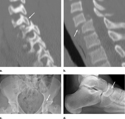
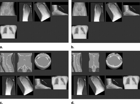
Each of 70 simulated multitrauma patients presented examinations of three anatomic areas. Readers evaluated each patient under two experimental conditions: when the images of the first anatomic area included a fracture (the SOS condition), and when it did not (the control condition). The SOS effect was measured on detection accuracy for subtle test fractures presented on examinations of the second and third anatomic areas. In an experiment with 12 radiology readers, the initial SOS radiographs showed nondisplaced fractures of extremities, fractures associated with low morbidity. In another experiment with 12 different radiology readers, the initial examination, usually a computed tomography scan, showed cervical and pelvic fractures of the type associated with high morbidity. Because of their more direct role in patient care, the experiment using high morbidity SOS fractures was repeated with 17 orthopedic readers.
Results
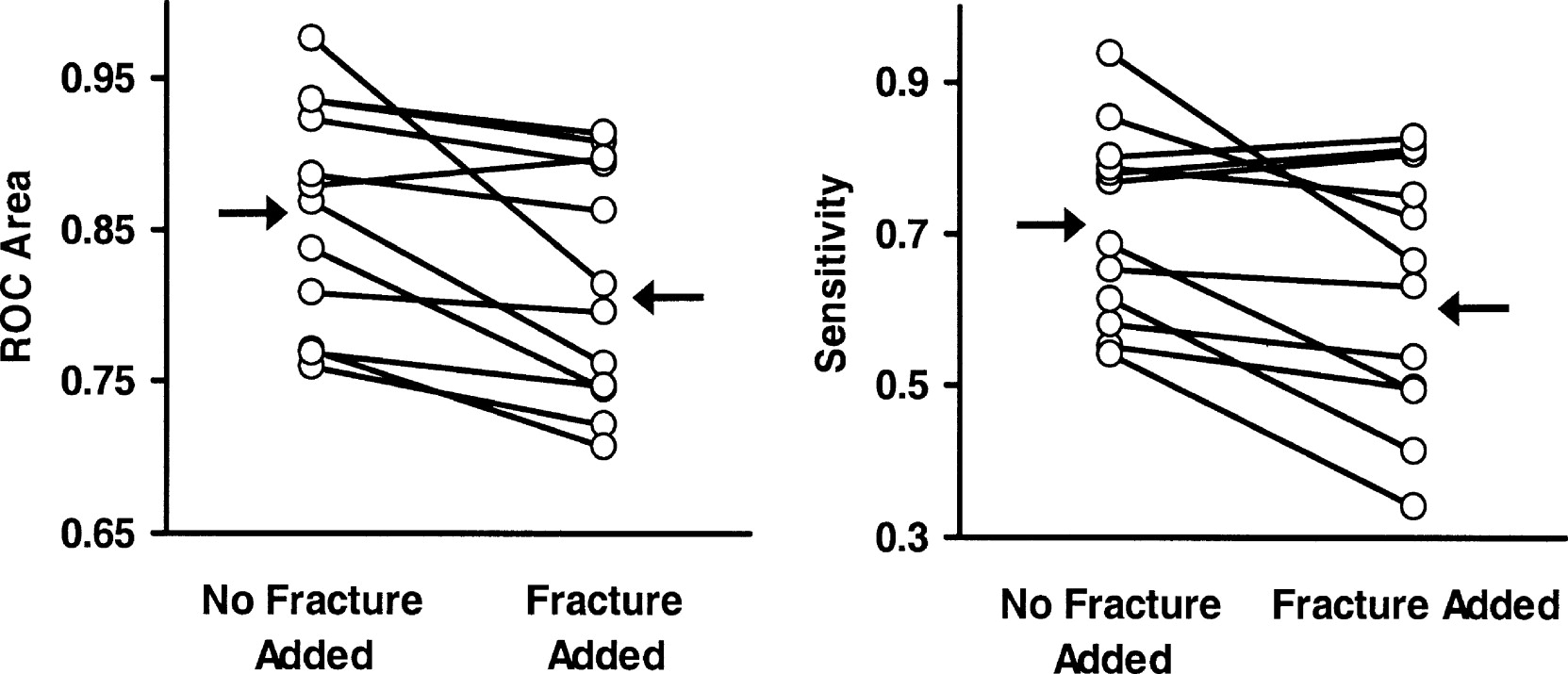
Detection of subtle test fractures was substantially reduced when fractures of low morbidity were added ( P < .01). No similar SOS effect was observed in either experiment in which added fractures were associated with high morbidity.
Conclusions
The satisfaction of search effect in skeletal radiology was replicated, essentially doubling the evidence for SOS in musculoskeletal radiology, and providing an essential contrast to the absence of SOS from high-morbidity fractures.
Physicians have long been aware that an injury may draw and hold their attention, diverting it from other injuries ( ). A “satisfaction of search” (SOS) effect has been demonstrated in which the discovery of a fracture on one image interfered with the detection of a subtle fracture on another image of the same patient ( ). Detection of subtle test fractures was substantially reduced when additional fractures were included in other images of each multitrauma series. The clinical practice of radiology has changed greatly since this experiment was performed in 1994. Whereas the previous experiment used radiographs interpreted at a film viewer, current practice relies heavily on computed tomography and direct digital radiography with interpretation at workstations equipped with high-resolution displays. The first experiment reported here attempts to replicate the SOS effect of finding “minor” added fractures on subsequent test fractures in a patient’s multitrauma series using modern images and displays.
In 2001, gaze time on fractures was measured in an attempt to determine whether test fractures were missed in this SOS effect because of misdirection of attention ( ). Although readers spent somewhat less time inspecting subsequent radiographs of a patient’s trauma series after viewing initial radiographs that contained added fractures, they generally did look at the test fractures that they failed to report. When the experiment was repeated to examine whether the severity of the added fracture affected search, test fractures were missed more often when they appeared with major added fractures than with minor added fractures. Because there were only 10 cases and only one that included no test fracture, true- and false-positive rates could not be estimated with acceptable precision or certainty. We address this question in the second and third experiments using receiver operating characteristic (ROC) methodology, testing whether added fractures with major morbidity yield greater SOS.
Get Radiology Tree app to read full this article<
Get Radiology Tree app to read full this article<
Materials and methods
Get Radiology Tree app to read full this article<
Imaging Material
Get Radiology Tree app to read full this article<
Get Radiology Tree app to read full this article<
Get Radiology Tree app to read full this article<
Simulated Multitrauma Patients
Get Radiology Tree app to read full this article<
Get Radiology Tree app to read full this article<
Get Radiology Tree app to read full this article<
Get Radiology Tree app to read full this article<
Get Radiology Tree app to read full this article<
Get Radiology Tree app to read full this article<
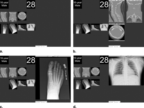
Display
Get Radiology Tree app to read full this article<
Get Radiology Tree app to read full this article<
Get Radiology Tree app to read full this article<
Readers
Get Radiology Tree app to read full this article<
Procedure
Get Radiology Tree app to read full this article<
Get Radiology Tree app to read full this article<
Get Radiology Tree app to read full this article<
Get Radiology Tree app to read full this article<
ROC Analysis and Statistical Analysis
Get Radiology Tree app to read full this article<
Get Radiology Tree app to read full this article<
Results
Get Radiology Tree app to read full this article<
Experiment 1: Radiology Readers and Minor Added Fractures
Get Radiology Tree app to read full this article<
Get Radiology Tree app to read full this article<
Get Radiology Tree app to read full this article<
Experiment 2: Radiology Residents and Fellows and Major Added Fractures
Get Radiology Tree app to read full this article<
Get Radiology Tree app to read full this article<
Get Radiology Tree app to read full this article<
Experiment 3: Orthopedic Surgery Residents and Fellows and Major Distractors
Get Radiology Tree app to read full this article<
Get Radiology Tree app to read full this article<
Get Radiology Tree app to read full this article<
Detectability of Added Fractures
Get Radiology Tree app to read full this article<
Get Radiology Tree app to read full this article<
Discussion
Get Radiology Tree app to read full this article<
Get Radiology Tree app to read full this article<
Get Radiology Tree app to read full this article<
Get Radiology Tree app to read full this article<
Get Radiology Tree app to read full this article<
Get Radiology Tree app to read full this article<
Get Radiology Tree app to read full this article<
Appendix
Quantifying the Magnitude of the SOS Effect
Get Radiology Tree app to read full this article<
Get Radiology Tree app to read full this article<
Get Radiology Tree app to read full this article<
References
1. Rogers L.F., Hendrix R.W.: Evaluating the multiply injured patient radiologically. Orthop Clin North Am 1990; 21: pp. 437-447.
2. Berbaum K.S., El-Khoury G.Y., Franken E.A., et. al.: Missed fractures resulting from satisfaction of search effect. Emerg Radiol 1994; 1: pp. 242-249.
3. Berbaum K.S., Brandser E.A., Franken E.A., et. al.: Gaze dwell times on acute trauma injuries missed because of satisfaction of search. Acad Radiol 2001; 8: pp. 304-314.
4. Rogers L.F.: 1982.Churchill-LivingstoneNew York
5. Rogers L.F.: Common oversights in the evaluation of the patient with multiple injuries. Skel Radiol 1984; 12: pp. 103-111.
6. LeBlang S.D., Nunez D.B.: Helical CT of cervical spine and soft tissue injuries of the neck. Radiol Clin North Am 1999; 37: pp. 515-532.
7. Nunez D.B., Quencer R.M.: The role of helical CT in the assessment of cervical spine injuries. Am J Roentgenol 1998; 171: pp. 951-957.
8. Berne J.D., Velmahos G.C., El-Tawil Q., et. al.: Value of complete cervical helical computed tomographic scanning in identifying cervical spine injury in the unevaluable blunt trauma patient with multiple injuries: a prospective study. J Trauma Inj Infect Crit Care 1999; 47: pp. 896-903.
9. Novelline R.A., Rhea J.T., Rao P.M., et. al.: Helical CT in emergency radiology. Radiology 1999; 213: pp. 321-339.
10. Blackmore C.C., Mann F.A., Wilson A.J.: Helical CT in the primary trauma evaluation of the cervical spine: an evidence-based approach. Skel Radiol 2000; 29: pp. 632-639.
11. Dorfman D.D., Berbaum K.S., Metz C.E.: Receiver operating characteristic rating analysis: generalization to the population of readers and patients with the jackknife method. Invest Radiol 1992; 27: pp. 723-731.
12. Dorfman D.D., Berbaum K.S., Lenth R.V., et. al.: Monte Carlo validation of a multireader method for receiver operating characteristic discrete rating data: Factorial experimental design. Acad Radiol 1998; 5: pp. 591-602.
13. Hillis S.L., Berbaum K.S.: Power estimation for the Dorfman-Berbaum-Metz method. Acad Radiol 2004; 11: pp. 1260-1273.
14. Hillis S.L., Obuchowski N.A., Schartz K.M., et. al.: A comparison of the Dorfman-Berbaum-Metz and Obuchowski-Rockette methods for receiver operating characteristic (ROC) data. Stat Med 2005; 24: pp. 1579-1607.
15. Hillis S.L.: Monte Carlo validation of the Dorfman-Berbaum-Metz method using normalized pseudovalues and less data-based model simplification. Acad Radiol 2005; 12: pp. 1534-1541.
16. Hillis S.L.: A comparison of denominator degrees of freedom methods for multiple observer ROC analysis. Stat Med 2007; 26: pp. 596-619.
17. Dorfman D.D., Berbaum K.S.: A contaminated binormal model for ROC data. Acad Radiol 2000; 7: pp. 427-437.
18. Dixon W.J.: BMDP statistical software manual1992.University of California PressBerkeley, CA 155–174; 201–227; 521–564
19. BMDP3D, BMDP7D, and BMDP2V, release: 8.0. Copyright 1993 by BMDP Statistical Software, Inc. Statistical Solutions Ltd., Cork, Ireland ( http://www.statsol.ie ).
20. Berbaum K.S., Franken E.A., Dorfman D.D., et. al.: Cause of satisfaction of search effects in contrast studies of the abdomen. Acad Radiol 1996; 3: pp. 815-826.
21. Madsen M.T., Berbaum K.S., Ellingson A.N., et. al.: A new software tool for removing, storing and adding abnormalities to medical images for perception research studies. Acad Radiol 2006; 13: pp. 305-312.
22. McNemar Q.: Psychological statistics.1969.WileyNew York:pp. 54-58.