
Because the four letters of correspondence describe a variety of concerns, I have organized them according to their general topic and in some instances re-emphasized data from the original reports .
Background Annual Percent Change (APC) in Breast Cancer Incidence
Drs. Baker and Kopans cite Helvie et al. in contending that the background APC since 1980 has been ≥1. Reasons why this is an overestimate include:
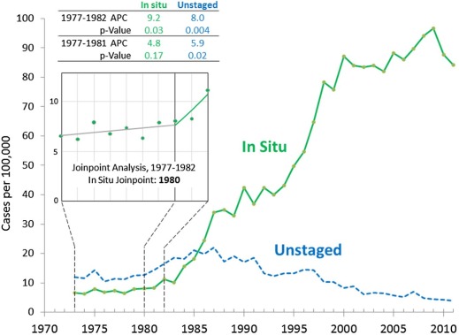
- 1. Helvie et al.’s central APC estimate of 1.3 from the Connecticut data was based on only 6 years, 1977–1982. Visual inspection of the in situ incidence shows that the incidence has already begun to rise in 1982 ( Fig 1 , green curve). Joinpoint analysis demonstrates 1980 as the year of change in slope ( Fig 1 , inset). When the last year, 1982, is not included, the APC for in situ cancer decreases 48% from a statistically significant rate of 9.2 ( P = 0.03) for 1977–1982 to a nonstatistically significant rate of 4.8 for 1977–1981 ( Fig 1 , table).
2. The Health Insurance Plan of Greater New York, next door to Connecticut, had been studying screening mammography since 1963 and favorable results have been reported by 1973 . By 1982, 5–10% of Connecticut’s women were already undergoing screening .
3. The incidence of unstaged breast cancer also increased by 1982 ( Fig 1 , blue curve), as expected from the uncertainty of the pathologic findings that occurred in the initial group of screened women. A few years later, when ductal carcinoma in situ (DCIS) became an established pathologic entity, the incidence of unstaged cancers declined. When 1982 is not included in the 1977–1982 calculation, the APC in unstaged cancer decreases from 8.0 ( P = 0.00) to 5.9 ( P = 0.02) ( Fig 1 , table).
Get Radiology Tree app to read full this article<
Get Radiology Tree app to read full this article<
Get Radiology Tree app to read full this article<
Diversity of Overdiagnosis Estimates
Get Radiology Tree app to read full this article<
Proportion of Women Screened
Get Radiology Tree app to read full this article<
Nonexistence of Overdiagnosis
Get Radiology Tree app to read full this article<
Get Radiology Tree app to read full this article<
women per year of screening age who are diagnosed in the United States to have DCIS and treated for breast cancer. On one hand, Drs. Kopans and Arleo would have us eliminate DCIS from any consideration of overdiagnosis. On the other hand, Drs. Arleo and Baker and other mammographers want all women with DCIS treated for cancer because we cannot (as yet) distinguish women with preinvasive cancer from those with indolent, nonmalignant lesions. As the saying goes, you cannot have it both ways (eliminate DCIS and treat all as cancer). David Seidenwurm, MD, of the Diagnostic Division of Radiologic Associates of Sacramento may have said it best: “We cannot discuss mammography without DCIS any more than we can discuss DCIS without mammography” .
Get Radiology Tree app to read full this article<
Impact of Screening Mammography on Late-stage Disease
Get Radiology Tree app to read full this article<
Get Radiology Tree app to read full this article<
Get Radiology Tree app to read full this article<
RCTs of Screening Mammography
Get Radiology Tree app to read full this article<
Get Radiology Tree app to read full this article<
Get Radiology Tree app to read full this article<
Role of Spontaneous Regression
Get Radiology Tree app to read full this article<
Get Radiology Tree app to read full this article<
Final Comments
Get Radiology Tree app to read full this article<
Get Radiology Tree app to read full this article<
Get Radiology Tree app to read full this article<
Get Radiology Tree app to read full this article<
Acknowledgment(s)
Get Radiology Tree app to read full this article<
Get Radiology Tree app to read full this article<
References
1. Bleyer A., Welch H.G.: Effect of three decades of screening mammography on the incidence of breast cancer. NEJM 2012; 367: pp. 1998-2005.
2. Bleyer A.: Were our estimates of overdiagnosis with mammography screening in the United States “based on faulty science?”. Oncologist 2014; 19: pp. 113-126.
3. Bleyer A.: Screening mammography: update and review of publications since our report in the New England Journal of Medicine on the magnitude of the problem in the United States. Acad Radiol 2015; 22: pp. 949-960.
4. Helvie M.A., Chang J.T., Hendrick R.E.: Reply to flawed assumptions used to defend screening mammography. Cancer 2015; 121: pp. 321-323.
5. Heston J.F., Cusano M.M., Young J.L., et. al.: Forty-five years of cancer incidence in Connecticut: 1935–1979. Natl Cancer Inst Monogr 1986; 70: pp. 1-706.
6. Howard J.: Using mammography for cancer control: an unrealized potential. CA Cancer J Clin 1987; 37: pp. 33-48.
7. Shapiro S.: The status of breast cancer screening: a quarter of a century of research. World J Surg 1989; 13: pp. 9-18.
8. Devesa S.S., Silverman D.T., Young J.L., et. al.: Cancer incidence and mortality trends among whites in the United States, 1947-84. J Natl Cancer Inst 1987; 79: pp. 701-770.
9. Christiansen P., Vejborg I., Kroman N., et. al.: Position paper: breast cancer screening, diagnosis, and treatment in Denmark. Acta Oncol 2014; 53: pp. 433-444.
10. Sigurdsson K., Olafsdottir E.J.: Population-based service mammography screening: the Icelandic experience. Breast Cancer (Dove Med Press) 2013; 5: pp. 17.
11. Demographics of Iceland. Available at: http://en.wikipedia.org/wiki/Demographics_of_Iceland Accessed August 3, 2015
12. Cronin K.A., Feuer E.J., Clarke L.D., et. al.: Impact of adjuvant therapy and mammography on U.S. mortality from 1975 to 2000: comparison of mortality results from the CISNET breast cancer base case analysis. J Natl Cancer Inst Monogr 2006; pp. 112-121.
13. Yankaskas B.C.: Epidemiology of breast cancer in young women. Breast Dis 2005-2006; 23: pp. 3-8.
14. Collaborative Group on Hormonal Factors in Breast Cancer: Breast cancer and breastfeeding: collaborative reanalysis of individual data from 47 epidemiological studies in 30 countries, including 50302 women with breast cancer and 96973 women without the disease. Lancet 2002; 360: pp. 187-195.
15. Helewa M., Lévesque P., Provencher D., et. al.: Breast cancer, pregnancy, and breastfeeding. J Obstet Gynaecol Can 2002; 24: pp. 164-180.
16. Etzioni R., Xia J., Hubbard R., et. al.: A reality check for overdiagnosis estimates associated with breast cancer screening. J Natl Cancer Inst 2014; 106:
17. Baker S.G., Prorok C., Kramer B.S.: Lead time and overdiagnosis. J Natl Cancer Inst 2014; 106: pp. dju346.
18. Use of mammography among women 40 years of age and over, by selected characteristics: United States, selected years 1987–2008. Table 86 (page 1 of 3). Atlanta: Centers for Disease Control and Prevention. Available at: http://www.cdc.gov/nchs/data/hus/2010/086.pdf Accessed August 4, 2015
19. Breast Cancer Surveillance Consortium mammography data. Available at: http://breastscreening.cancer.gov/statistics/mammography_data.html Accessed August 4, 2015
20. Seidenwurm D.: Counterpoint: the New England Journal of Medicine article suggesting overdiagnosis from mammography screening is scientifically correct and should not be withdrawn. J Am Coll Radiol 2013; 10: pp. 320-323.
21. Surveillance, Epidemiology, and End Results (SEER) Program. SEER*Stat Database: Incidence—SEER 18 Regs Research Data + Hurricane Katrina Impacted Louisiana Cases, Nov 2014 Sub (1973-2012 varying)—Linked To County Attributes—Total U.S., 1969-2013 Counties, National Cancer Institute, DCCPS, Surveillance Research Program, Surveillance Systems Branch, released April 2015, based on the November 2014 submission. Available at: www.seer.cancer.gov Accessed August 4, 2015
22. Brodersen J., Jørgensen K.J.: The balance is shifting away from screening. Clin Adv Hematol Oncol 2014; 12: pp. 407-409.
23. Harding C., Pompei F., Burmistrov D., et. al.: Breast cancer screening, incidence, and mortality across U.S. counties. JAMA Intern Med 2015; 175: pp. 1483-1489. Published online July 6, 2015
24. Appendix of randomized controlled trials. Available at http://www.cancer.gov/types/breast/hp/breast-screening-pdq/#section/_377 Accessed August 4, 2015
25. Miller A.B., Wall C., Baines C.J., et. al.: Twenty five year follow-up for breast cancer incidence and mortality of the Canadian National Breast Screening Study: randomised screening trial. BMJ 2014; 348: pp. g366.
26. Gøtzsche P.C., Nielsen M.: Screening for breast cancer with mammography (Review). Cochrane Database Syst Rev 2011; CD001877
27. Marmot M.G.: Sorting through the arguments on breast screening. JAMA 2013; 309: pp. 2553-2554.
28. Newman D.H.: Screening for breast and prostate cancers. Moving toward transparency. J Natl Cancer Inst 2010; 102: pp. 1008-1011.
29. Breast cancer screening for health professionals: Benefits of screening: Randomized controlled trials; Summary. Available at: http://www.cancer.gov/types/breast/hp/breast-screening-pdq#link/_101_toc Accessed August 4, 2015
30. Walter L.C., Schonberg M.A.: Screening mammography in older women: a review. JAMA 2014; 311: pp. 1336-1347.
31. Lee S.J., Boscardin W.J., Stijacic-Cenzer I., et. al.: Time lag to benefit after screening for breast and colorectal cancer: meta-analysis of survival data from the United States, Sweden, United Kingdom, and Denmark. BMJ 2013; 346: pp. e8441.
32. Osler W.: The medical aspects of carcinoma of the breast, with a note on the spontaneous disappearance of secondary growths. Amer Med Phila 1901; 17: pp. 63-66.
33. Osler W.: An address on the medical aspects of carcinoma of the breast. Br Med J 1906; 1: pp. 1-4.
34. Rohdenburg G.L.: Fluctuations in the growth energy of tumors in man, with special reference to spontaneous recession. J Cancer Res 1918; 3: pp. 193-225.
35. Fauvet J., Roujeau J., Piet R.: Spontaneous cancer cures and regressions. Rev Prat 1964; 14: pp. 2177-2180. in French
36. Challis G.B., Stam H.J.: The spontaneous regression of cancer. A review of cases from 1900 to 1987. Acta Oncol 1990; 29: pp. 545-550.
37. Nielsen M., Thomsen J.L., Primdahl S., et. al.: Breast cancer and atypia among young and middle-aged women: a study of 110 medicolegal autopsies. Br J Cancer 1987; 56: pp. 814-819.
38. Cancer Query System: probability of developing or dying of cancer. Available at: http://surveillance.cancer.gov/devcan/canques.html Accessed August 3, 2015
39. Bhathal P.S., Brown R.W., Lesueur G.C., et. al.: Frequency of benign and malignant breast lesions in 207 consecutive autopsies in Australian women. Br J Cancer 1985; 51: pp. 271-278.
40. Alpers C.E., Wellings S.R.: The prevalence of carcinoma in situ in normal and cancer-associated breasts. Hum Pathol 1985; 16: pp. 796-807.
41. Breast screening: helping you decide. National Health Service. NHS Cancer Screening Programmes 2901195, pp. 9–11. Available at: https://www.orderline.dh.gov.uk/ecom_dh/public/saleproduct.jsf?catalogueCode=2901195 Accessed August 4, 2015
42. Research Council of Norway : Research-based evaluation of the Norwegian Breast Cancer Screening Program Final report. The Research Council of Norway, Oslo, May; p. 9. Available at: http://www.forskningsradet.no/servlet/Satellite?pagename=ForskningsradetEngelsk%2FHovedsidemal&cid=1214551683682&c=InnholdsKontainer&p=1178541060225&querystring=Breast+Cancer+Screening+Program&sortby=title&sortorder=asc&hits=30&configuration=nfrcspublikasjonsppublished&publicationType=ALLE Accessed August 4, 2015
43. Woods W.G.: Substitute “prostate cancer” for “neuroblastoma?”. J Clin Oncol 2002; 20: pp. 1154-1155.
44. Patz E.F., Pinsky P., Gatsonis C., et. al.: Overdiagnosis in low-dose computed tomography screening for lung cancer. JAMA Intern Med 2014; 174: pp. 269-274.
45. Weyers W.: The “epidemic” of melanoma between under- and overdiagnosis. J Cutan Pathol 2012; 39: pp. 9-16.
46. Spix C., Michaelis J., Berthold F., et. al.: Lead-time and overdiagnosis estimation in neuroblastoma screening. Stat Med 2003; 22: pp. 2877-2892.
47. Loeb S., Bjurlin M.A., Nicholson J., et. al.: Overdiagnosis and overtreatment of prostate cancer. Eur Urol 2014; 65: pp. 1046-1055.
48. O’Grady T.J., Gates M.A., Boscoe F.P.: Thyroid cancer incidence attributable to overdiagnosis in the United States 1981-2011. Int J Cancer 2015; 137: pp. 2664-2673.