
Rationale and Objectives
The aim of this study was to determine whether a lateral chest radiograph provides additional diagnostic information to a posteroanterior (PA) radiograph in the screening of asymptomatic children with positive purified protein derivative (PPD) skin tests in a nonendemic area.
Materials and Methods
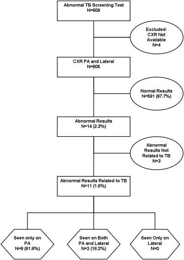
This was an Institutional Review Board–approved, Health Insurance Portability and Accountability Act–compliant, retrospective study of 605 consecutive pediatric patients (294 males, 311 females; mean age, 10.8 ± 5.2 years) with positive PPD skin test results, who underwent PA and lateral chest radiographs between July 2003 and May 2009 at a tertiary care pediatric hospital in a nonendemic area for tuberculosis (TB). Two pediatric radiologists independently reviewed each chest radiograph for evidence of abnormalities that may be indicative of acute or chronic TB infection. The reviewers first analyzed the PA radiograph alone and subsequently evaluated the PA and the lateral radiograph together to determine whether any observed abnormality was identified only on the lateral radiograph. When an abnormality was detected on both PA and lateral radiographs, the reviewers determined whether the abnormality on the lateral radiograph changed the reviewer’s decision based on the PA radiograph alone. Assessment of nonconcordance between PA and lateral chest radiographs for each reviewer was evaluated by the McNemar test of matched binary pairs. Agreement between reviewers for detecting abnormalities on radiographs was evaluated by using the kappa (κ) statistic.
Results
The frequency of an abnormal chest radiograph related to TB was 1.8% (11/605). The PA radiograph showed abnormalities in all 11 (100%) children with radiographic abnormalities. Lateral radiographs showed abnormalities related to TB in 2 (18.2%) of 11 cases found to be abnormal on PA radiographs. Nine (81.8%) of 11 abnormalities on PA radiographs were not detected on the lateral chest radiographs. There was statistical evidence of nonconcordance between PA and lateral chest radiographs in detecting TB-related abnormalities for reviewer 1 ( P < .001) and reviewer 2 ( P = .004). In cases with abnormalities observed on both PA and lateral radiographs, there were no cases in which information obtained from the lateral chest radiograph resulted in a change in interpretation based on the PA radiograph alone. A high level of agreement was observed between the two independent reviewers in detecting TB-related abnormalities on PA radiographs (κ = 0.84, P < .001).
Conclusions
A PA radiograph alone is sufficient for TB screening of asymptomatic pediatric patients with positive PPD skin test results in an area non-endemic for TB.
Despite increasing prevention efforts, tuberculosis (TB) continues to be a major cause of morbidity and mortality in children . Although it is widely recognized that chest radiographs are indicated for children with positive purified protein derivative (PPD) skin test results , there is controversy regarding how the examination should be performed.
Both the Centers for Disease Control and Prevention and the American Thoracic Society recommend that pediatric TB radiographic screening should consist of both posteroanterior (PA) and lateral radiographs in children <5 years of age and a PA radiograph alone in children >5 years of age, with additional views “at the physician’s discretion” . However, at many centers, both views are commonly obtained in children of all ages. Whereas studies of adults have shown that a PA radiograph alone is sufficient for TB screening of individuals with positive PPD skin test results , a previous study of children in an area endemic for TB recommended that a lateral radiograph be obtained routinely, regardless of the patient’s age .
Get Radiology Tree app to read full this article<
Materials and methods
Institutional Review Board Approval
Get Radiology Tree app to read full this article<
Patient Population
Get Radiology Tree app to read full this article<
Get Radiology Tree app to read full this article<
Get Radiology Tree app to read full this article<
Imaging Technique
Get Radiology Tree app to read full this article<
Chest Radiograph Review and Evaluation
Chest radiograph review
Get Radiology Tree app to read full this article<
Chest radiograph evaluation
Get Radiology Tree app to read full this article<
Get Radiology Tree app to read full this article<
Get Radiology Tree app to read full this article<
Statistical Analysis
Get Radiology Tree app to read full this article<
Results
Imaging Findings on Posteroanterior Radiographs
Get Radiology Tree app to read full this article<
Get Radiology Tree app to read full this article<
Get Radiology Tree app to read full this article<
Get Radiology Tree app to read full this article<
Imaging Findings on Lateral Radiographs
Get Radiology Tree app to read full this article<
Get Radiology Tree app to read full this article<
Comparison of PA and Lateral Radiographs
Get Radiology Tree app to read full this article<
Get Radiology Tree app to read full this article<
Table 1
Assessment of Concordance Between PA and Lateral Chest Radiographs
Reviewer 1 Reviewer 2 Concordance of negative cases 590 589 Concordance of positive cases 3 7 Nonconcordance 12 9 PA negative, lateral positive 0 0 PA positive, lateral negative 12 9
PA, posteroanterior.
There was a highly significant level of nonconcordance between PA and lateral chest radiographs for reviewer 1 ( P < .001) and reviewer 2 ( P = .004) as evaluated by the McNemar test for binary matched pairs.
Get Radiology Tree app to read full this article<
Interobserver Agreement
Get Radiology Tree app to read full this article<
Table 2
Interobserver Agreement for Detecting TB-related Abnormalities on CXR ∗
PA Chest Radiographs Lateral Chest Radiographs Original readings κ value (95% CI) 0.84 (0.72–0.96) 0.40 (0.10–0.70) Agreement on negative cases 587 597 Agreement on positive cases 13 2 Nonagreement 5 6 Postarbitration readings Compared with reviewer 1 κ value (95% CI) 0.89 (0.79–0.99) 0.57 (0.17–0.97) Agreement on negative cases 589 600 Agreement on positive cases 13 2 Nonagreement 3 3 Compared with reviewer 2 κ value (95% CI) 0.93 (0.85–1.00) 0.55 (0.20–0.90) Agreement on negative cases 589 597 Agreement on positive cases 14 3 Nonagreement 2 5
CI, confidence interval; CXR, chest x-ray; κ, kappa coefficient; PA, posteroanterior; TB, tuberculosis.
Get Radiology Tree app to read full this article<
Get Radiology Tree app to read full this article<
Discussion
Get Radiology Tree app to read full this article<
Get Radiology Tree app to read full this article<
Get Radiology Tree app to read full this article<
Get Radiology Tree app to read full this article<
Get Radiology Tree app to read full this article<
Get Radiology Tree app to read full this article<
Get Radiology Tree app to read full this article<
References
1. Dye C., Lonnroth K., Jaramillo E., et. al.: Trends in tuberculosis incidence and their determinants in 134 countries. Bull World Health Organ 2009; 87: pp. 683-691.
2. Kabra S.K., Ladha R., Seth V.: Tuberculosis in children—what has changed in last 20 years?. Indian J Pediatr 2002; 69: pp. S5-S10.
3. Kabra S.K., Lodha R., Seth V.: Some current concepts on childhood tuberculosis. Indian J Med Res 2004; 120: pp. 387-389.
4. Marais B.J., Gie R.P., Schaaf H.S., et. al.: Childhood pulmonary tuberculosis: old wisdom and new challenges. Am J Respir Crit Care Med 2006; 173: pp. 1078-1090.
5. Marais B.J., Obihara C.C., Warren R.M., et. al.: The burden of childhood tuberculosis: a public health perspective. Int J Tuberc Lung Dis 2005; 9: pp. 1305-1313.
6. Communicable Diseases, Tuberculosis, Factsheets, TB and Children. World Health Organization. Available online at: http://www.searo.who.int/en/Section10/Section2097/Section2106_10681.htm . Updated April 27, 2006. Accessed March 23, 2010.
7. MMWR. Recommendations and Reports, Screening for Tuberculosis and Tuberculosis Infection in High-Risk Populations Recommendations of the Advisory Council for the Elimination of Tuberculosis. Centers for Disease Control and Prevention. Available online at: http://www.cdc.gov/mmwr/preview/mmwrhtml/00038873.htm . Published September 8, 1995. Updated September 9, 1998. Accessed March 23, 2010.
8. MMWR. Recommendations and Reports, Targeted Tuberculin Testing and Treatment of Latent Tuberculosis Infection. Centers for Disease Control and Prevention Website. Available online at: http://www.cdc.gov/mmwr/preview/mmwrhtml%5Crr4906a1.htm . Published June 9, 2000. Updated June 9, 2000. Accessed March 23, 2010.
9. Targeted tuberculin testing and treatment of latent tuberculosis infection. Am J Respir Crit Care Med 2000; 161: pp. S221-S247.
10. Eisenberg R.L., Romero J., Litmanovich D., et. al.: Tuberculosis: value of lateral chest radiography in pre-employment screening of patients with positive purified protein derivative skin test results. Radiology 2009; 252: pp. 882-887.
11. Meyer M., Clarke P., O’Regan A.W.: Utility of the lateral chest radiograph in the evaluation of patients with a positive tuberculin skin test results. Chest 2003; 124: pp. 1824-1827.
12. Smuts N.A., Beyers N., Gie R.P., et. al.: Value of lateral chest radiograph in tuberculosis in children. Pediatr Radiol 1994; 24: pp. 478-480.
13. Hansell D.M., Bankier A.A., MacMahon H., et. al.: Fleischner Society: glossary of terms for thoracic imaging. Radiology 2008; 246: pp. 697-722.
14. Muller N.L., Silva C.I.S.: Mediastinum.Muller N.L.Silva C.I.S.Imaging of the chest.2008.Elsevier SaundersPhiladelphia, PA:pp. 1447-1572.
15. Kundel H.L., Polansky M.: Measurement of observer agreement. Radiology 2003; 228: pp. 303-308.
16. Landis J.R., Koch G.G.: The measurement of observer agreement for categorical data. Biometrics 1977; 33: pp. 159-174.
17. National Center for HIV/AIDS, viral hepatitis, STD, and TB prevention. Massachusetts–2008 Profile. Centers for Disease Control and Prevention. Available online at: http://www.cdc.gov/nchhstp/stateprofiles/pdf/massachusetts_profile.pdf Published March 23, 2009. Updated June 24, 2009. Accessed March 30, 2010.
18. Kim W.S., Choi J.I., Cheon J.E., et. al.: Pulmonary tuberculosis in infants: radiographic and CT findings. AJR Am J Roentgenol 2006; 187: pp. 1024-1033.