
Rationale and Objectives
To explore whether strain ratio measurement could semi-quantitatively evaluate the stiffness of breast lesions.
Materials and Methods
From January 2008 to May 2008, 148 patients with 254 solid lesions (183 benign, 71 malignant) in the breast were included in the study. Ultrasound sonography found the lesions and ultrasonic elastography obtained the strain images. By using the strain ratio measurement method together with the ultrasound machine, the strain index of the lesion was calculated. Different depths of breast tissue were selected as the reference. The strain indexes of malignant and benign solid lesions were calculated with the same level of breast tissue as the reference.
Results
The strain indexes of breast lesions were different compared to the same depth of breast tissue and the superior level of fat tissue ( P = 0.000). The strain indexes of breast lesions were different compared to different depths of breast glandular tissues ( P = 0.003). At the same level of the breast lesions, 212 lesions were glandular tissue, 11 were fat tissue, and 40 were both. In the lesion plane, six lesions had almost no glandular tissue and 20 had almost no superior fat tissue. Compared to the same depth of breast tissue, the strain indexes of benign lesions (range, 0.62–11.07) and malignant lesions (range, 3.12–39.28) were different ( P = 0.000).
Conclusion
Using the strain ratio measurement, stiffness of breast lesions could be semi-quantitated with the same depth of breast tissue as the reference. This method may provide another diagnostic method in addition to the 5-point scoring system used with ultrasonic elastography in the future.
An important property of tissues is their intrinsic elasticity, which may change under the influence of pathophysiologic processes, such as tumor development ( ). This property serves as the basis for the oldest and most frequently used physical examination method for detecting breast cancers—palpation. Harder and less mobile lesions are considered more likely to be malignant ( ). Unfortunately, palpation is a highly subjective method and depends on the size and location of the lesion and on the skill of the practitioner. These changes in elasticity drove the development of elasticity measurement, which began in the 1980s ( ).
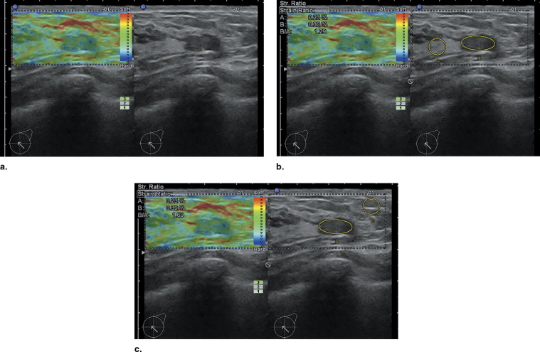
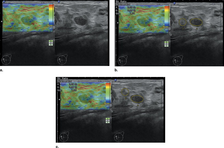
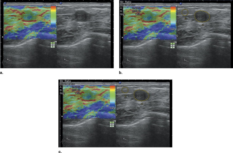
Elastography is a newly developed dynamic technique that uses ultrasound (US) to provide an estimation of tissue elasticity by measuring the degree of distortion under the application of an external force ( ). The principle of elastography is that tissue compression produces displacement within the tissue and that the displacement is smaller in harder tissue than in softer tissue. Real-time ultrasonic elastography (UE) allows calculation of tissue elasticity in real time and, similar to color Doppler, superimposes the information in color on the B-mode image. Different color represents different elasticity. Blue is hard, red is soft, and green is median. Although UE is not yet used in routine clinical practice, it has been shown to be useful in the differential diagnosis of breast cancer ( ), thyroid cancer ( ), and prostate cancer ( ), and attempts have been made to use it in the diagnosis of liver cirrhosis ( ).
Get Radiology Tree app to read full this article<
Get Radiology Tree app to read full this article<
Materials and methods
Patients
Get Radiology Tree app to read full this article<
Study Design
Get Radiology Tree app to read full this article<
Statistical Analysis
Get Radiology Tree app to read full this article<
Results
Get Radiology Tree app to read full this article<
Table 1
Histologic Diagnoses of Benign and Malignant Breast Lesions in Patients with Breast Lesions
Benign Lesions ( n = 183) Malignant Lesions ( n = 71) Histopathologic Diagnosis_n_ Histopathologic Diagnosis_n_ Fibroadenoma 117 Invasive ductal carcinoma 65 Fibrocystic mastopathy 47 Papillocarcinama 2 Papilloma 10 Invasive lobular carcinoma 1 Hyperplasia 3 Mucinous carcinoma 2 Lipoma 6 Lymphoma 1
Get Radiology Tree app to read full this article<
Get Radiology Tree app to read full this article<
Get Radiology Tree app to read full this article<
Get Radiology Tree app to read full this article<
Get Radiology Tree app to read full this article<
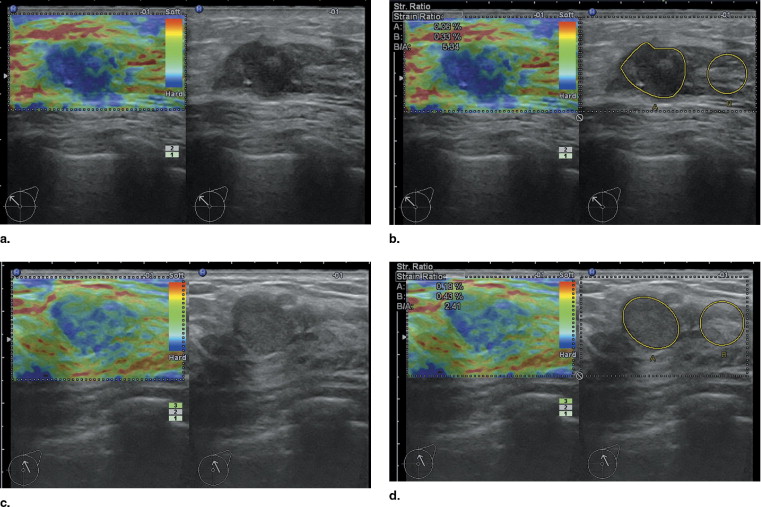
Table 2
Histopathology of 16 Benign Lesions Stiffer Than the Minimum of Malignancy
Histopathologic Diagnosis_N_ Fibroadenoma 6 Fibrocystic mastopathy 5 Papilloma 3 Lipoma 2
Get Radiology Tree app to read full this article<
Discussion
Get Radiology Tree app to read full this article<
Get Radiology Tree app to read full this article<
Get Radiology Tree app to read full this article<
Get Radiology Tree app to read full this article<
Get Radiology Tree app to read full this article<
Get Radiology Tree app to read full this article<
References
1. Bogonoletz W.V.: Elastosis in breast cancer. Pathol Annu 1986; 21: pp. 347-366.
2. Andersion W.A.D.: Pathology.1953.MosbySt Louis, MO
3. Ophir J., Barra B., Kallel F., et. al.: Elastographic imaging. Ultrasound Med Biol 2000; 26: pp. 23-29.
4. Ophir J., Céspedes I., Ponnekanti H., et. al.: Elastography: a quantitative method for imaging the elasticity of biological tissues. Ultrason Imaging 1991; 13: pp. 111-134.
5. O’Donnell M., Skovoroda A., Shapo B., Emelianov S.: Internal displacement and strain imaging using ultrasonic speckle tracking. IEEE Trans Ultrason Ferroelectr Freq Control 1994; 41: pp. 314-325.
6. Zhi H., Ou B., Luo B.M., et. al.: Comparison of ultrasound elastography, mammography, and sonography in the diagnosis of solid breast lesions. J Ultrasound Med 2007; 26: pp. 807-815.
7. Cho N., Moon W.K., Park J.S., et. al.: Nonpalpable breast masses: evaluation by US elastography. Korean J Radiol 2008; 9: pp. 111-118.
8. Asteria C., Giovanardi A., Pizzocaro A., et. al.: US-elastography in the differential diagnosis of benign and malignant thyroid nodules. Thyroid 2008; 18: pp. 523-531.
9. Rago T., Santini F., Scutari M., et. al.: Elastography: new developments in ultrasound for predicting malignancy in thyroid nodules. J Clin Endocrinol Metab 2007; 92: pp. 2917-2922.
10. Kamoi K., Okihara K., Ochiai A., et. al.: The utility of transrectal real-time elastography in the diagnosis of prostate cancer. Ultrasound Med Biol 2008; 34: pp. 1025-1032.
11. Nelson E.D., Slotoroff C.B., Gomella L.G., Halpern E.J.: Targeted biopsy of the prostate: the impact of color Doppler imaging and elastography on prostate cancer detection and Gleason score. Urology 2007; 70: pp. 1136-1140.
12. Friedrich-Rust M., Ong M.F., Herrmann E., et. al.: Real-time elastography for noninvasive assessment of liver fibrosis in chronic viral hepatitis. AJR Am J Roentgenol 2007; 188: pp. 758-764.
13. Itoh A., Ueno E., Tohno E., et. al.: Breast disease: clinical application of US elastography for diagnosis. Radiology 2006; 239: pp. 341-350.
14. Waki K., Murayama N., Matsumura T., Mitake T.: Investigation of strain ratio using ultrasound elastography technique.2007.pp. 449-452. In: Proceedings of ISICE: The First International Symposium on Information and Computer Elements, Kitakyushu, Japan, Sept. 12–14.
15. Jemal A., Murray T., Ward E., et. al.: Cancer statistics, 2005. CA Cancer J Clin 2005; 55: pp. 10-30.
16. Tabar L., Yen M.F., Vitak B., et. al.: Mammography service screening and mortality in breast cancer patients: 20-year follow-up before and after introduction of screening. Lancet 2003; 361: pp. 1405-1410.
17. Maskarinec G., Meng L., Ursin G.: Ethnic differences in mammographic densities. Int J Epidemiol 2001; 30: pp. 959-965.
18. Shiina T., Doyley M.M., Bamber J.C.: Strain imaging using combined RF and envelope autocorrelation processing.1996.Institute of Electrical and Electronics Engineers Ultrasonics, Ferroelectrics, and Frequency Control Digital ArchiveSavoy, Ill:pp. 1331-1336. In: Proceedings of the IEEE Ultrasonics Symposium. 2.
19. Lyshchik A., Higashi T., Asato R., et. al.: Thyroid gland tumor diagnosis at US elastography. Radiology 2005; 237: pp. 202-211.
20. Samani A., Zubovits J., Plewes D.: Elastic moduli of normal and pathological human breast tissues: an inversion-technique-based investigation of 169 samples. Phys Med Biol 2007; 52: pp. 1565-1576.
21. Bodian C.A., Perzin K.H., Lattes R., et. al.: Prognostic significance of benign proliferative breast disease. Cancer 1993; 71: pp. 3798-3807.
22. Hiltawsky K.M., Kruger M., Starke C., Ermert H., Jensen A.: Freehand ultrasound elastography of breast lesions: clinical results. Ultrasound Med Biol 2001; 27: pp. 1461-1469.