
Rationale and Objectives
To assess the feasibility of 70-kVp high-pitch non–ECG-gated thoracic aortic computed tomography angiography (CTA) with 40-mL contrast agent compared to 100-kVp standard-pitch CTA with 60-mL contrast agent.
Materials and Methods
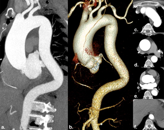
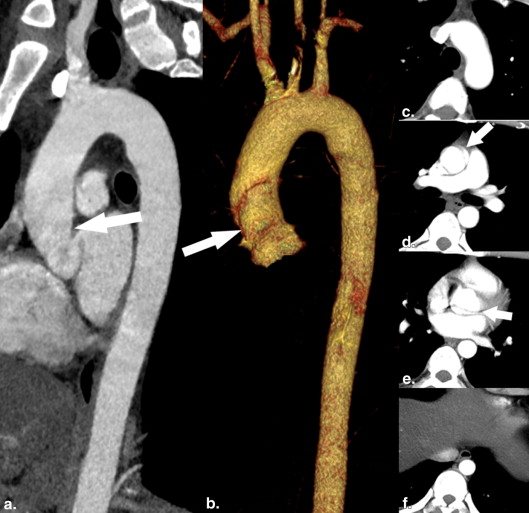
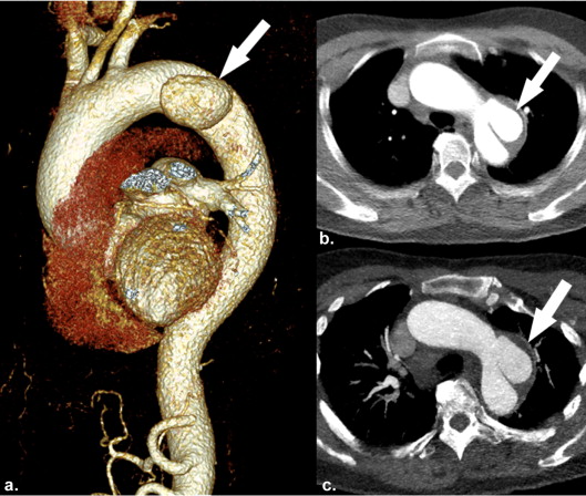
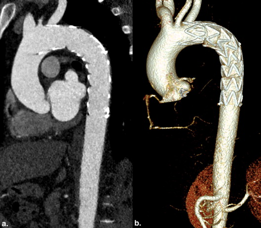
Sixty-seven patients (51 men and 16 women; mean age, 55 ± 14 years) received non–ECG-gated aortic CTA at 70 kVp, high pitch of 3.4, and 40-mL contrast agent (group A, n = 31) or CTA at 100-kVp, pitch of 1.2, and 60-mL contrast agent (group B, n = 36). Iterative reconstruction was used in all patients. For image quality assessment, CTA images were evaluated on a three-point scale and signal-to-noise ratio (SNR) and contrast-to-noise ratio (CNR) were calculated and compared. Furthermore, computed tomography (CT) dose index was recorded.
Results
Mean CT values and noise levels were higher in group A compared to group B (all P < .001), whereas SNR and CNR were lower than those in group B (all P < .001). Furthermore, the image quality of the aorta at the level of the diaphragm was lower in group A than that in group B ( P < .05). However, image quality was graded as diagnostic in all patients, and motion artifacts of the aortic arch were significantly decreased in group A ( P <.05). Interreader agreement was good or excellent for image quality assessment ( k = 0.625–0.835). The 70-kVp CTA protocol, which allows dose reduction of 85%, was considered diagnostic in all instances by two readers.
Conclusions
Our proposed thoracic aortic CTA protocol provides diagnostic information with substantial reduction of both radiation and contrast agent doses compared to standard-pitch CTA at 100 kVp.
Aortic disease accounts for significant cardiovascular morbidity and mortality worldwide . With high temporal and spatial resolution, versatile postprocessing techniques, and widespread availability, computed tomography angiography (CTA) has become the gold standard in the assessment of aortic diseases, especially in acute aortic syndrome . Furthermore, CTA is the preferred imaging modality for follow-up examinations in patients with thoracic aortic diseases . However, long-range scanning coverage of the aorta over multiple follow-up examinations results in a high cumulative radiation dose. Thus, reducing the radiation dose of aortic CTA is of great interest. Several CTA protocols that reduce radiation dose without compromising diagnostic image quality have been described in the literature. These include using lower tube voltage , automatic tube voltage selection , high-pitch or iterative reconstruction (IR) .
However, the potential to reduce radiation dose of aortic CTA examinations should be further explored by combining the previously mentioned radiation dose saving techniques. For example, the study by Farrelly et al. demonstrated a significant reduction of the effective dose (ED) from 26.2 ± 6.0 to 2.9 ± 0.5 mSv by performing a prospectively ECG-gated CTA of the thoracic aorta at 100 kVp instead of the standard retrospectively ECG-gated CTA at 120 kVp. In addition, CTA examinations at 70 kVp have recently been shown to provide diagnostic image quality with an ultralow radiation dose in selected patients undergoing imaging of peripheral, cerebral, and coronary arteries . However, to the best of our knowledge, there has been no published report on a 70-kVp CTA protocol of the thoracic aorta. Additionally, it is known that lower tube voltages allow the reduction of contrast agent as the attenuation of iodinated contrast agent increases at low tube voltages . This proves advantageous in certain situations, especially when scanning patients with preexisting renal dysfunction. Therefore, the aim of this study was to assess the feasibility of high-pitch 70-kVp CTA of the thoracic aorta with 40-mL contrast agent in comparison to the standard-pitch CTA of the aorta at 120 kVp with 60-mL contrast agent.
Materials and methods
Subject Population
Get Radiology Tree app to read full this article<
CT Imaging Protocol
Get Radiology Tree app to read full this article<
Get Radiology Tree app to read full this article<
Image Reconstruction and Analysis
Get Radiology Tree app to read full this article<
Get Radiology Tree app to read full this article<
Objective Analysis of Image Quality
Get Radiology Tree app to read full this article<
Subjective Analysis of Image Quality
Get Radiology Tree app to read full this article<
Estimation of Radiation Dose
Get Radiology Tree app to read full this article<
Statistical Analysis
Get Radiology Tree app to read full this article<
Results
Study Population
Get Radiology Tree app to read full this article<
Quantitative Image Analysis
Get Radiology Tree app to read full this article<
Table 1
Objective Measurements in Different Segments of the Aorta
Location Group A Group B_P_ Value Aortic root CT value (HU) 491.2 ± 124.5 352.7 ± 58.3 <.001 Noise (HU) 58.9 ± 10.9 24.9 ± 3.7 <.001 SNR 8.5 ± 2.3 14.3 ± 2.5 <.001 CNR 10.1 ± 4.6 17.3 ± 4.6 <.001 Ascending aorta CT value (HU) 508.4 ± 120.1 364.6 ± 55.0 <.001 Noise (HU) 49.7 ± 10.7 21.0 ± 5.0 <.001 SNR 10.5 ± 2.9 18.9 ± 9.0 <.001 CNR 9.3 ± 4.1 15.2 ± 4.1 <.001 Aortic arch CT value (HU) 510.0 ± 110.0 343.2 ± 67.4 <.001 Noise (HU) 51.0 ± 9.1 19.8 ± 3.9 <.001 SNR 10.3 ± 2.7 18.0 ± 5.0 <.001 CNR 10.6 ± 4.5 16.7 ± 4.7 <.001 Descending thoracic aorta CT value (HU) 508.3 ± 99.0 347.9 ± 49.9 <.001 Noise (HU) 59.7 ± 12.3 22.8 ± 4.6 <.001 SNR 8.9 ± 2.4 15.7 ± 3.1 <.001 CNR 9.3 ± 3.9 14.4 ± 3.8 <.001 Diaphragmatic aorta CT value (HU) 484.2 ± 92.4 337.6 ± 51.2 <.001 Noise (HU) 70.0 ± 13.4 22.7 ± 4.2 <.001 SNR 7.2 ± 2.0 15.4 ± 3.9 <.001 CNR 8.7 ± 3.6 13.9 ± 3.8 <.001 Paraspinal muscle CT value (HU) 102.4 ± 22.5 53.4 ± 8.3 <.001 Noise (HU) 48.0 ± 13.3 21.3 ± 4.7 <.001
CNR, contrast-to-noise ratio; CT, computed tomography; HU, Hounsfield unit; SNR, signal-to-noise ratio.
Get Radiology Tree app to read full this article<
Get Radiology Tree app to read full this article<
Table 2
Subjective Image Quality of Aortic CTA in Different Groups
Location Group A Group B_P_ Value Reader 1 Aortic root 1.10 ± 0.40 1.39 ± 0.50 .010 Aortic arch 1.06 ± 0.25 1.03 ± 0.17 .476 Pulmonary trunk 1.06 ± 0.25 1.03 ± 0.17 .476 Diaphragmatic aorta 1.26 ± 0.44 1.00 ± 0.00 .001 Reader 2 Aortic root 1.06 ± 0.25 1.44 ± 0.50 <.001 Aortic arch 1.06 ± 0.25 1.03 ± 0.17 .476 Pulmonary trunk 1.06 ± 0.25 1.00 ± 0.00 .126 Diaphragmatic aorta 1.19 ± 0.40 1.03 ± 0.17 .027 Both readers Aortic root 1.09 ± 0.40 1.36 ± 0.49 .019 Aortic arch 1.06 ± 0.25 1.00 ± 0.00 .126 Pulmonary trunk 1.06 ± 0.25 1.00 ± 0.00 .126 Diaphragmatic aorta 1.19 ± 0.40 1.00 ± 0.00 .005
CTA, computed tomography angiography.
Get Radiology Tree app to read full this article<
Get Radiology Tree app to read full this article<
Get Radiology Tree app to read full this article<
Radiation Dose
Get Radiology Tree app to read full this article<
Table 3
Radiation Dose Comparison Between Two Groups
Parameter Group A Group B_P_ Value CTDIvol (mGy) 0.8 ± 0.0 5.6 ± 1.0 <.001 DLP (mGy × cm) 31.3 ± 2.9 191.3 ± 42.4 <.001 ED (mSv) 0.4 ± 0.0 2.7 ± 0.6 <.001
CTDIvol, volume CT dose index; DLP, dose–length product; ED, effective dose.
Get Radiology Tree app to read full this article<
Discussion
Get Radiology Tree app to read full this article<
Get Radiology Tree app to read full this article<
Get Radiology Tree app to read full this article<
Get Radiology Tree app to read full this article<
Get Radiology Tree app to read full this article<
Get Radiology Tree app to read full this article<
Get Radiology Tree app to read full this article<
Get Radiology Tree app to read full this article<
Get Radiology Tree app to read full this article<
References
1. Hiratzka L.F., Bakris G.L., Beckman J.A., et. al., American College of Cardiology Foundation/American Heart Association Task Force on Practice Guidelines, American Association for Thoracic Surgery, American College of Radiology, American Stroke Association, Society of Cardiovascular Anesthesiologists, Society for Cardiovascular Angiography and Interventions, Society of Interventional Radiology, Society of Thoracic Surgeons, Society for Vascular Medicine: 2010 ACCF/AHA/AATS/ACR/ASA/SCA/SCAI/SIR/STS/SVM Guidelines for the diagnosis and management of patients with thoracic aortic disease. A report of the American College of Cardiology Foundation/American Heart Association Task Force on Practice Guidelines, American Association for Thoracic Surgery, American College of Radiology, American Stroke Association, Society of Cardiovascular Anesthesiologists, Society for Cardiovascular Angiography and Interventions, Society of Interventional Radiology, Society of Thoracic Surgeons, and Society for Vascular Medicine. J Am Coll Cardiol 2010; 55: pp. e27-e129.
2. Evangelista A.: Imaging aortic aneurysmal disease. Heart 2014; 100: pp. 909-915.
3. Chin A.S., Fleischmann D.: State-of-the-art computed tomography angiography of acute aortic syndrome. Semin Ultrasound CT MRI 2012; 33: pp. 222-234.
4. Stein E., Mueller G.C., Sundaram B.: Thoracic aorta (multidetector computed tomography and magnetic resonance evaluation). Radiol Clin N Am 2014; 52: pp. 195-217.
5. Demehri S., Signorelli J., Kumamaru K.K., et. al.: Volumetric quantification of type II endoleaks: an indicator for aneurysm sac growth following endovascular abdominal aortic aneurysm repair. Radiology 2014; 271: pp. 282-290.
6. Krissak R., Henzler T., Prechel A., et. al.: Triple-rule-out dual-source CT angiography of patients with acute chest pain: dose reduction potential of 100 kV scanning. Eur J Radiol 2012; 81: pp. 3691-3696.
7. Farrelly C., Davarpanah A., Keeling A.N., et. al.: Low dose dual-source CT angiography of the thoracic aorta. Int J Cardiovasc Imaging 2011; 27: pp. 1025-1034.
8. Krazinski A.W., Meinel F.G., Schoepf U.J., et. al.: Reduced radiation dose and improved image quality at cardiovascular CT angiography by automated attenuation-based tube voltage selection: intra-individual comparison. Eur Radiol 2014; 24: pp. 2677-2684.
9. Karlo C., Leschka S., Goetti R.P., et. al.: High-pitch dual-source CT angiography of the aortic valve-aortic root complex without ECG-synchronization. Eur Radiol 2011; 21: pp. 205-212.
10. Bolen M.A., Popovic Z.B., Tandon N., et. al.: Image quality, contrast enhancement, and radiation dose of ECG-triggered high-pitch CT versus non-ECG-triggered standard-pitch CT of the thoracoabdominal aorta. AJR Am J Roentgenol 2012; 198: pp. 931-938.
11. Bischoff B., Meinel F.G., Reiser M., et. al.: Novel single-source high-pitch protocol for CT angiography of the aorta: comparison to high-pitch dual-source protocol in the context of TAVI planning. Int J Cardiovasc Imaging 2013; 29: pp. 1159-1165.
12. Apfaltrer P., Hanna E.L., Schoepf U.J., et. al.: Radiation dose and image quality at high-pitch CT angiography of the aorta: intraindividual and interindividual comparisons with conventional CT angiography. AJR Am J Roentgenol 2012; 199: pp. 1402-1409.
13. Rajiah P., Schoenhagen P., Mehta D., et. al.: Low-dose, wide-detector array thoracic aortic CT angiography using an iterative reconstruction technique results in improved image quality with lower noise and fewer artifacts. J Cardiovasc Comput Tomogr 2012; 6: pp. 205-213.
14. Pontana F., Pagniez J., Duhamel A., et. al.: Reduced-dose low-voltage chest CT angiography with sinogram-affirmed iterative reconstruction versus standard-dose filtered back projection. Radiology 2013; 267: pp. 609-618.
15. Qi L., Zhao Y., Zhou C.S., et. al.: Image quality and radiation dose of lower extremity CT angiography at 70 kVp on an integrated circuit detector dual-source computed tomography. Acta Radiol 2014 Jun 11; pii: 0284185114535391
16. Zhang L.J., Qi L., Wang J., et. al.: Feasibility of prospectively ECG-triggered high-pitch coronary CT angiography with 30 mL iodinated contrast agent at 70 kVp: initial experience. Eur Radiol 2014; 24: pp. 1537-1546.
17. Zhang L.J., Qi L., De Cecco C.N., et. al.: High-pitch coronary CT angiography at 70 kVp with low contrast medium volume: comparison of 80 and 100 kVp high-pitch protocols. Medicine 2014; 93: pp. e92.
18. Meinel F.G., Canstein C., Schoepf U.J., et. al.: Image quality and radiation dose of low tube voltage 3rd generation dual-source coronary CT angiography in obese patients: a phantom study. Eur Radiol 2014; 24: pp. 1643-1650.
19. Meyer M., Haubenreisser H., Schoepf U.J., et. al.: Closing in on the K Edge: coronary CT angiography at 100, 80, and 70 kV-initial comparison of a second- versus a third-generation dual-source CT system. Radiology 2014; 273: pp. 373-382.
20. Chen G., Zhang L.J., Schoepf U.J., et. al.: Radiation dose and image quality of 70 kVp cerebral CT angiography with optimized sinogram-affirmed iterative reconstruction: comparison with 120 kVp cerebral CT angiography. Eur Radiol 2015;
21. Korn A., Fenchel M., Bender B., et. al.: High-pitch dual-source CT angiography of supra-aortic arteries: assessment of image quality and radiation dose. Neuroradiology 2013; 55: pp. 423-430.
22. Blanke P., Baumann T., Bulla S., et. al.: Prospective ECG-triggered CT angiography of the thoracic aorta in patients with atrial fibrillation or accelerated heart rates: feasibility and image quality. AJR Am J Roentgenol 2010; 194: pp. W111-W114.
23. Luo S., Zhang L.J., Meinel F.G., et. al.: Low tube voltage and low contrast material volume cerebral CT angiography. Eur Radiol 2014; 24: pp. 1677-1685.
24. Leipsic J., Heilbron B.G., Hague C.: Iterative reconstruction for coronary CT angiography: finding its way. Int J Cardiovasc Imaging 2012; 28: pp. 613-620.