
Rationale and Objectives
This study aimed to determine the optimal tube potential for unenhanced chest computed tomographies (CTs) with age-related phantoms.
Materials and Methods
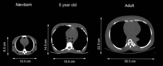
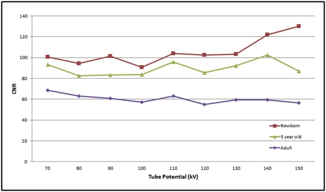
Three physical anthropomorphic phantoms (newborn, 5-year-old child, and adult) were scanned on a third-generation dual-source CT using CAREkV in semi-mode and CAREDose4D (ref. KV: 120; ref. mAs 50). Scans were performed with all available tube potentials (70–150 kV and Sn150 kV). The lowest volume computed tomography dose index (CTDI vol ) was selected to perform additional Sn100-kV scans with matched and half (Sn100-half) CTDI vol value. Image quality was evaluated on the basis of contrast-to-noise ratio (CNR).
Results
For the newborn phantom, 70–110 kV was selected as the optimal range (0.36–0.37 mGy). Using Sn150 kV led to an increase in radiation dose (0.75 mGy) without improving CNR (96.9 vs 101.5). Sn100-half showed a decrease in CNR (73.1 vs 101.5). The lowest CTDI vol for the child phantom was achieved between 100 and 120 kV (0.78–0.80 mGy). Using Sn150 kV increased radiation dose (1.02 mGy) without improvement of CNR (92.4 vs 95.8). At Sn100-half CNR was decreased (61.4 vs 95.8). For adults, 140 and 150 kV revealed the lowest CTDI vol (2.68 and 2.67 mGy). The Sn150 kV scan delivered comparable CNR (54.4 vs 56.6), but a lower CTDI vol (2.08 mGy). At Sn100-half CNR was comparable to the 150 kV scan (58.1 vs 56.6).
Conclusion
Unenhanced chest CT performed at 100 kV or 150 kV with tin filtration enables radiation dose reduction for the adult phantom, but not for the pediatric phantoms.
Introduction
For the evaluation of pulmonary pathologies, computed tomography (CT) is the method of choice due to its high spatial and inherent contrast resolution. Especially for patients with dyspnea or for pediatric patients, high-pitch protocols can provide high-quality images despite the presence of patient motion . Based on the ALARA (“as low as reasonably achievable”) principle, notably pediatric patients should receive the lowest achievable radiation dose owing to the increased radiosensitivity of children. To minimize the potential risks associated with radiation exposure, different techniques have been established to reduce the radiation dose , including automatic tube current modulation, lower tube voltage settings , and iterative image reconstruction techniques .
The third generation dual-source computed tomography (DSCT) system is able to adjust the tube voltage in narrow 10-kV steps ranging from 70 kV to150 kV and is additionally equipped with increased generator power. The scanner also includes Stellar Infinity detectors, which incorporate the advantages of integrated circuit design in the original Stellar detector while increasing spatial resolution because of a 25% increase in the number of detector channels and the addition of a two-dimensional antiscatter grid. Due to the resulting noise reduction in low-dose CTs, low-voltage examinations can be applied in a wider range, especially in contrast-enhanced angiographic studies . Furthermore, it is endued with a novel protocol using a tin-filtered 100 kVp (Sn100 kVp) and 150 kVp spectrum (Sn150 kVp) in single-energy dual-source acquisition mode. This thin absorber tin filter with a thickness of 0.6 mm absorbs primarily low-energy photons that contribute little to noncontrast image quality but increase the radiation dose burden for the patient.
Get Radiology Tree app to read full this article<
Get Radiology Tree app to read full this article<
Materials and Methods
Phantoms
Get Radiology Tree app to read full this article<
Get Radiology Tree app to read full this article<
CT Acquisition Protocol
Get Radiology Tree app to read full this article<
Get Radiology Tree app to read full this article<
Phantom Analysis
Get Radiology Tree app to read full this article<
Get Radiology Tree app to read full this article<
Get Radiology Tree app to read full this article<
Radiation Dose
Get Radiology Tree app to read full this article<
Results
Get Radiology Tree app to read full this article<
Get Radiology Tree app to read full this article<
Get Radiology Tree app to read full this article<
TABLE 1
CTDI vol and Tube Current (mAs) for all Phantom Sizes and all Kilovoltage Settings
Phantom Size kV CTDI vol (mGy) Effective mAs Newborn 70 0.37 31 80 0.36 18 90 0.36 12 100 0.37 9 110 0.37 7 120 0.45 6 130 0.58 7 140 0.69 7 150 0.81 7 Sn150 0.75 25 5-Year-old 70 0.91 77 80 0.85 44 90 0.82 28 100 0.80 20 110 0.78 15 120 0.78 11 130 0.85 10 140 0.89 9 150 0.95 8 Sn150 1.02 35 Adult 70 5.74 491 80 4.40 229 90 3.59 124 100 3.19 79 110 2.94 56 120 2.81 42 130 2.76 33 140 2.68 27 150 2.67 23 Sn150 2.08 71
CTDI vol , volume computed tomography dose index.
CAREDose4D and CAREkV in semi-mode were used for all the scans.
Get Radiology Tree app to read full this article<
Get Radiology Tree app to read full this article<
Get Radiology Tree app to read full this article<
Get Radiology Tree app to read full this article<
TABLE 2
Comparison of CTDI vol , CNR, and σ M Between the Different Tube Potentials, Which Provide the Lowest Dose for the Newborn Phantom and the Two Different Tube Voltages Using Spectral Shaping (Sn100 and Sn150)
Tube Potential (kV) CTDI vol (mGy) CNR σ M 70 0.37 100.7 8.0 80 0.36 94.5 8.6 90 0.36 101.5 7.9 100 0.37 90.8 9.0 110 0.37 104.1 8.0 Sn150 0.75 96.9 8.4 Sn100-full \* 0.36 112.5 6.8 Sn100-half \* 0.18 73.1 11.3
CNR, contrast-to-noise ratio; CTDI vol , volume computed tomography dose index.
Get Radiology Tree app to read full this article<
TABLE 3
Comparison of CTDI vol , CNR, and σ M Between the Different Tube Potentials, Which Provide the Lowest Dose for the 5-Year-Old Phantom and the Two Different Tube Voltages Using Spectral Shaping (Sn100 and Sn150)
Tube Potential (kV) CTDI vol (mGy) CNR σ M 100 0.80 83.5 10.2 110 0.78 95.8 8.2 120 0.78 85.4 9.6 Sn150 1.02 92.4 9.4 Sn100-full \* 0.78 92.1 9.6 Sn100-half \* 0.39 61.4 13.9
CNR, contrast-to-noise ratio; CTDI vol , volume computed tomography dose index.
Get Radiology Tree app to read full this article<
Get Radiology Tree app to read full this article<
Get Radiology Tree app to read full this article<
TABLE 4
Comparison of CTDI vol , CNR, and σ M Between the Different Tube Potentials, Which Provide the Lowest Dose for the Adult Phantom and the Two Different Tube Voltages Using Spectral Shaping (Sn100 and Sn150)
Tube Potential (kV) CTDI vol (mGy) CNR σ M 140 2.68 59.3 14.6 150 2.67 56.6 15.0 Sn150 2.08 54.4 16.3 Sn100-full \* 2.67 73.3 11.4 Sn100-half \* 1.33 58.1 14.6
CNR, contrast-to-noise ratio; CTDI vol , volume computed tomography dose index.
Get Radiology Tree app to read full this article<
Get Radiology Tree app to read full this article<
Discussion
Get Radiology Tree app to read full this article<
Get Radiology Tree app to read full this article<
Get Radiology Tree app to read full this article<
Get Radiology Tree app to read full this article<
Get Radiology Tree app to read full this article<
Get Radiology Tree app to read full this article<
Get Radiology Tree app to read full this article<
Get Radiology Tree app to read full this article<
Acknowledgments
Get Radiology Tree app to read full this article<
Get Radiology Tree app to read full this article<
Get Radiology Tree app to read full this article<
Get Radiology Tree app to read full this article<
References
1. Lell M.M., May M., Deak P., et. al.: High-pitch spiral computed tomography: effect on image quality and radiation dose in pediatric chest computed tomography. Invest Radiol 2011; 46: pp. 116-123.
2. Raman S.P., Johnson P.T., Deshmukh S., et. al.: CT dose reduction applications: available tools on the latest generation of CT scanners. J Am Coll Radiol 2013; 10: pp. 37-41.
3. Niemann T., Henry S., Duhamel A., et. al.: Pediatric chest CT at 70 kVp: a feasibility study in 129 children. Pediatr Radiol 2014; 44: pp. 1347-1357.
4. Kordolaimi S.D., Argentos S., Pantos I., et. al.: A new era in computed tomographic dose optimization: the impact of iterative reconstruction on image quality and radiation dose. J Comput Assist Tomogr 2013; 37: pp. 924-931.
5. Winklehner A., Karlo C., Puippe G., et. al.: Raw data-based iterative reconstruction in body CTA: evaluation of radiation dose saving potential. Eur Radiol 2011; 21: pp. 2521-2526.
6. Meinel F.G., Canstein C., Schoepf U.J., et. al.: Image quality and radiation dose of low tube voltage 3rd generation dual-source coronary CT angiography in obese patients: a phantom study. Eur Radiol 2014; 24: pp. 1643-1650.
7. Hausleiter J., Martinoff S., Hadamitzky M., et. al.: Image quality and radiation exposure with a low tube voltage protocol for coronary CT angiography results of the PROTECTION II Trial. JACC Cardiovasc Imaging 2010; 3: pp. 1113-1123.
8. Haubenreisser H., Meyer M., Sudarski S., et. al.: Unenhanced third-generation dual-source chest CT using a tin filter for spectral shaping at 100 kVp. Eur J Radiol 2015; 84: pp. 1608-1613.
9. Braun F.M., Johnson T.R., Sommer W.H., et. al.: Chest CT using spectral filtration: radiation dose, image quality, and spectrum of clinical utility. Eur Radiol 2015; 25: pp. 1598-1606.
10. Weis M., Henzler T., Nance J.W., et. al.: Radiation dose comparison between 70 kVp and 100 kVp with spectral beam shaping for non-contrast-enhanced pediatric chest computed tomography: a prospective randomized controlled study. Invest Radiol 2017; 52: pp. 155-162.
11. Farrance I., Frenkel R.: Uncertainty of measurement: a review of the rules for calculating uncertainty components through functional relationships. Clin Biochem Rev 2012; 33: pp. 49-75.
12. Glasbey C.A., Horgan G.W.: Image analysis for the biological sciences.1995.WileyChichester
13. Bodelle B., Fischbach C., Booz C., et. al.: Single-energy pediatric chest computed tomography with spectral filtration at 100 kVp: effects on radiation parameters and image quality. Pediatr Radiol 2017; 47: pp. 831-837.
14. Yu L., Bruesewitz M.R., Thomas K.B., et. al.: Optimal tube potential for radiation dose reduction in pediatric CT: principles, clinical implementations, and pitfalls. Radiographics 2011; 31: pp. 835-848.
15. Siegel M.J., Schmidt B., Bradley D., et. al.: Radiation dose and image quality in pediatric CT: effect of technical factors and phantom size and shape. Radiology 2004; 233: pp. 515-522.
16. Guimaraes L.S., Fletcher J.G., Harmsen W.S., et. al.: Appropriate patient selection at abdominal dual-energy CT using 80 kV: relationship between patient size, image noise, and image quality. Radiology 2010; 257: pp. 732-742.
17. Borggrefe J., Giravent S., Campbell G., et. al.: Association of osteolytic lesions, bone mineral loss and trabecular sclerosis with prevalent vertebral fractures in patients with multiple myeloma. Eur J Radiol 2015; 84: pp. 2269-2274.
18. Li X., Ni Q.Q., Schoepf U.J., et. al.: 70-kVp High-pitch computed tomography pulmonary angiography with 40 mL contrast agent: initial experience. Acad Radiol 2015; 22: pp. 1562-1570.
19. Wichmann J.L., Hu X., Kerl J.M., et. al.: 70 kVp Computed tomography pulmonary angiography: potential for reduction of iodine load and radiation dose. J Thorac Imaging 2015; 30: pp. 69-76.
20. Lu G.M., Luo S., Meinel F.G., et. al.: High-pitch computed tomography pulmonary angiography with iterative reconstruction at 80 kVp and 20 mL contrast agent volume. Eur Radiol 2014; 24: pp. 3260-3268.