
Rationale and Objectives
To investigate the performance of spectral computed tomography (CT) in depiction of the superior mesenteric artery (SMA) compared to conventional polychromatic CT.
Materials and Methods
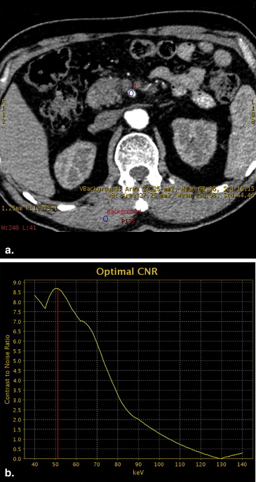
This prospective study had institutional review board approval, and written informed consent was obtained. Fifty patients underwent spectral CT examination using gemstone spectral imaging with a single-tube, fast dual-tube voltage-switching technique. Spectrum analysis was used to select the monochromatic images that provide the optimal contrast-to-noise ratio (CNR) for SMA angiography. The CNR for SMA at the selected monochromatic level was compared with that from the conventional polychromatic images. Image quality and visibility of the branch order of SMA were also assessed and compared.
Results
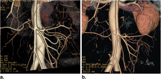
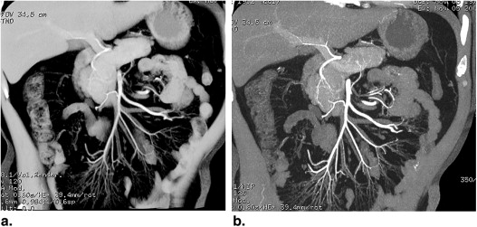
The monochromatic images at 50 keV (mean 50.09 ± 1.98) provided the optimal CNR for SMA angiography. At this energy level, the monochromatic images had higher (20.8 vs 9.2) CNR than the polychromatic images, and the image quality was superior to conventional polychromatic images ( P < .05). Fourth to fifth (mean 4.3) and third to fourth (mean 3.5) order branches of SMA were demonstrated at monochromatic and polychromatic images, respectively.
Conclusions
Gemstone spectral imaging with monochromatic images at 50 keV by spectral CT could improve the CTA image quality and demonstrate more branch order in depiction of normal SMA compared to conventional polychromatic imaging.
Computed tomography (CT) angiography (CTA) has rapidly emerged as a noninvasive imaging modality of choice to evaluate vasculature and vascular conditions in the abdomen . Multidetector CT (MDCT) has played a crucial role in the widespread acceptance of CTA as a noninvasive alterative to catheter angiography. The key to improve CTA imaging quality is to enhance the contrast between the artery and soft tissues. The image quality of CTA could be improved by increasing the contrast media injection rate or dosage , whereas increasing contrast could also increase the beam hardening artifacts with the conventional imaging using a polychromatic X-ray beam. Recently, one of the advancements of CT technology is the introduction of spectral CT, which not only provides material decomposed images but also generates a set of single-energy images from 40 to 140 keV. Since the attenuation of iodine, which is the active ingredient of the contrast media in the vessel, increases rapidly as photon energy decreases, it is thus feasible to optimize imaging condition for displaying arteries without increasing dosage of contrast media. The superior mesenteric artery (SMA) is one main branch of the abdominal aorta with many fine branches; high-order SMA branch depiction may contribute to the early diagnosis of vascular diseases such as atherosclerosis or arteritis. Spectral CT using a monochromatic X-ray beam may help to improve the image quality of SMA and show the smaller and deeper branches of the mesenteric artery. Therefore, the purpose of the study was to evaluate the effect of spectral CT on improving CTA image quality of the SMA.
Materials and methods
Patient Information
From October 2010 to May 2012, 50 consecutive patients who required mesenteric CTA scanned with gemstone spectral imaging (GSI) mode by spectral CT were included in our study. There were 22 men and 28 women with an age range of 18–73 years (mean age 41.8 years). These patients presented for a variety of reasons, including suspected aortic disease, primary or secondary cancer of the liver, and persistent abdominal pain. Subjects with any pathological conditions that may cause modifications of the vascular anatomy of SMA were excluded. Based on this criterion, 15 patients were excluded (parasitic flow in hepatocellular carcinoma [ n = 8], dissection of SMA [ n = 3], and thrombosis of SMA [ n = 4]). Therefore, 35 patients were included in this study. This prospectively study was approved by our institutional review board, and written informed consent was obtained from each participant before the study.
CT Technique
Get Radiology Tree app to read full this article<
Get Radiology Tree app to read full this article<
GSI Image Postprocessing
Get Radiology Tree app to read full this article<
Get Radiology Tree app to read full this article<
Image Reconstruction
Get Radiology Tree app to read full this article<
Image Evaluation
Get Radiology Tree app to read full this article<
Statistical Analysis
Get Radiology Tree app to read full this article<
Results
Get Radiology Tree app to read full this article<
Table 1
CNRs, Scores of Image Quality, and Visibility of the Branch Order of SMA at Monochromatic and Polychromatic Images with Different Imaging
Monochromatic Images Polychromatic Images_P_ Value CNR 20.8 ± 4.1 9.2 ± 1.0 .031 Scores 4.33 ± 0.48 3.67 ± 0.73 .012 Visualized branch order from SMA 4.3 ± 0.36 3.5 ± 0.42 .027
CNR, contrast-to-noise ratio; SMA, superior mesenteric artery.
Data were expressed as mean ± SD.
Get Radiology Tree app to read full this article<
Get Radiology Tree app to read full this article<
Get Radiology Tree app to read full this article<
Get Radiology Tree app to read full this article<
Get Radiology Tree app to read full this article<
Discussion
Get Radiology Tree app to read full this article<
Get Radiology Tree app to read full this article<
Get Radiology Tree app to read full this article<
Get Radiology Tree app to read full this article<
Get Radiology Tree app to read full this article<
Conclusions
Get Radiology Tree app to read full this article<
Acknowledgments
Get Radiology Tree app to read full this article<
References
1. Napoli A., Fleischmann D., Chan F.P., et. al.: Computed tomography angiography: state-of-the art imaging using multidetector-row technology. J Comput Assist Tomogr 2004; 28: pp. S32-S45.
2. Rosow D.E., Sahani D., Strobel O., et. al.: Imaging of acute mesenteric ischemia using multidetector CT and CT angiography in a porcine model. J Gastrointest Surg 2005; 9: pp. 1262-1274.
3. Willmann J.K., Baumert B., Schertler T., et. al.: Aortoiliac and lower extremity arteries assessed with 16-detector row CT angiography: prospective comparison with digital subtraction angiography. Radiology 2005; 236: pp. 1083-1093.
4. Nikolaou K., Flohr T., Knez A., et. al.: Advances in cardiac CT imaging: 64-slice scanner. Int Cardiovasc Imaging 2004; 20: pp. 535-540.
5. Awai K., Hiraishi K., Hori S.: Effect of contrast material injection duration and rate on aortic peak time and peak enhancement at dynamic CT involving injection protocol with dose tailored to patient weight. Radiology 2004; 230: pp. 142-150.
6. Erturk S.M., Ichikawa T., Sou H., et. al.: Effect of duration of contrast material injection on peak enhancement times and values of the aorta, main portal vein, and liver at dynamic MDCT with the dose of contrast medium tailored to patient weight. Clin Radiol 2008; 63: pp. 263-271.
7. Fleischmann D., Kamaya A.: Optimal vascular and parenchymal contrast enhancement: the current state of the art. Radiol Clin North Am 2009; 47: pp. 13-26.
8. Sahani D., Saini S., Pena C., et. al.: Using multidetector CT for preoperative vascular evaluation of liver neoplasms: technique and results. AJR Am J Roentgenol 2002; 179: pp. 53-59.
9. Awai K., Inoue M., Yagyu Y., et. al.: Moderate versus high concentration of contrast material for aortic and hepatic enhancement and tumor-to-liver contrast at multi-detector row CT. Radiology 2004; 233: pp. 682-688.
10. Roos J.E., Desbiolles L.M., Weishaupt D., et. al.: Multi-detector row CT: effect of iodine dose reduction on hepatic and vascular enhancement. Rofo 2004; 176: pp. 556-563.
11. Cademartiri F., de Monye C., Pugliese F., et. al.: High iodine concentration contrast material for noninvasive multislice computed tomography coronary angiography: iopromide 370 versus iomeprol 400. Invest Radiol 2006; 41: pp. 349-353.
12. Hurrell M.A., Butler A.P., Cook N.J., et. al.: Spectral Hounsfield units: a new radiological concept. Eur Radiol 2012; 22: pp. 1008-1013.
13. Lv P., Lin X.Z., Chen K., et. al.: Spectral CT in patients with small HCC: investigation of image quality and diagnostic accuracy. Eur Radiol 2012; 22: pp. 2117-2124.
14. Zhao L.Q., He W., Li J.Y., et. al.: Improving image quality in portal venography with spectral CT imaging. Eur J Radiol 2012; 81: pp. 1677-1681.
15. Lv P., Lin X.Z., Li J., et. al.: Differentiation of small hepatic hemangioma from small hepatocellular carcinoma: recently introduced spectral CT method. Radiology 2011; 259: pp. 720-729.
16. Martı′nez-Isla A., Griffith P.S., Markogiannakis H., et. al.: A novel laparoscopic approach to lesions related to the posterior aspect of the pancreatic head. Am J Surg 2009; 197: pp. e51-e53.
17. Matsumoto K., Jinzaki M., Tanami Y., et. al.: Virtual monochromatic spectral imaging with fast kilovoltage switching: improved image quality as compared with that obtained with conventional 120-kVp CT. Radiology 2011; 259: pp. 257-262.
18. Silva A.C., Morse B.G., Hara A.K., et. al.: Dual-energy (spectral) CT: applications in abdominal imaging. Radiographics 2011; 31: pp. 1031-1046.
19. Jung D.C., Oh Y.T., Kim M.D., et. al.: Usefulness of the virtual monochromatic image in dual-energy spectral CT for decreasing renal cyst pseudoenhancement: a phantom study. AJR Am J Roentgenol 2012; 199: pp. 1316-1319.
20. Deng K., Zhang C.Q., Li W., et. al.: Preliminary application of high-definition CT Gemstone Spectral Imaging in hand and foot tendons. Kor J Radiol 2012; 13: pp. 743-751.
21. Pinho D.F., Kulkarni N.M., Krishnaraj A., et. al.: Initial experience with single-source dual-energy CT abdominal angiography and comparison with single-energy CT angiography: image quality, enhancement, diagnosis and radiation dose. Eur Radiol 2013; 23: pp. 351-359.