
Rationale and Objectives
This study summarizes the literature on the detection of cancer among indeterminate extracolonic findings on computed tomographic (CT) colonography in five targeted organs.
Materials and Methods
We searched PubMed for English-language literature published between January 1, 1994, and December 31, 2010. We describe extracolonic findings in the kidney, lung, liver, pancreas, and ovary suspect for malignancy as they are associated with high mortality. For each organ, we calculated the median prevalence, positive predictive value (PPV), and false positive rate of malignancy and a pooled false-positive rate across studies.
Results
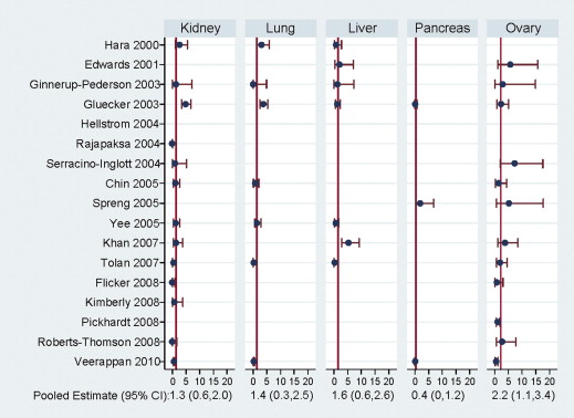
Of 91 publications initially identified, 24 were eligible for review. Indeterminate renal masses on CT colonography had 20.5% median PPV and low pooled false positive rate of 1.3% (95% confidence interval 0.6–2.0). In contrast, indeterminate masses of the lung, liver, pancreas, and ovary had low PPV (median values ranged from 0% to 3.8%). Indeterminate masses of the ovary resulted in the highest pooled false-positive rate of 2.2%. Results were similar in studies of both screening and nonscreening populations. We estimated the probability of false positive results through the detection of significant extracolonic findings as 46 per 1000 for men and 68 per 1000 for women.
Conclusions
Indeterminate renal masses newly detected on CT colonography have an estimated one in five chance of malignancy and therefore warrant further follow-up to provide a definitive diagnosis. Conversely, indeterminate masses of the lung, liver, pancreas, and ovary are associated with high false positive rates and merit more conservative clinical follow-up.
Computed tomographic (CT) colonography was first described as a method to assess colon neoplasia in 1994 . CT colonography involves insufflation of the colon and rectum with gas and the acquisition of thin-section CT images to visualize polyps and masses using both two-dimensional and three-dimensional interpretation . Multiple studies have demonstrated that the accuracy of CT colonography is similar to that of traditional (optical) colonoscopy for the detection of adenomas and colorectal cancer (ie, sensitivity of 91.3% and specificity of 93.1% for lesions >5 mm) . Both of these screening techniques require cathartic bowel preparation, which is a major deterrent to colorectal cancer screening for patients. However, because CT colonography does not require sedation, it has the potential to increase overall adherence to colorectal cancer screening and is associated with fewer risks than optical colonoscopy.
Unlike traditional colonoscopy, CT colonography can identify extracolonic findings (ie, outside the colon or rectal lumen) because the lung bases, abdomen, and pelvis are included in the examination. To guide management of extracolonic findings, the Working Group on Virtual Colonoscopy developed a rating of extracolonic findings using a scale of E1 to E4 . Under this system, E3 and E4 findings are potentially significant to the patient’s health but incompletely characterized on CT colonography and usually require further imaging and medical follow-up for definitive characterization. E3 findings (eg, pulmonary nodules <1 cm or cystic renal or adnexal masses) are likely insignificant and might require nonurgent follow-up. E4 findings (eg, solid renal masses or pulmonary nodules >1 cm) are likely significant and require urgent follow-up.
Get Radiology Tree app to read full this article<
Materials and methods
Literature Review
Get Radiology Tree app to read full this article<

Get Radiology Tree app to read full this article<
Get Radiology Tree app to read full this article<
Get Radiology Tree app to read full this article<
Targeted Extracolonic Findings
Get Radiology Tree app to read full this article<
Statistical Analysis
Get Radiology Tree app to read full this article<
Get Radiology Tree app to read full this article<
Results
Get Radiology Tree app to read full this article<
Table 1
Summary of Reviewed Published Literature of CT Colonography by Calculation of Median Test Properties ∗ Across Five Target Organ Sites, Stratified by Screening and Nonscreening Populations, through 2010
Target Organ No. of Studies FPR (/100) Prevalence (/1000) PPV (/100) Kidneys Screening 6 1.0 0.6 1.5 Nonscreening 13 1.0 4.2 25.0 Mixed/unknown 3 † † † Overall 22 1.1 2.2 20.5 Lung Screening 6 0.2 0.1 20.0 Nonscreening 6 0.7 0.7 0 Mixed/unknown 3 † † † Overall 15 0.7 0.1 3.8 Liver Screening 4 0 1.2 0 Nonscreening 8 1.8 0 0 Mixed/unknown 2 † † † Overall 14 1.3 0 0 Pancreas Screening 2 0.1 0 0 Nonscreening 5 1.4 1.2 25.0 Mixed/unknown 1 † † † Overall 8 0.5 0.5 0 Ovary Screening 5 1.5 0 0 Nonscreening 9 3.7 0 10.0 Mixed/unknown 1 † † † Overall 15 2.5 0 0
Get Radiology Tree app to read full this article<
Get Radiology Tree app to read full this article<
Get Radiology Tree app to read full this article<
Get Radiology Tree app to read full this article<
Table 2
Description of Reviewed Published Articles Reporting Extracolonic Findings for Renal Cancer
First Author (Reference) Year Country Sample Size Age Range, y Mean/Median Age. y Men No. of ECFs No. of True Positives FPR (/100) Prevalence (/1000) PPV (/100) Screening populations Ginnerup Pederson B ∗ 2003 Denmark 75 33–78 61 53.3 1 0 1.3 0 0 Gluecker TM 2003 United States 681 41–80 64 62.6 34 1 4.9 1.5 2.9 Chin M ∗ 2005 Australia 432 50–69 † 53.2 6 1 1.2 2.3 16.7 Kimberly JR 2008 United States 143 44–83 57 48 1 0 0.7 0 0 Pickhardt PJ 2010 United States 10,286 † 59.8 52.3 NA 11 † 1.1 † Veerappan GR 2010 United States 2277 † 59 56 16 3 0.6 0.1 18.8 Nonscreening populations Edwards JT 2001 Australia 100 65 47 1 NA † † † Hellstrom M 2004 Sweden 111 19–86 66 59.0 4 1 2.7 9.0 25.0 Pilch-Kowalczyk J 2004 Poland 71 20–85 † 40.8 NA 1 † 14.1 † Rajapaksa RC 2004 United States 250 † 62.5 98 1 1 0 0.4 100 Serracino-Inglott F ∗ 2004 United Kingdom 103 43–88 68 45.6 1 0 1.0 0 † Spreng A 2005 Switzerland 102 20–91 66 61.7 NA 1 † 9.8 † Yee J 2005 United States 500 30–90 62.5 100 8 2 1.2 4.0 25.0 Tolan D 2007 United Kingdom 400 70–96 79.6 37.5 4 2 0.5 5.0 50.0 Khan KY ∗ 2007 United Kingdom 225 62–81 74 40.4 3 0 1.3 0 0 Flicker MS 2008 United States 376 26–89 61 36.7 2 2 0 5.3 100.0 Roberts-Thomson IC 2008 Australia 225 25–85 60 51.0 1 1 0 4.4 100.0 Yucel C 2008 United States 42 60–87 71 38.0 4 NA † † † White TJ 2009 United States 150 40–83 60.9 48.7 NA 3 † 2.0 † Mixed/unknown Hara AK 2000 United States 264 33–88 64 55.3 9 2 2.7 7.6 22.2 Iafrate F 2008 Italy 136 70–92 81 NA 1 NA † † † Park SK 2009 South Korea 920 34–87 57.3 58.1 3 NA † † †
ECF, extracolonic finding; NA, not available; CI, confidence interval.
Get Radiology Tree app to read full this article<
Get Radiology Tree app to read full this article<
Get Radiology Tree app to read full this article<
Get Radiology Tree app to read full this article<
Table 3
Description of Reviewed Published Articles Reporting Extracolonic Findings for Lung Cancer
First Author (Reference) Year Country Sample Size Age Range, y Mean/Median Age, y Men No. of ECFs No. of True Positives FPR (/100) Prevalence (/1000) PPV (/100) Screening populations Ginnerup Pederson B 2003 Denmark 75 33–78 61 53.3 1 1 0 1.3 100.0 Gluecker TM 2003 United States 681 41–80 64 62.6 26 1 3.7 0.1 3.8 Chin M 2005 Australia 432 50–69 † 53.2 3 0 0.7 0 0 Kimberly JR 2008 United States 143 44–83 57 48 23 NA † † † Pickhardt PJ 2010 United States 10,286 † 59.8 52.3 NA 8 † 0.1 † Veerappan GR ∗ 2010 United States 2277 † 59 56 5 1 0.2 0.0 20.0 Nonscreening populations Rajapaksa RC 2004 United States 250 † 62.5 98 3 NA † † † Spreng A 2005 Switzerland 102 20–91 66 61.7 NA 1 † 1.0 † Yee J 2005 United States 500 30–90 62.5 100 7 0 1.4 0.0 0.0 Tolan D 2007 United Kingdom 400 70–96 79.6 37.5 7 6 0.3 1.5 85.7 Roberts-Thomson IC 2008 Australia 225 25–85 60 51.0 2 NA † † † White TJ 2009 United States 150 40–83 60.9 48.7 NA 1 † 0.7 † Unknown/mixed Hara AK 2000 United States 264 33–88 64 55.3 8 0 3.0 0.0 0 Iafrate F 2008 Italy 136 70–92 81 NA 1 NA † † † Park SK 2009 South Korea 920 34–87 57.3 58.1 9 NA † † †
ECF, extracolonic finding; NA, not available.
Get Radiology Tree app to read full this article<
Get Radiology Tree app to read full this article<
Table 4
Description of Reviewed Published Articles Reporting Extracolonic Findings for Ovarian Cancer
First Author (Reference) Year Country Sample Size Age Range, y Mean/Median Age, y No. of ECFs No. of True Positives FPR (/100) Prevalence (/1000) PPV (/100) Screening populations Ginnerup Pederson B 2003 Denmark 35 33–78 61 1 0 2.9 0 0 Gluecker TM 2003 United States 255 41–80 64 6 0 2.4 0 0 Chin M 2005 Australia 202 50–69 3 0 1.5 0 0 Pickhardt PJ 2008 United States 1199 40–90 58 13 0 1.1 0 0 Veerappan GR 2010 United States 1002 59 5 0 0.5 0 0 Nonscreening populations Morrin MM 1999 United States 28 22–96 1 NA ∗ ∗ ∗ Edwards JT 2001 Australia 53 65 3 0 5.7 0 0 Serracino-Inglott F 2004 United Kingdom 56 43–88 68 5 1 7.3 17.9 20.0 Spreng A 2005 Switzerland 39 20–91 66 3 1 5.3 25.6 33.3 Khan KY 2007 United Kingdom 134 62–81 74 5 0 3.7 0 0 Tolan D 2007 United Kingdom 250 70–96 79.6 5 0 2.0 0 0 Flicker MS 2008 United States 238 26–89 61 3 1 0.8 4.2 33.3 Roberts-Thomson IC 2008 Australia 110 25–85 60 3 0 2.7 0 0 Yucel C 2008 United States 26 60–87 71 1 NA ∗ ∗ ∗ Unknown/mixed Park SK 2009 South Korea 385 34–87 57.3 1 NA ∗ ∗ ∗
ECF, extracolonic finding; NA, not available; CI, confidence interval.
Get Radiology Tree app to read full this article<
Get Radiology Tree app to read full this article<
Get Radiology Tree app to read full this article<
Get Radiology Tree app to read full this article<
Get Radiology Tree app to read full this article<
Discussion
Get Radiology Tree app to read full this article<
Get Radiology Tree app to read full this article<
Get Radiology Tree app to read full this article<
Get Radiology Tree app to read full this article<
Get Radiology Tree app to read full this article<
Get Radiology Tree app to read full this article<
Get Radiology Tree app to read full this article<
Get Radiology Tree app to read full this article<
References
1. Vining D.J., Gelfand D.W., Bechthold R.E., et. al.: Technical feasibility of colon imaging with helical CT and virtual reality. AJR Am J Roentgenol 1994; 162: pp. 104.
2. Parkins T.: Computer lets doctor fly through the virtual colon. J Natl Cancer Inst 1994; 86: pp. 1046-1047.
3. Graser A., Stieber P., Nagel D., et. al.: Comparison of CT colonography, colonoscopy, sigmoidoscopy and faecal occult blood tests for the detection of advanced adenoma in an average risk population. Gut 2009; 58: pp. 241-248.
4. Moawad F.J., Maydonovitch C.L., Cullen P.A., et. al.: CT colonography may improve colorectal cancer screening compliance. AJR Am J Roentgenol 2010; 195: pp. 1118-1123.
5. Zalis M.E., Barish M.A., Choi J.R., et. al.: CT colonography reporting and data system: a consensus proposal. Radiology 2005; 236: pp. 3-9.
6. Casarella W.J.: A patient’s viewpoint on a current controversy. Radiology 2002; 224: pp. 927.
7. Siddiki H., Fletcher J.G., McFarland B., et. al.: Incidental findings in CT colonography: literature review and survey of current research practice. J Law Med Ethics 2008; 36: pp. 320-331. 213
8. Outwater E.K., Siegelman E.S., Hunt J.L.: Ovarian teratomas: tumor types and imaging characteristics. Radiographics 2001; 21: pp. 475-490.
9. Comerci J.T., Licciardi F., Bergh P.A., et. al.: Mature cystic teratoma: a clinicopathologic evaluation of 517 cases and review of the literature. Obstet Gynecol 1994; 84: pp. 22-28.
10. Talerman A.: Germ cell tumors of the ovary.Kurman R.J.Blaustein’s pathology of the female genital tract.1994.Springer-VerlagNew York, NY:pp. 849-914.
11. Young-Xu Y., Chan K.A.: Pooling overdispersed binomial data to estimate event rate. BMC Med Res Methodol 2008; 8: pp. 58.
12. Guimaraes P.: A simple approach to fit the beta-binomial model. Stata J 2005; 5: pp. 385-394.
13. StataCorp : Stata Statistical Software: Release 12.2011.StataCorp LPCollege Station, TX
14. Altekruse S.F., Kosary F.L., Krapcho M., et. al.: SEER Cancer Statistics Review, 1975–2007.2010.National Cancer InstituteBethesda, MD
15. Chow W.H., Devesa S.S., Warren J.L., et. al.: Rising incidence of renal cell cancer in the United States. JAMA 1999; 281: pp. 1628-1631.
16. Gordis L.: Epidemiology.Third Edition ed.2004.WB SaundersPhiladelphia, PA
17. Pickhardt P.J., Hanson M.E., Vanness D.J., et. al.: Unsuspected extracolonic findings at screening CT colonography: clinical and economic impact. Radiology 2008; 249: pp. 151-159.
18. Veerappan G.R., Ally M.R., Choi J.H., et. al.: Extracolonic findings on CT colonography increases yield of colorectal cancer screening. AJR Am J Roentgenol 2010; 195: pp. 677-686.
19. Edwards J.T., Wood C.J., Mendelson R.M., et. al.: Extracolonic findings at virtual colonoscopy: implications for screening programs. Am J Gastroenterol 2001; 96: pp. 3009-3012.
20. Flicker M.S., Tsoukas A.T., Hazra A., et. al.: Economic impact of extracolonic findings at computed tomographic colonography. J Comput Assist Tomogr 2008; 32: pp. 497-503.
21. Gluecker T.M., Johnson C.D., Wilson L.A., et. al.: Extracolonic findings at CT colonography: evaluation of prevalence and cost in a screening population. Gastroenterology 2003; 124: pp. 911-916.
22. Spreng A., Netzer P., Mattich J., et. al.: Importance of extracolonic findings at IV contrast medium-enhanced CT colonography versus those at non-enhanced CT colonography. Eur Radiol 2005; 15: pp. 2088-2095.
23. Chin M., Mendelson R., Edwards J., et. al.: Computed tomographic colonography: prevalence, nature, and clinical significance of extracolonic findings in a community screening program. Am J Gastroenterol 2005; 100: pp. 2771-2776.
24. Khan K.Y., Xiong T., McCafferty I., et. al.: Frequency and impact of extracolonic findings detected at computed tomographic colonography in a symptomatic population. Br J Surg 2007; 94: pp. 355-361.
25. Roberts-Thomson I.C., Tucker G.R., Hewett P.J., et. al.: Single-center study comparing computed tomography colonography with conventional colonoscopy. World J Gastroenterol 2008; 14: pp. 469-473.
26. Serracino-Inglott F., Atkinson H.D., Jha P., et. al.: Early experiences with computed axial tomography colonography. Am J Surg 2004; 187: pp. 511-514.
27. Yucel C., Lev-Toaff A.S., Moussa N., et. al.: CT colonography for incomplete or contraindicated optical colonoscopy in older patients. AJR Am J Roentgenol 2008; 190: pp. 145-150.
28. Tolan D.J., Armstrong E.M., Chapman A.H.: Replacing barium enema with CT colonography in patients older than 70 years: the importance of detecting extracolonic abnormalities. AJR Am J Roentgenol 2007; 189: pp. 1104-1111.
29. Levine D., Brown D.L., Andreotti R.F., et. al.: Management of asymptomatic ovarian and other adnexal cysts imaged at US: Society of Radiologists in Ultrasound Consensus Conference Statement. Radiology 2010; 256: pp. 943-954.
30. Johnson P.T., Horton K.M., Megibow A.J., et. al.: Common incidental findings on MDCT: survey of radiologist recommendations for patient management. J Am Coll Radiol 2011; 8: pp. 762-767.
31. Volpe A., Panzarella T., Rendon R.A., et. al.: The natural history of incidentally detected small renal masses. Cancer 2004; 100: pp. 738-745.
32. Yee J., Rosen M.P., Blake M.A., et. al.: ACR Appropriateness Criteria on colorectal cancer screening. J Am Coll Radiol 2010; 7: pp. 670-678.
33. Ginnerup Pedersen B., Rosenkilde M., Christiansen T.E., et. al.: Extracolonic findings at computed tomography colonography are a challenge. Gut 2003; 52: pp. 1744-1747.
34. Kimberly J.R., Phillips K.C., Santago P., et. al.: Extracolonic findings at virtual colonoscopy: an important consideration in asymptomatic colorectal cancer screening. J Gen Intern Med 2009; 24: pp. 69-73.
35. Pickhardt P.J., Kim D.H., Meiners R.J., et. al.: Colorectal and extracolonic cancers detected at screening CT colonography in 10,286 asymptomatic adults. Radiology 2010; 255: pp. 83-88.
36. Hellstrom M., Svensson M.H., Lasson A.: Extracolonic and incidental findings on CT colonography (virtual colonoscopy). AJR Am J Roentgenol 2004; 182: pp. 631-638.
37. Pilch-Kowalczyk J., Konopka M., Gibinska J., et. al.: Extracolonic findings at CT colonography - additional advantage of the method. Med Sci Monit 2004; 10: pp. 22-25.
38. Rajapaksa R.C., Macari M., Bini E.J.: Prevalence and impact of extracolonic findings in patients undergoing CT colonography. J Clin Gastroenterol 2004; 38: pp. 767-771.
39. Yee J., Kumar N.N., Godara S., et. al.: Extracolonic abnormalities discovered incidentally at CT colonography in a male population. Radiology 2005; 236: pp. 519-526.
40. White T.J., Avery G.R., Kennan N., et. al.: Virtual colonoscopy vs conventional colonoscopy in patients at high risk of colorectal cancer–a prospective trial of 150 patients. Colorectal Dis 2009; 11: pp. 138-145.
41. Hara A.K., Johnson C.D., MacCarty R.L., et. al.: Incidental extracolonic findings at CT colonography. Radiology 2000; 215: pp. 353-357.
42. Iafrate F., Hassan C., Zullo A., et. al.: CT colonography with reduced bowel preparation after incomplete colonoscopy in the elderly. Eur Radiol 2008; 18: pp. 1385-1395.
43. Park S.K., Park D.I., Lee S.Y., et. al.: Extracolonic findings of computed tomographic colonography in Koreans. World J Gastroenterol 2009; 15: pp. 1487-1492.