
Rationale and Objectives
Computed tomographic colonography (CTC) is a robust tool for evaluating colorectal lesions in both screening and diagnostic settings. The purpose of this study was to assess the relationship between colorectal cancer (CRC) tumor characteristics and patient symptomatology.
Materials and Methods
This is a retrospective analysis of all pathology-confirmed cases of CRC evaluated with CTC at our institution from October 2004 to October 2012. Cases were reviewed to determine tumor size, morphology, and degree of luminal narrowing. An electronic medical record review was performed to delineate specific patient symptomatology and determine depth of invasion.
Results
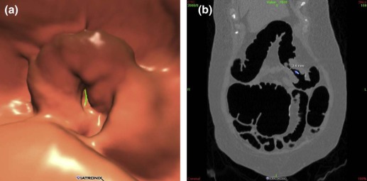
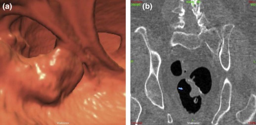
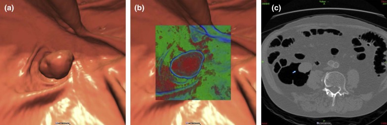
A total of 55 patients (36 symptomatic and 19 asymptomatic) with a total of 63 CRCs were evaluated by CTC during the study time period. The most common symptoms were gastrointestinal (GI) bleeding/anemia ( n = 26), followed by obstructive symptoms ( n = 23), and constitutional symptoms ( n = 5). Symptomatic cancers were more likely to have annular morphology ( n = 30/43, 70% vs. n = 3/20, 15%; odds ratio [OR] = 13.1, P = 0.0003), whereas asymptomatic cancers were more likely to be polypoid ( n = 11/20, 55% vs. n = 6/43, 14%, OR = 7.5, P = 0.001). Symptomatic cancers were also larger (46.1 ± 22.4 vs. 38.8 ± 18.4 mm, P = 0.005) and resulted in greater luminal narrowing (8.7 ± 8.5 mm vs. 35.8 ± 18.8 mm, P < 0.0001) with deeper invasion ( n = 29/35 [invasion unknown for 8 cases], 83% vs. n = 6/20, 30%, OR = 11.3, P = 0.0003). Invasive cancers were more likely to have annular morphology (66%, 23/25, P = 0.002).
Conclusions
There is an intuitive and predictable relationship between tumor characteristics on CTC and patient symptoms. Annular morphology, tumor size, degree of luminal narrowing, and invasive disease all correlate with the presence of symptoms.
Introduction
Colorectal cancer (CRC) is the third most frequently diagnosed cancer in the United States and is the second deadliest cancer, accounting for 50,310 cancer-related deaths in 2014 . Despite current evidence-based screening guidelines, CRC remains a prominent cause of cancer-related mortality, owing in part to suboptimal adherence to recommended screening protocols within the average-risk patient population .
Early in the time course of disease when prognosis is most favorable, CRC presents in completely asymptomatic individuals. However, as the disease progresses and prognosis becomes worse, symptoms may become evident. Symptoms of obstruction, nausea and vomiting, and abdominal distension are of prognostic significance in CRC patients in terms of both overall survival and disease-free survival . Abdominal pain, change in bowel habits, bleeding/anemia, malaise/fatigue, and weight loss are among the most common clinical manifestations of CRC prompting medical evaluation. Symptoms of CRC are often attributed to tumor size, location, degree of obstruction, and extent of disease .
Get Radiology Tree app to read full this article<
Get Radiology Tree app to read full this article<
Materials and Methods
Study Group
Get Radiology Tree app to read full this article<
CTC Technique
Get Radiology Tree app to read full this article<
Get Radiology Tree app to read full this article<
Procedures, Classification, and Definitions
Get Radiology Tree app to read full this article<
Get Radiology Tree app to read full this article<
Get Radiology Tree app to read full this article<
Get Radiology Tree app to read full this article<
Get Radiology Tree app to read full this article<
Statistical Analysis
Get Radiology Tree app to read full this article<
Results
Get Radiology Tree app to read full this article<
Table 1
Patient Demographics
Symptomatic Asymptomatic No. of patients 36 19 Male : female 15:21 (42%) 10:9 (53%) Mean age (years) 67 60 Colorectal cancer history 6% 0% Screening 0 19 No. of cancers 43 20 No. of patients with synchronous cancers 5 1
Get Radiology Tree app to read full this article<
Get Radiology Tree app to read full this article<
Get Radiology Tree app to read full this article<
Table 2
Tumor Morphology
Symptomatic Asymptomatic Annular 72% (31/43) 15% (3/20) Plaque like 14% (6/43) 30% (6/20) Polypoid 14% (6/43) 55% (11/20)
Get Radiology Tree app to read full this article<
Get Radiology Tree app to read full this article<
Table 3
Symptom Category by Morphological Type
Annular Plaque Like Polypoid Obstructive 85% (17/20, P = 0.07) 5% (1/20, P = 0.07) 10% (2/20, P = 1.00) Gastrointestinal bleeding/anemia 73% (15/23, P = 0.27) 15% (5/23, P = 0.39) 12% (3/23, P = 1.00) Constitutional 100% (5/5, P = 0.29) 0 0
Get Radiology Tree app to read full this article<
Get Radiology Tree app to read full this article<
Get Radiology Tree app to read full this article<
Table 4
Number of Symptomatic and Asymptomatic CRCs by Location
Colonic Location Symptomatic (Total: 43) Asymptomatic (Total: 20) Rectum 6 (14%) 5 (25%) Sigmoid 16 (37%) 4 (20%) Descending 7 (16%) 1 (5%) Transverse 4 (9%) 4 (20%) Ascending 5 (12%) 2 (10%) Cecum 5 (12%) 4 (20%)
Get Radiology Tree app to read full this article<
Get Radiology Tree app to read full this article<
Discussion
Get Radiology Tree app to read full this article<
Get Radiology Tree app to read full this article<
Get Radiology Tree app to read full this article<
Get Radiology Tree app to read full this article<
Get Radiology Tree app to read full this article<
Get Radiology Tree app to read full this article<
Author disclosures
Get Radiology Tree app to read full this article<
Get Radiology Tree app to read full this article<
Get Radiology Tree app to read full this article<
Get Radiology Tree app to read full this article<
References
1. Siegel R., DeSantis C., Jemal A.: Colorectal cancer statistics, 2014. CA Cancer J Clin 2014; 64: pp. 104-117.
2. Winawer S.J., Zauber A.G., Ho M.N., et. al.: Prevention of colorectal cancer by colonoscopic polypectomy. The National Polyp Study Workgroup. N Engl J Med 1993; 329: pp. 1977-1981.
3. Rim S.H., Joseph D.A., Steele C.B., et. al.: Colorectal cancer screening—United States, 2002, 2004, 2006, and 2008. MMWR Surveill Summ 2011; 60: pp. 42-46.
4. Steinberg S.M., Barkin J.S., Kaplan R.S., et. al.: Prognostic indicators of colon tumors. The Gastrointestinal Tumor Study Group experience. Cancer 1986; 57: pp. 1866-1870.
5. Cappell M.S., Goldberg E.S.: The relationship between the clinical presentation and spread of colon cancer in 315 consecutive patients. A significant trend of earlier cancer detection from 1982 through 1988 at a university hospital. J Clin Gastroenterol 1992; 14: pp. 227-235.
6. Majumdar S.R., Flecher R.H., Evans A.T.: How does colorectal cancer present? Symptoms, duration, and clues to location. Am J Gastroenterol 1999; 94: pp. 3039-3045.
7. Cappell M.S.: Pathophysiology, clinical presentation, and management of colon cancer. Gastroenterol Clin North Am 2008; 37: pp. 1-24. v
8. Pickhardt P.J., Choi J.R., Hwang I., et. al.: Computed tomographic virtual colonoscopy to screen for colorectal neoplasia in asymptomatic adults. N Engl J Med 2003; 349: pp. 2191-2200.
9. Kim D.H., Pickhardt P.J., Taylor A.J., et. al.: CT colonography versus colonoscopy for the detection of advanced neoplasia. N Engl J Med 2007; 357: pp. 1403-1412.
10. Johnson C.D., Chen M.H., Toledano A.Y., et. al.: Accuracy of CT colonography for detection of large adenomas and cancers. N Engl J Med 2008; 359: pp. 1207-1217.
11. Pickhardt P.J.: Noninvasive radiologic imaging of the large intestine: a valuable complement to optical colonoscopy. Curr Opin Gastroenterol 2010; 26: pp. 61-68.
12. Pickhardt P.J.: Screening CT colonography: how I do it. AJR Am J Roentgenol 2007; 189: pp. 290-298.
13. Alexiusdottir K.K., Moller P.H., Snaebjornsson P., et. al.: Association of symptoms of colon cancer patients with tumor location and TNM tumor stage. Scand J Gastroenterol 2012; 47: pp. 795-801.
14. Lee G.H., Malietzis G., Askari A., et. al.: Is right-sided colon cancer different to left-sided colorectal cancer?—a systematic review. Eur J Surg Oncol 2015; 41: pp. 300-308.
15. Snaebjornsson P., Jonasson L., Jonsson T., et. al.: Colon cancer in Iceland-a nationwide comparative study on various pathology parameters with respect to right and left tumor location and patients age. Int J Cancer 2010; 127: pp. 2645-2653.
16. Nawa T., Kato J., Kawamoto H., et. al.: Differences between right- and left-sided colon cancer in patient characteristics, cancer morphology and histology. J Gastroenterol Hepatol 2008; 23: pp. 418-423.
17. Soetikno R.M., Kaltenbach T., Rouse R.V., et. al.: Prevalence of nonpolypoid (flat and depressed) colorectal neoplasms in asymptomatic and symptomatic adults. JAMA 2008; 299: pp. 1027-1035.
18. Bose M., Bell J., Jackson L., et. al.: Virtual vs. optical colonoscopy in symptomatic gastroenterology out-patients: the case for virtual imaging followed by targeted diagnostic or therapeutic colonoscopy. Aliment Pharmacol Ther 2007; 26: pp. 727-736.
19. Atkin W., Dadswell E., Wooldrage K., et. al.: Computed tomographic colonography versus colonoscopy for investigation of patients with symptoms suggestive of colorectal cancer (SIGGAR): a multicentre randomised trial. Lancet 2013; 381: pp. 1194-1202.
20. Copel L., Sosna J., Kruskal J.B., et. al.: CT colonography in 546 patients with incomplete colonoscopy. Radiology 2007; 244: pp. 471-478.