
Rationale and Objectives
Long circulating core-encapsulated gadolinium (CE-Gd) liposomal nanoparticles that have surface conjugated polyethylene glycol are a promising platform technology for use as blood pool T1-based magnetic resonance (MR) contrast agents. The objective of this study was to investigate the effect of liposome size and internal (core) Gd concentration on the T1 relaxivity of CE-Gd liposomes.
Materials and Methods
Twelve different liposomal formulations were synthesized and characterized, resulting in a size (50, 100, 200, and 400 nm) and core Gd-concentration (200, 350, and 500 mM) “matrix” of test samples. Subsequently, CE-Gd liposomes were diluted in deionized water (four diluted samples) and molar T1 relaxivity (r1) measurements were performed at 2- and 7-T MR field strengths.
Results
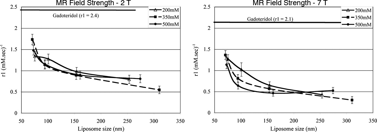
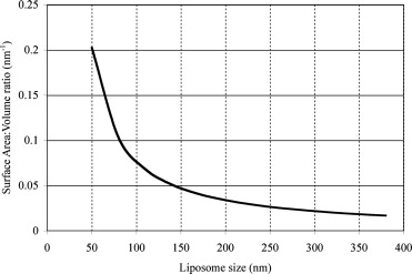
The r1 of CE-Gd liposomes was inversely related to the liposome size. The largest change in r1 was observed between liposomes that were extruded through 50- and 100-nm filter membranes. At both field strengths, the variation in internal gadolinium concentration did not show any significant correlation (α ≤ 0.05) with r1.
Conclusions
The size of CE-Gd liposomal nanoparticles significantly affects the T1 relaxivity. An inverse relation was observed between liposome size and T1 relaxivity. The T1 relaxivity did not change significantly with core Gd concentration over the measured concentration range.
The utility of blood pool contrast agents is gradually increasing in the field of magnetic resonance imaging (MRI) ( ). Blood pool contrast agents have longer in vivo circulation half-lives compared to traditional MRI contrast agents. As a result, these agents provide for optimal imaging over an extended time in comparison to conventional agents. This also enables the ability to image over a larger anatomic region and at higher spatial resolution, both of which increase the duration of scanning. Blood pool contrast agents fall into two major classes: macromolecular-based and nanoparticle-based. Liposomes are spherical vesicles of between 50 and 400 nm, composed of a lipid bilayer surrounding an aqueous internal core. They represent one specific “platform technology” within the nanoparticle class of blood pool agents and have begun to demonstrate utility as blood pool MRI contrast agents in preclinical imaging ( ).
Traditionally, liposomal-based contrast agents contain the signal generating moiety (eg, paramagnetic metal chelates in the case of MRI) within the central core of the liposome ( ). This core encapsulation alters their pharmacokinetic properties of the signal molecules. For example, free gadolinium chelates (Gd-chelates) are rapidly eliminated from systemic circulation via the renal system. The Gd-chelates within the core of a liposome are protected from glomerular filtration and are therefore not rapidly filtered by the kidneys. Elimination of the Gd-chelates is dictated by the biodistribution and elimination of the carrier particle, the liposome, which is eliminated by the reticuloendothelial system. The conventional core-encapsulated Gd (CE-Gd) liposomal nanoparticles initially served well as T1-based contrast agents for hepatic imaging because of rapid sequestration by the reticuloendothelial system ( ). With the development of Stealth technology (modification of liposome by conjugation of polyethylene glycol to the surface), extended intravascular circulatory half-lives of up to 18 hours were obtained. This facilitated the concept of the blood pool contrast agent as opposed to hepatic contrast agent. Long-circulating CE-Gd nanoparticles (liposomes) have been used as blood pool contrast agents in small animal imaging ( ).
Get Radiology Tree app to read full this article<
Materials and methods
Synthesis of CE-Gd Liposomes
Get Radiology Tree app to read full this article<
Get Radiology Tree app to read full this article<
Characterization of CE-Gd Liposomes
Get Radiology Tree app to read full this article<
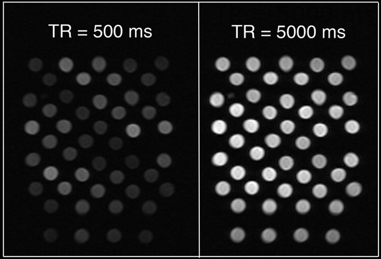
Measurement of T1 Relaxation Times
Preparation of samples for MRI experiment
Get Radiology Tree app to read full this article<
MRI system
Get Radiology Tree app to read full this article<
MRI pulse sequence
Get Radiology Tree app to read full this article<
Data analysis
Get Radiology Tree app to read full this article<
Results
Get Radiology Tree app to read full this article<
Table 1
Dynamic Light Scattering Determined Diameter (nanometers) of Core-encapsulated Gadolinium (CE-Gd) Liposomes
Nuclepore Size (nm) CE-Gd Concentration (mM) 200 350 500 50 75.3 70.5 72.6 100 100.7 94.1 94.5 200 154.1 153.4 160.8 400 253.5 310.3 274.2

Get Radiology Tree app to read full this article<
Get Radiology Tree app to read full this article<
Discussion
Get Radiology Tree app to read full this article<
Get Radiology Tree app to read full this article<
Get Radiology Tree app to read full this article<
Get Radiology Tree app to read full this article<
Get Radiology Tree app to read full this article<
Get Radiology Tree app to read full this article<
Get Radiology Tree app to read full this article<
References
1. Klessen C., Hein P.A., Huppertz A., et. al.: First-pass whole-body magnetic resonance angiography (MRA) using the blood-pool contrast medium gadofosveset trisodium: comparison to gadopentetate dimeglumine. Invest Radiol 2007; 42: pp. 659-664.
2. Goyen M., Edelman M., Perreault P., et. al.: MR angiography of aortoiliac occlusive disease: a phase III study of the safety and effectiveness of the blood-pool contrast agent MS-325. Radiology 2005; 236: pp. 825-833.
3. Mohs A.M., Lu Z.R.: Gadolinium(III)-based blood-pool contrast agents for magnetic resonance imaging: status and clinical potential. Expert Opin Drug Deliv 2007; 4: pp. 149-164.
4. Unger E., Needleman P., Cullis P., et. al.: Gadolinium-DTPA liposomes as a potential MRI contrast agent. Invest Radiol 1988; 23: pp. 928-932.
5. Weissig V.V., Babich J., Torchilin V.V.: Long-circulating gadolinium-loaded liposomes: potential use for magnetic resonance imaging of the blood pool. Colloids Surf B Biointerfaces 2000; 18: pp. 293-299.
6. Ayyagari A.L., Zhang X., Ghaghada K.B., et. al.: Long-circulating liposomal contrast agents for magnetic resonance imaging. Magn Reson Med 2006; 55: pp. 1023-1029.
7. Ghaghada K.B., Bockhorst K.H., Mukundan S., et. al.: High-resolution vascular imaging of the rat spine using liposomal blood pool MR agent. AJNR Am J Neuroradiol 2007; 28: pp. 48-53.
8. Tilcock C., Unger E., Cullis P., et. al.: Liposomal Gd-DTPA: preparation and characterization of relaxivity. Radiology 1989; 171: pp. 77-80.
9. Unger E.C., Winokur T., MacDougall P., et. al.: Hepatic metastases: liposomal Gd-DTPA-enhanced MR imaging. Radiology 1989; 171: pp. 81-85.
10. Tilcock C., MacDougall P., Unger E., et. al.: The effect of lipid composition on the relaxivity of Gd-DTPA entrapped in lipid vesicles of defined size. Biochim Biophys Acta 1990; 1022: pp. 181-186.
11. Fossheim S.L., Fahlvik A.K., Klaveness J., et. al.: Paramagnetic liposomes as MRI contrast agents: influence of liposomal physicochemical properties on the in vitro relaxivity. Magn Reson Imaging 1999; 17: pp. 83-89.
12. Rohrer M., Bauer H., Mintorovitch J., et. al.: Comparison of magnetic properties of MRI contrast media solutions at different magnetic field strengths. Invest Radiol 2005; 40: pp. 715-724.
13. Barsky D., Putz B., Schulten K., et. al.: Theory of paramagnetic contrast agents in liposome systems. Magn Reson Med 1992; 24: pp. 1-13.
14. Huang S.K., Lee K.D., Hong K., et. al.: Microscopic localization of sterically stabilized liposomes in colon carcinoma-bearing mice. Cancer Res 1992; 52: pp. 5135-5143.
15. Litzinger D.C., Buiting A.M., van Rooijen N., et. al.: Effect of liposome size on the circulation time and intraorgan distribution of amphipathic poly(ethylene glycol)-containing liposomes. Biochim Biophys Acta 1994; 1190: pp. 99-107.