
Rationale and Objectives
This study was designed to develop a novel magnetic resonance (MR) probe for the antigen OC183B2 in ovarian cancer cells and investigate its imaging features in vitro and in vivo.
Materials and Methods
Molecular probes were achieved through ultrasmall superparamagnetic iron oxide nanoparticles (USPIOs) conjugated to ovarian cancer monoclonal antibodies 183B2 (OCMab183B2) using a chemical method. In the control group, USPIOs were coupled with murine immunoglobulin G (mIgG) and conjugated the same way. Native polyacrylamide gel electrophoresis was used to evaluate the conjugation reaction. The cytotoxicity of the probe was measured using the methyl thiazolyl tetrazolium assay, and its cell-labeling efficiency was evaluated by Prussian blue staining. In vitro cell MR imaging was performed to evaluate the targeting of the probe to the cells. After that, the OCMab183B2 USPIOs and mIgG USPIOs were injected intravenously into nude mice implanted with ovarian cancer xenograft tumors, respectively. T2-weighted imaging and T2 mapping were then performed on a 3.0-T MR imaging system equipped with an animal birdcage coil at different times. Finally, the nude mice were sacrificed for histologic examination to confirm the imaging results.
Results
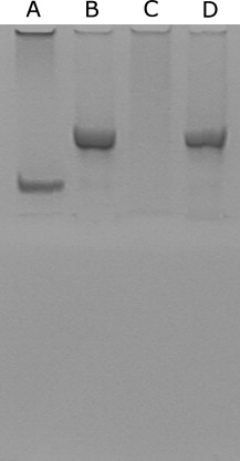
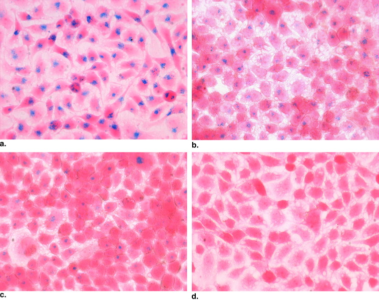
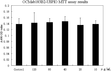
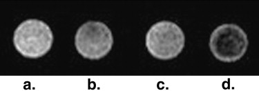
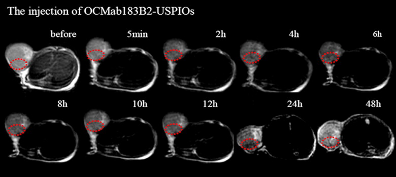
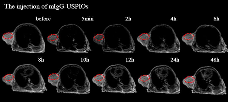
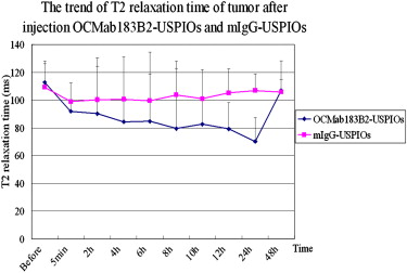
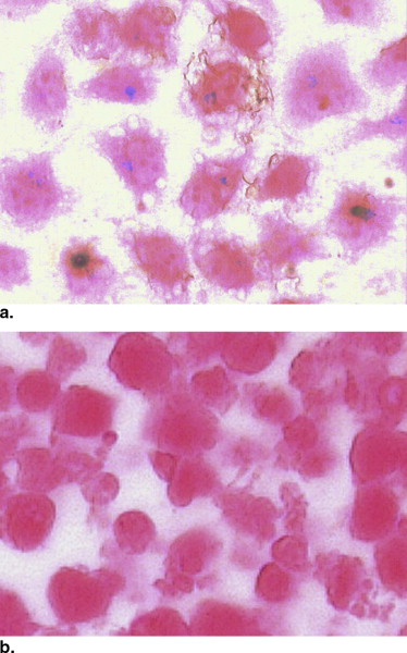
Native polyacrylamide gel electrophoresis displayed an optimal conjugation of USPIOs to OCMab183B2 and mIgG. Various blue-staining particles were found in the cells labeled with the molecular probe at different iron concentrations, and the density of particles was positively related to the iron concentration. Its labeling rate was 96.06%, which was higher than that of USPIOs (62.5%) at the same iron concentration (20 μg/mL). The methyl thiazolyl tetrazolium assay showed that there was no difference in cellular bioactivity between OCMab183B2 USPIO–labeled and nonlabeled cells ( P > .05). In vitro cell MR imaging showed that there was an obvious decrease in signal intensity for the probe-labeled cells compared to mIgG USPIO–labeled cells. For in vivo MR imaging, distinct changes of signal intensities and T2 values of ovarian cancers were detected after the injection of OCMab183B2 USPIOs compared to mIgG USPIOs. The histologic analysis showed that iron depositions were visualized in the experimental group but not in the control group.
Conclusion
OCMab183B2 USPIO conjugates have the potential to be useful as OC183B2-targeted MR imaging agents for the early detection of ovarian cancers.
Ovarian cancer accounts for 4% of all cancers in women and is the leading cause of death from gynecologic malignancies. Early detection and accurate diagnosis are crucial for its successful treatment . Magnetic resonance (MR) imaging has emerged as an important contributor to its diagnosis, but current conventional MR imaging has several limitations in early diagnosis due to its imaging mechanism, which is based on anatomic changes . Recent developments in molecular imaging have provided new views for cancer imaging and treatment. This new imaging technology focuses on molecular changes of diseases and enables MR imaging to detect these changes at “predisease states.” In the process of MR molecular imaging, an important step is to develop a targeted imaging probe, which is made of MR contrast agents, mostly including superparamagnetic iron oxide nanoparticles or ultrasmall superparamagnetic iron oxide nanoparticles (USPIOs), and various ligands or antibodies. This imaging strategy relies on targeted imaging of biomarker molecules coupled with imaging probes guided by ligands or antibodies that can recognize and interact with target molecules .
In our previous studies, we have discovered an ovarian cancer antigen 183B2 (OC183B2), which is expressed on both cytoplasm and cytomembrane of SKOV-3 cells, one kind of epithelial ovarian carcinoma cells, and we successfully obtained its monoclonal antibody (OCMab183B2) from mouse ascites . The murine monoclonal antibody OCMab183B2 was raised against human epithelial ovarian adenocarcinoma-associated antigen by the fusion of murine myeloma cells with spleen lymphocytes from BALB/c nude mice immunized with soluble antigens prepared from ovarian papillary serous cyst adenocarcinoma. The monoclonal antibody was stable after culture, and its subclass was immunoglobulin G. In the current study, we aimed to develop novel MR molecular probe OCMab183B2 USPIO targeting to the antigen OC183B2 and then investigate its imaging features in vitro and in vivo.
Materials and methods
Cell Culture
Get Radiology Tree app to read full this article<
Immunocytochemistry
Get Radiology Tree app to read full this article<
Conjugation of Antibody with Biocompatible Magnetic Nanoparticles
Get Radiology Tree app to read full this article<
Native Polyacrylamide Gel Electrophoresis
Get Radiology Tree app to read full this article<
Labeling of OC183B2 USPIOs and USPIOs
Get Radiology Tree app to read full this article<
Methyl Thiazolyl Tetrazolium (MTT) Assay
Get Radiology Tree app to read full this article<
In Vitro MR Imaging
Get Radiology Tree app to read full this article<
Animals and Tumor Models
Get Radiology Tree app to read full this article<
In Vivo MR Imaging
Get Radiology Tree app to read full this article<
Statistical Analysis
Get Radiology Tree app to read full this article<
Results
Cell Culture
Get Radiology Tree app to read full this article<
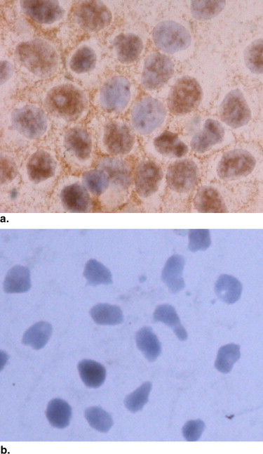
Expression of the Antigen OC183B2 in SKOV-3 Cells
Get Radiology Tree app to read full this article<
Get Radiology Tree app to read full this article<
Native Polyacrylamide Gel Electrophoresis
Get Radiology Tree app to read full this article<
Get Radiology Tree app to read full this article<
Comparing the Labeling Ability Between OC183B2 USPIOs and USPIOs
Get Radiology Tree app to read full this article<
Get Radiology Tree app to read full this article<
MTT Results
Get Radiology Tree app to read full this article<
Get Radiology Tree app to read full this article<
In Vitro MR Imaging
Get Radiology Tree app to read full this article<
Get Radiology Tree app to read full this article<
In Vivo MR Imaging
Get Radiology Tree app to read full this article<
Get Radiology Tree app to read full this article<
Histologic Analysis
Get Radiology Tree app to read full this article<
Get Radiology Tree app to read full this article<
Discussion
Get Radiology Tree app to read full this article<
Get Radiology Tree app to read full this article<
Get Radiology Tree app to read full this article<
Get Radiology Tree app to read full this article<
Get Radiology Tree app to read full this article<
Get Radiology Tree app to read full this article<
Conclusions
Get Radiology Tree app to read full this article<
Acknowledgments
Get Radiology Tree app to read full this article<
Get Radiology Tree app to read full this article<
References
1. Young R.C., Perez C.A., Hoskins W.J.: Cancer of the ovary.DeVita V.T.Hellman S.Rosenberg S.A.Cancer: principles and practice of oncology.1993.LippincottPhiladelphia, PA:pp. 1226-1263.
2. Nicoletta C., Toon V.G., Gabriella P., et. al.: Ovarian cancer. Crit Rev Oncol Hematol 2006; 60: pp. 159-179.
3. Hanna S., Maarit A., Johanna N., et. al.: A highly reproducible xenograft model for human ovarian carcinoma and application of MRI and ultrasound in longitudinal follow-up. Gynecol Oncol 2006; 103: pp. 315-320.
4. Outwater E.K., Siegelman E.S., Kim B., et. al.: Ovarian Brenner tumors: MR imaging characteristics. Magn Reson Imaging 1998; 16: pp. 1147-1153.
5. Tanaka Y.O., Yoshizako T., Nishida M., et. al.: Ovarian carcinoma in patients with endometriosis: MR imaging findings. AJR Am J Roentgenol 2000; 175: pp. 1423-1430.
6. Togashi K.: Ovarian cancer: the clinical role of US, CT, and MRI. Eur Radiol 2003; 13: pp. L87-L104.
7. Katayama M., Masui T., Kobayashi S., et. al.: Diffusion-weighted echo planar imaging of ovarian tumors: is it useful to measure apparent diffusion coefficients. J Comput Assist Tomogr 2002; 26: pp. 250-256.
8. Weissleder R., Mahmood U.: Molecular imaging. Radiology 2001; 219: pp. 316-333.
9. Weissleder R.: Molecular imaging: exploring the next frontier. Radiology 1999; 212: pp. 609-614.
10. Weissleder R., Pittet M.J.: Imaging in the era of molecular oncology. Nature 2008; 452: pp. 580-589.
11. Chaudhuri T.R., Rogers B.E., Buchsbaum D.J., et. al.: A Noninvasive reporter system to image adenoviral-mediated gene transfer to ovarian cancer xenografts. Gynecol Oncol 2001; 83: pp. 432-438.
12. Tiefenauer L.X., Tschirky A., Kühne G., et. al.: In vivo evaluation of magnetite nanoparticles for use as a tumor contrast agent in MRI. Magn Reson Imaging 1996; 14: pp. 391-402.
13. Huang G., Diakur J., Xu Z., et. al.: Asialoglycoprotein receptor-targeted superparamagnetic iron oxide nanoparticles. Int J Pharm 2008; 360: pp. 197-203.
14. Qian H.N., Feng J., Cui H., et. al.: Generation and characterization of three monoclonal antibodies to human ovarian epithelial adenocarcinomas. Chin Med J (Engl) 1989; 102: pp. 839-843.
15. Li Z., Wei L., Gao M.Y., et. al.: One-pot reaction to synthesize biocompatible magnetite nanoparticles. Adv Mater 2005; 17: pp. 1001-1005.
16. Hu F.Q., Li Z., Tu C.F., et. al.: Preparation of magnetite nanocrystals with surface reactive moieties by one-pot reaction. J Colloid Interface Sci 2007; 311: pp. 469-474.
17. Hu F.Q., Li Z., Tu C.F., et. al.: Preparation of biocompatible magnetite nanocrystals for in vivo magnetic resonance detection of cancer. Adv Mater 2006; 18: pp. 2553-2556.
18. Loyer E.M., Whiteman G.J., Fenstermacher M.J.: Imaging of ovarian carcinoma. Int J Gynecol Cancer 1999; 9: pp. 351-361.
19. Kuszyk B.S., Corl F.M., Franano N.F., et. al.: Tumor transport physiology: implications for imaging and imaging-guided therapy. AJR Am J Roentgenol 2001; 177: pp. 747.
20. Rakesh K.J.: Delivery of molecular medicine to solid tumors. Science 1996; 271: pp. 1079.
21. Jain R.K.: Delivery of molecular medicine to solid tumors: lessons from in vivo imaging of gene expression and function. J Control Rel 2001; 74: pp. 7-25.
22. Saccavini J.C., Curtet C., Bohy J., et. al.: Magnetic resonance imaging studies on nude mice grafted with colorectal adenocarcinoma using gadolinium-labelled monoclonal antibody. Invest Radiol 1988; 23: pp. 292-293.
23. Matumura A., Shibata Y., Nakagawa K., et. al.: MRI contrast enhancement by Gd-DTPA-monoclonal antibody in 9L glioma rats. Acta Neurchir Suppl (Wien) 1994; 60: pp. 356-358.
24. Toma A., Otsuji E., Kuriu Y., et. al.: Monoclonal antibody A7-superparamagnetic iron oxide as contrast agent of MR imaging of rectal carcinoma. Br J Cancer 2005; 93: pp. 131-136.
25. Yoshiaki K., Eigo O., Syuichi K., et. al.: Monoclonal antibody conjugated to gadolinium as a contrast agent for magnetic resonance imaging of human rectal carcinoma. J Surg Oncol 2006; 94: pp. 144-148.
26. Marie-France B, M Vasile, S. Morel P. Currently used non-specific extracellular MR contrast media. Eur Radiol 2003; 3:2688–2698.
27. Marie-France B.: MR contrast agents, the old and the new. Eur Radiol 2006; 60: pp. 314-323.
28. Bremer C., Ntziachristos V., Weissleder R.: Optical-based molecular imaging contrast agents and potential medical applications. Eur Radiol 2003; 13: pp. 231-243.