
Rationale and Objectives
To investigate image quality characteristics of abdominal computed tomography (CT) scans reconstructed with adaptive statistical iterative reconstruction V (ASIR-V) vs currently using applied adaptive statistical iterative reconstruction (ASIR).
Materials and Method
This institutional review board-approved study included 35 consecutive patients who underwent CT of the abdomen. Among these 35 patients, 27 with focal liver lesions underwent abdomen CT with a 128-slice multidetector unit using the following parameters: fixed noise index of 30, 1.25 mm slice thickness, 120 kVp, and a gantry rotation time of 0.5 seconds. CT images were analyzed depending on the method of reconstruction: ASIR (30%, 50%, and 70%) vs ASIR-V (30%, 50%, and 70%). Three radiologists independently assessed randomized images in a blinded manner. Imaging sets were compared to focal lesion detection numbers, overall image quality, and objective noise with a paired sample t test. Interobserver agreement was assessed with the intraclass correlation coefficient.
Results
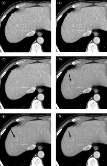
The detection of small focal liver lesions (<10 mm) was significantly higher when ASIR-V was used when compared to ASIR ( P < 0.001). Subjective image noise, artifact, and objective image noise in liver were generally significantly better for ASIR-V compared to ASIR, especially in 50% ASIR-V. Image sharpness and diagnostic acceptability were significantly worse in 70% ASIR-V compared to various levels of ASIR.
Conclusion
Images analyzed using 50% ASIR-V were significantly better than three different series of ASIR or other ASIR-V conditions at providing diagnostically acceptable CT scans without compromising image quality and in the detection of focal liver lesions.
Introduction
The increase in CT utilization has raised concerns of the increased risk of cancer from medical radiation exposures because organ doses with CT are often higher than those with other imaging tests . For this reason, radiation dose reduction in body CT has become a top priority . However, dose reduction must be balanced by an acceptable level of image quality, and above all, diagnostic accuracy. Excessive dose reduction can compromise diagnostic image quality for lesion identification owing to substantial increase in image noise . Lesion detection is also affected by tissue contrast and lesion size. Variable dose reduction techniques, such as tube current modulation, low tube voltage, and noise reduction filters, have been successfully implemented; most promising are the iterative reconstruction algorithms that have evolved beyond the traditional reconstruction method of filtered back projection (FBP) . The adaptive statistical iterative reconstruction (ASIR; GE Healthcare, Waukesha, WI, USA) technique is one of the most widely studied iterative reconstruction techniques providing clinically acceptable image quality with an estimated dose reduction in the range of 25%–40% . Currently, many radiologists and manufacturers offer this technology as a combination of classic FBP and 20%~40% ASIR because of quality problems such as artificial texture or blotchy appearance in a high percentage of iterative reconstruction used.
Model-based iterative reconstruction (MBIR; Veo) has become available as a fully iterative method. MBIR uses a more complex system to predict optical factors, the noise of the system, X-ray physics and objects, and therefore offers considerably better image quality than FBP and ASIR, even at ultralow doses . However, because MBIR requires a long processing time, it has not been widely used yet in routine clinical practice . The recently developed adaptive statistical iterative reconstruction-V (ASIR-V) is a hybrid technique; it includes an algorithm that takes into account the noise of the system (photon statistics and electronic noise), X-ray physics, and objects with omitting optical factors requiring long processing time. Therefore, ASIR-V has the potential for clinically feasible dose reduction with better image quality than conventional ASIR and a shorter imaging processing time than MBIR. So it is conceptually “Augmented ASIR” or “Simplified MBIR” .
Get Radiology Tree app to read full this article<
Materials and Methods
Get Radiology Tree app to read full this article<
Get Radiology Tree app to read full this article<
Patient Population
Get Radiology Tree app to read full this article<
CT Scanning Technique
Get Radiology Tree app to read full this article<
Postprocessing and Image Reconstruction
Get Radiology Tree app to read full this article<
Analysis of Image Quality
Get Radiology Tree app to read full this article<
Qualitative Analysis of ASIR and ASIR-V
Get Radiology Tree app to read full this article<
Get Radiology Tree app to read full this article<
Get Radiology Tree app to read full this article<
TABLE 1
Grading Score for Qualitative Image Analysis of ASIR-V and ASIR Images
Grading Score Qualitative Analysis Subjective Noise Sharpness Artifact Diagnostic Confidence 1 Unacceptable noisy Blurry Major Unacceptable 2 Above average Poor than average Minor Poor 3 Average Average Very little Average 4 Less than average Better than average None High 5 Minimal Sharpest Excellent
Get Radiology Tree app to read full this article<
Interobserver Agreement of Qualitative Analysis
Get Radiology Tree app to read full this article<
Quantitative Analysis of ASIR and ASIR-V
Get Radiology Tree app to read full this article<
Results
Get Radiology Tree app to read full this article<
TABLE 2
Scoring of Lesion Detection and Subjective Image Quality
30% ASIR 50% ASIR 70% ASIR R1 R2 R3 R1 R2 R3 R1 R2 R3 Lesion detection 45 43 44 44 45 45 45 45 45 Subjective noise 3.10 ± 0.20 3.08 ± 0.18 3.10 ± 0.20 4.04 ± 0.24 4.04 ± 0.20 4.04 ± 0.24 4.88 ± 0.21 4.92 ± 0.18 4.92 ± 0.18 Sharpness 3.78 ± 0.25 3.76 ± 0.29 3.76 ± 0.25 3.16 ± 0.23 3.18 ± 0.24 3.18 ± 0.24 2.56 ± 0.41 2.52 ± 0.44 2.56 ± 0.38 Artifact 3.74 ± 0.40 3.74 ± 0.43 3.74 ± 0.35 3.94 ± 0.16 3.90 ± 0.25 3.92 ± 0.18 4.00 ± 0.00 3.98 ± 0.01 4.00 ± 0.00 Diagnostic confidence 4.78 ± 0.26 4.80 ± 0.28 4.66 ± 0.31 4.84 ± 0.27 4.94 ± 0.16 4.86 ± 0.27 4.42 ± 0.39 4.36 ± 0.39 4.38 ± 0.38
30% ASIR-V 50% ASIR-V 70% ASIR-V R1 R2 R3 R1 R2 R3 R1 R2 R3 Lesion detection 53 53 51 62 62 62 61 60 61 Subjective noise 3.96 ± 0.14 3.98 ± 0.10 3.98 ± 0.10 4.82 ± 0.24 4.86 ± 0.22 4.80 ± 0.25 5.00 ± 0.00 5.00 ± 0.00 5.00 ± 0.00 Sharpness 3.90 ± 0.20 3.86 ± 0.22 3.80 ± 0.24 3.86 ± 0.22 3.84 ± 0.23 3.86 ± 0.22 2.40 ± 0.42 2.20 ± 0.32 2.38 ± 0.41 Artifact 3.96 ± 0.14 3.94 ± 0.22 3.98 ± 0.01 4.00 ± 0.00 4.00 ± 0.00 4.00 ± 0.00 4.00 ± 0.00 4.00 ± 0.00 4.00 ± 0.00 Diagnostic confidence 5.00 ± 0.00 4.98 ± 0.10 5.00 ± .0.00 5.00 ± 0.00 5.00 ± 0.00 5.00 ± 0.00 4.00 ± 0.37 3.88 ± 0.29 4.00 ± 0.37
ASIR, adaptive statistical iterative reconstruction; ASIR-V, adaptive statistical iterative reconstruction V; R, radiologist.
Scoring of lesion detection is the sum of the number of lesions.
Scoring of subjective image quality is expressed as mean value and standard deviation.
TABLE 3
Paired Sample t Test for Qualitative Analysis of Subjective Image Quality
Open full size image
ASIR, adaptive statistical iterative reconstruction; ASIR-V, adaptive statistical iterative reconstruction V.
Light grey shading: statistically significant positive relationship.
No shading without negative sign: statistically nonsignificant positive relationship.
No shading with negative sign: statistically nonsignificant negative relationship.
Dark grey shading with negative sign: statistically significant negative relationship.
TABLE 4
Mean Values and Standard Deviations of Image Noise in All Series of ASIR and ASIR-V
Body Site 30% ASIR 50% ASIR 70% ASIR 30% ASIR-V 50% ASIR-V 70% ASIR-V Liver 23.36 ± 4.24 17.90 ± 3.43 14.21 ± 2.73 18.93 ± 3.48 15.33 ± 2.62 12.81 ± 2.28 Gluteal fat tissue 21.40 ± 3.56 15.67 ± 3.43 13.11 ± 2.66 17.41 ± 3.23 14.59 ± 3.15 13.16 ± 3.79 Bladder 22.33 ± 4.59 17.52 ± 3.54 13.85 ± 3.00 18.96 ± 3.77 15.12 ± 2.85 12.18 ± 2.43
ASIR, adaptive statistical iterative reconstruction; ASIR-V, adaptive statistical iterative reconstruction V.
TABLE 5
Paired Simple t Test for Quantitative Image Noise
Open full size image
ASIR, adaptive statistical iterative reconstruction; ASIR-V, adaptive statistical iterative reconstruction V.
Light grey shading: statistically significant positive relationship.
No shading without negative sign: statistically nonsignificant positive relationship.
Dark grey shading with negative sign: statistically significant negative relationship.
Get Radiology Tree app to read full this article<
Qualitative Analysis
Lesion Detection
Get Radiology Tree app to read full this article<
Overall Image Quality (Subjective Image Noise, Sharpness, Artifacts, and Diagnostic Acceptability)
Get Radiology Tree app to read full this article<
Get Radiology Tree app to read full this article<
Get Radiology Tree app to read full this article<
Get Radiology Tree app to read full this article<
Get Radiology Tree app to read full this article<
Interobserver Agreement
Get Radiology Tree app to read full this article<
Quantitative Analysis
Get Radiology Tree app to read full this article<
Discussion
Get Radiology Tree app to read full this article<
Get Radiology Tree app to read full this article<
Get Radiology Tree app to read full this article<
Get Radiology Tree app to read full this article<
Get Radiology Tree app to read full this article<
Get Radiology Tree app to read full this article<
Get Radiology Tree app to read full this article<
Acknowledgements
Get Radiology Tree app to read full this article<
References
1. Brenner D.J., Hall E.J.: Computed tomography: an increasing source of radiation exposure. N Engl J Med 2007; 357: pp. 2277-2284.
2. Pearce M.S., Salotti J.A., Little M.P., et. al.: Radiation exposure from CT scans in childhood and subsequent risk of leukemia and brain tumors: a retrospective cohort study. Lancet 2012; 380: pp. 499-505.
3. Blake S.P., Weisinger K., Atkins M.B., et. al.: Liver metastases from melanoma: detection with multiphasic contrast-enhanced CT. Radiology 1999; 213: pp. 92-96.
4. Singh S., Kalra M.K., Hsieh J., et. al.: Abdominal CT: comparison of adaptive statistical iterative and filtered back projection reconstruction techniques. Radiology 2010; 257: pp. 373-383.
5. Sagara Y., Hara A.K., Pavlicek W., et. al.: Comparison of low-dose CT with adaptive statistical iterative reconstruction and routine-dose CT with filtered back projection in 53 patients. AJR Am J Roentgenol 2010; 195: pp. 713-719.
6. Flicek K.T., Hara A.K., Silva A.C., et. al.: Reducing the radiation dose for CT colonography using adaptive statistical iterative reconstruction; a pilot study. AJR Am J Roentgenol 2010; 195:
7. Pickhardt P.J., Lubner M.G., Kim D.H., et. al.: Abdominal CT with model-based iterative reconstruction (MBIR): initial results of a prospective trial comparing ultralow-dose with standard-dose imaging. AJR Am J Roentgenol 2012; 199: pp. 1266-1274.
8. Cornfeld D., Israel G., Detroy E., et. al.: Impact of Adaptive Statistical Iterative Reconstruction (ASIR) on radiation dose and image quality in aortic dissection studies: a qualitative and quantitative analysis. AJR Am J Roentgenol 2011; 196: pp. W336-W340.
9. Singh S., Kalra M.K., Gilman M.D., et. al.: Adaptive statistical iterative reconstruction technique for radiation dose reduction in chest CT: a pilot study. Radiology 2011; 259: pp. 565-573.
10. Yu Z., Thibault J.B., Bouman C.A., et. al.: Fast model-based x-ray CT reconstruction using spatially nonhomogeneous ICD optimization. IEEE Trans Image Process 2011; 20: pp. 161-175.
11. Deák Z., Grimm J.M., Treitl M., et. al.: Filtered back projection, adaptive statistical iterative reconstruction, and a model-based iterative reconstruction in abdominal CT: an experimental clinical study. Radiology 2013; 266: pp. 197-206.
12. Lim K., Kwon H., Cho J., et. al.: Initial phantom study comparing image quality in computed tomography using adaptive statistical iterative reconstruction and new adaptive statistical iterative reconstruction V. J Comput Assist Tomogr 2015; 39: pp. 443-448.
13. Bartko J.J.: The intraclass correlation coefficient as a measure of reliability. Psychol Rep 1966; 19: pp. 3-11.
14. Leipsic J., Nguyen G., Brown J., et. al.: A prospective evaluation of dose reduction and image quality in chest CT using adaptive statistical iterative reconstruction. AJR Am J Roentgenol 2010; 195: pp. 1095-1099.
15. Pontana F., Duhamel A., Pagniez J., et. al.: Chest computed tomography using iterative reconstruction vs filtered back projection (Part 2): image quality of low-dose CT examinations in 80 patients. Eur Radiol 2011; 21: pp. 636-643.
16. May M.S., Wüst W., Brand M., et. al.: Dose reduction in abdominal computed tomography: intraindividual comparison of image quality of full-dose standard and half-dose iterative reconstructions with dual-source computed tomography. Invest Radiol 2011; 46: pp. 465-470.
17. Hu X.H., Ding X.F., Wu R.Z., et. al.: Radiation dose of non-enhanced chest CT can be reduced 40% by using iterative reconstruction in image space. Clin Radiol 2011; 66: pp. 1023-1029.
18. Koc G., Courtier J.L., Phelps A., et. al.: Computed tomography depiction of small pediatric vessels with model-based iterative reconstruction. Pediatr Radiol 2014; 44: pp. 787-794.
19. Miéville F.A.1., Berteloot L., Grandjean A., et. al.: Model-based iterative reconstruction in pediatric chest CT: assessment of image quality in a prospective study of children with cystic fibrosis. Pediatr Radiol 2013; 43: pp. 558-567.
20. Brady S.L., Yee B.S., Kaufman R.A.: Characterization of adaptive statistical iterative reconstruction algorithm for dose reduction in CT: a pediatric oncology perspective. Med Phys 2012; 39: pp. 5520-5531.