
Rationale and Objectives
Radiology expertise is dependent on the use of efficient search strategies. The aim of this study is to investigate the effect of teaching search strategies on trainee’s accuracy in detecting lung nodules at computed tomography.
Materials and Methods
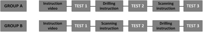
Two search strategies, “scanning” and “drilling,” were tested with a randomized crossover design. Nineteen junior radiology residents were randomized into two groups. Both groups first completed a baseline lung nodule detection test allowing a free search strategy, followed by a test after scanning instruction and drilling instruction or vice versa. True positive (TP) and false positive (FP) scores and scroll behavior were registered. A mixed-design analysis of variance was applied to compare the three search conditions.
Results
Search strategy instruction had a significant effect on scroll behavior, F (1.3) = 54.2, P < 0.001; TP score, F (2) = 16.1, P < 0.001; and FP score, F (1.3) = 15.3, P < 0.001. Scanning instruction resulted in significantly lower TP scores than drilling instruction (M = 10.7, SD = 5.0 versus M = 16.3, SD = 5.3), t (18) = 4.78, P < 0.001; or free search (M = 15.3, SD = 4.6), t (18) = 4.44, P < 0.001. TP scores for drilling did not significantly differ from free search. FP scores for drilling (M = 7.3, SD = 5.6) were significantly lower than for free search (M = 12.5, SD = 7.8), t (18) = 4.86, P < 0.001.
Conclusions
Teaching a drilling strategy is preferable to teaching a scanning strategy for finding lung nodules.
Introduction
Perceptual errors account for a substantial part of misdiagnoses in radiology and can be related to the search behavior of the observer . For educational purposes, it is important to identify which visual search patterns are most effective and to investigate if teaching search strategies improves perception.
Visual search characteristics that are related to expertise and high performance have been identified in various radiology perception tasks . For example experts tend to fixate on abnormalities faster and need less time and a smaller number of eye fixations to inspect the image . These characteristics derive from experience, and they lack an underlying structure that can be taught to novices.
Get Radiology Tree app to read full this article<
Get Radiology Tree app to read full this article<
Get Radiology Tree app to read full this article<
Materials and Methods
Design
Get Radiology Tree app to read full this article<
Get Radiology Tree app to read full this article<
Study Population and Procedure
Get Radiology Tree app to read full this article<
Get Radiology Tree app to read full this article<
Get Radiology Tree app to read full this article<
Get Radiology Tree app to read full this article<
Tests
Get Radiology Tree app to read full this article<
TABLE 1
Test Blueprints with Number, Size, and Location of Nodules
Number Per Case Size Location \* 0–2 3–6 >6 3–4 mm 5–6 mm Average Size (mm) Easy Difficult_Test 1_ 2 3 2 23 8 4.0 25 6Test 2 2 3 2 23 8 4.1 25 6Test 3 2 3 2 23 8 4.0 25 6
Get Radiology Tree app to read full this article<
Get Radiology Tree app to read full this article<
Questionnaire
Get Radiology Tree app to read full this article<
Analysis
Get Radiology Tree app to read full this article<
Get Radiology Tree app to read full this article<
Get Radiology Tree app to read full this article<
Get Radiology Tree app to read full this article<
Get Radiology Tree app to read full this article<
Get Radiology Tree app to read full this article<
Institutional Review Board Approval
Get Radiology Tree app to read full this article<
Results
Participants
Get Radiology Tree app to read full this article<
Test Performance
Get Radiology Tree app to read full this article<
Scroll Behavior
Get Radiology Tree app to read full this article<
TABLE 2
Scroll Behavior and Perceptual Performance Measures Per Search Strategy Condition
Free Search With Drilling Instruction With Scanning Instruction_Scroll behavior_ Number of runs per case, Mdn (IQR) 10(4) 10(4) 2(3) Scrolling time per case in seconds, Mdn(IQR) 219.6(35.9) 208.0(39.3) 167.0(96.6)Perceptual performance True positives, M(SD) 15.3(4.6) 16.3(5.3) 10.7(5.0) False positives, M(SD) 12.5(7.8) 7.3(5.6) 5.6(4.8)
IQR, interquartile range; M, mean; Mdn, median; SD, standard deviation.
Get Radiology Tree app to read full this article<
Get Radiology Tree app to read full this article<
TABLE 3
Mixed-Design ANOVA for Scroll Behavior Outcomes
Number of Runs Time_F__P__F__P__Between subjects_ Year of residency 1.0 0.33 0.4 0.55 Study group 3.5 0.08 0.7 0.41Within subjects Search strategy 54.2 <0.001 10.5 <0.001 Search strategy × Year of residency 0.1 0.88 0.7 0.49 Search strategy × Study group 28.3 0.74 0.9 0.43 Search strategy × Year of residency × Study group 0.4 0.37 1.8 0.18
ANOVA, analysis of variance.
Study group: intervention group A or B; search strategy: free search, drilling instruction, or scanning instruction; year of residency: first or second year.
Get Radiology Tree app to read full this article<
Get Radiology Tree app to read full this article<
Get Radiology Tree app to read full this article<
Perceptual Performance
Get Radiology Tree app to read full this article<
TABLE 4
Mixed-Design ANOVA for Perceptual Performance Outcomes
TP FP_F__P__F__P__Between subjects_ Year of residency 9.0 <0.01 1.4 0.26 Study group 0.12 0.74 0.04 0.85Within subjects Search strategy 16.1 <0.001 15.3 <0.001 Search strategy × Year of residency 1.5 0.85 0.3 0.66 Search strategy × Study group 28.3 0.07 0.9 0.84 Search strategy × Year of residency × Study group 0.4 0.96 0.9 0.38
ANOVA, analysis of variance; FP, false positives; TP, true positives.
Study group: intervention group A or B; search strategy: free search, drilling instruction, or scanning instruction; year of residency: first or second year.
Get Radiology Tree app to read full this article<
Get Radiology Tree app to read full this article<
Get Radiology Tree app to read full this article<
Questionnaire
Get Radiology Tree app to read full this article<
Get Radiology Tree app to read full this article<
Get Radiology Tree app to read full this article<
Discussion
Get Radiology Tree app to read full this article<
Get Radiology Tree app to read full this article<
Get Radiology Tree app to read full this article<
Get Radiology Tree app to read full this article<
Get Radiology Tree app to read full this article<
Get Radiology Tree app to read full this article<
Conclusion
Get Radiology Tree app to read full this article<
Appendix
Supplementary Data
Get Radiology Tree app to read full this article<
Appendix S1
Get Radiology Tree app to read full this article<
Get Radiology Tree app to read full this article<
References
1. Donald J.J., Barnard S.A.: Common patterns in 558 diagnostic radiology errors. J Med Imaging Radiat Oncol 2012; 56: pp. 173-178.
2. Kundel H.L., Nodine C.F., Carmody D.: Visual scanning, pattern recognition and decision-making in pulmonary nodule detection. Invest Radiol 1978; 13: pp. 175-181.
3. van der Gijp A., Ravesloot C.J., Jarodzka H., et. al.: How visual search relates to visual diagnostic performance: a narrative systematic review of eye-tracking research in radiology. Adv Health Sci Educ Theory Pract 2016; Epub ahead of print
4. Krupinski E.A.: Visual scanning patterns of radiologists searching mammograms. Acad Radiol 1996; 3: pp. 137-144.
5. Cooper L., Gale A., Saada J., et. al.: The assessment of stroke multidimensional CT and MR imaging using eye movement analysis: does modality preference enhance observer performance?.Manning D.J.Abbey C.K.Medical imaging 2010: image perception, observer performance, and technology assessment.2010.
6. Wood G., Knapp K.M., Rock B., et. al.: Visual expertise in detecting and diagnosing skeletal fractures. Skeletal Radiol 2013; 42: pp. 165-172.
7. Alzubaidi M.Black J.A.Patel A. et. al.Conscious vs. subconscious perception, as a function of radiological expertise.2009. Proceedings—IEEE Symposium on Computer-Based Medical Systems
8. Manning D., Ethell S., Donovan T., et. al.: How do radiologists do it? The influence of experience and training on searching for chest nodules. Radiography 2006; 12: pp. 134-142.
9. Kok E.M., de Bruin A.B.H., Robben S.G.F., et. al.: Looking in the same manner but seeing it differently: bottom-up and expertise effects in radiology. Appl Cogn Psychol 2012; 26: pp. 854-862.
10. Hu C.H., Kundel H.L., Nodine C.F., et. al.: Searching for bone fractures: a comparison with pulmonary nodule search. Acad Radiol 1994; 1: pp. 25-32.
11. Drew T., Le-Hoa Vo M., Olwal A., et. al.: Scanners and drillers: characterizing expert visual search through volumetric images. J Vis 2013; 13:
12. Baghdady M.T., Pharoah M.J., Regehr G., et. al.: The role of basic sciences in diagnostic oral radiology. J Dent Educ 2009; 73: pp. 1187-1193.
13. Nodine C.F., Kundel H.L., Mello-Thoms C., et. al.: How experience and training influence mammography expertise. Acad Radiol 1999; 6: pp. 575-585.
14. Nodine C.F., Kundel H.L., Lauver S.C., et. al.: Nature of expertise in searching mammograms for breast masses. Acad Radiol 1996; 3: pp. 1000-1006.
15. Elmore J.G., Wells C.K., Howard D.H.: Does diagnostic accuracy in mammography depend on radiologists’ experience?. J Womens Health 1998; 7: pp. 443-449.
16. Venjakob A., Marnitz T., Mahler J., et. al.: Radiologists’ eye gaze when reading cranial CT images. Medical Imaging 2012: Image Perception, Observer Performance, and Technology Assessment2012.pp. 8318.
17. Nodine C.F., Mello-Thoms C., Kundel H.L., et. al.: Time course of perception and decision making during mammographic interpretation. AJR Am J Roentgenol 2002; 179: pp. 917-923.
18. Valencia R., Denecke T., Lehmkuhl L., et. al.: Value of axial and coronal maximum intensity projection (MIP) images in the detection of pulmonary nodules by multislice spiral CT: comparison with axial 1-mm and 5-mm slices. Eur Radiol 2006; 16: pp. 325-332.