
Rationale and Objectives
The aim of this study was to investigate the effects of exogenous endothelial progenitor cells (EPCs) on the growth and invasiveness of glioma in vivo to provide an experimental basis for the value and safety of using magnetically labeled EPCs as target vectors to detect early infiltration of glioma.
Materials and Methods
EPCs were collected from the spleens of healthy Sprague-Dawley rats, made EPCs conditioned medium after identification. Four models of Sprague-Dawley rat glioma (60 rats in total) were established as a control and three experimental groups (group A, B, and C). In the control group, orthotopic transplantation of C6 glioma cells was performed. Compared to the control group, EPCs conditioned medium was added in group A and P7228-labeled EPCs were added in group B. In group C, P7228-labeled EPCs were transplanted via the tail vein. Magnetic resonance imaging and perfusion-weighted imaging were performed on several days. Tumor microvascular density and vascular endothelial growth factor expression were determined through immunohistochemistry.
Results
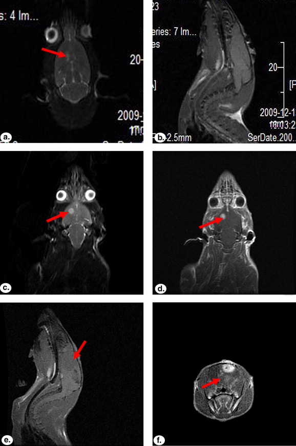
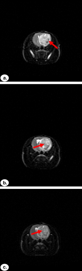
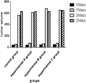
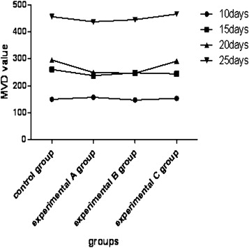
In group C, hypointense areas were detected at the periphery of the tumor on the first day after transplantation of EPCs, and more hypointense areas were found inside the tumor over time. Tumor size in all four groups developed significantly with increasing time ( P < .01), but there was no marked difference among these groups at the same time ( P > .05). No remarkable differences in microvascular density and cells positive for vascular endothelial growth factor were found at the same time among the four groups ( P > .05).
Conclusions
Both magnetic resonance imaging and immunohistochemical findings confirmed that exogenous EPCs could not affect the biologic behavior of C6 glioma cells in vivo through a paracrine effect or by direct cellular interaction. Therefore, exogenous EPCs could not exert significant promoting effects on glioma growth.
Endothelial progenitor cells (EPCs), one kind of precursor cells that can proliferate and differentiate into mature endothelial cells in vitro, have sound proliferating and differentiating capacity. They not only participate in vasculogenesis at the embryonic stage but also play an important role in postnatal neovasculogenesis . EPCs can home in to a tumor site and incorporate into the tumor’s vascular endothelium. They can secrete angiogenic factors that promote tumor neovasculature. In light of the homing feature of EPCs and the biocompatibility of iron oxide–labeled nanomaterial molecular probes, the use of magnetically labeled EPCs as a magnetic resonance contrast agent to detect early brain glioma is promising. However, whether transplanted exogenous EPCs can promote the development of glioma has not been clearly clarified.
In this study, we performed orthotopic transplantation and transplantation via the tail vein of P7228-labeled EPCs and used magnetic resonance imaging (MRI) to determine the distribution of EPCs in glioma tissue and their effects on the biologic behavior of tumors, such as growth and invasion, to provide an experimental basis for evaluating the clinical safety of using magnetically labeled EPCs in diagnosing early brain glioma.
Materials and methods
Get Radiology Tree app to read full this article<
Isolation, Culture, and Identification of Rat Spleen-originated EPCs and Preparation of EPCs Conditioned Medium
Get Radiology Tree app to read full this article<
Get Radiology Tree app to read full this article<
Labeling EPCs with P7228
Get Radiology Tree app to read full this article<
Establishment of Glioma Model
Get Radiology Tree app to read full this article<
Get Radiology Tree app to read full this article<
MRI Scanning for Rats with Glioma
Get Radiology Tree app to read full this article<
Image Analysis
Get Radiology Tree app to read full this article<
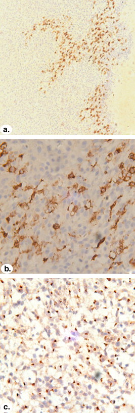
Pathologic Analysis
Get Radiology Tree app to read full this article<
Statistical Analysis
Get Radiology Tree app to read full this article<
Results
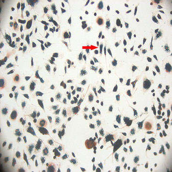
Observation of P7228-labeled EPCs with Inverted Microscopy
Get Radiology Tree app to read full this article<
Get Radiology Tree app to read full this article<
Establishment of Glioma Model
Get Radiology Tree app to read full this article<
MRI Scanning for Rats with Glioma
Get Radiology Tree app to read full this article<
Get Radiology Tree app to read full this article<
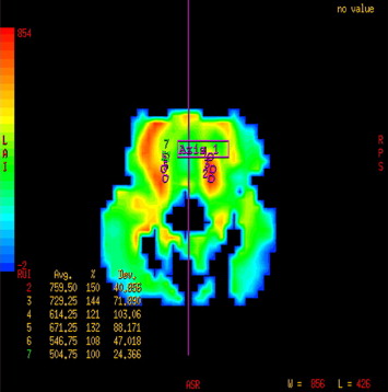
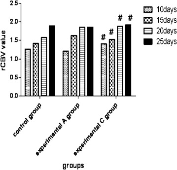
PWI Results and Statistical Analysis
Get Radiology Tree app to read full this article<
Get Radiology Tree app to read full this article<
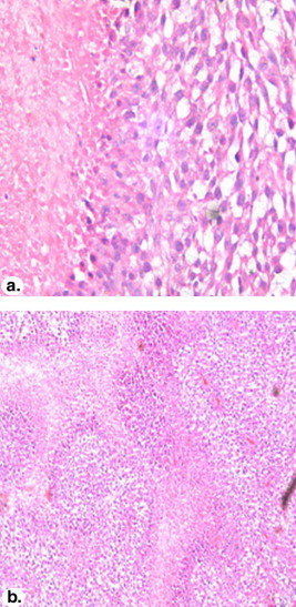
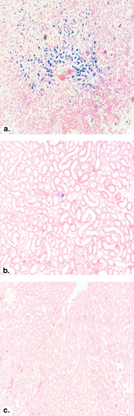
Pathologic Examination
Get Radiology Tree app to read full this article<
Get Radiology Tree app to read full this article<
Get Radiology Tree app to read full this article<
Correlation Analysis
Get Radiology Tree app to read full this article<
Discussion
Get Radiology Tree app to read full this article<
Get Radiology Tree app to read full this article<
Get Radiology Tree app to read full this article<
Get Radiology Tree app to read full this article<
Get Radiology Tree app to read full this article<
Get Radiology Tree app to read full this article<
Conclusions
Get Radiology Tree app to read full this article<
Get Radiology Tree app to read full this article<
References
1. Chade A.R., Zhu X., Lavi R., et. al.: Endothelial progenitor cells restore renal function in chronic experimental renovascular disease. Circulation 2009; 119: pp. 547-557.
2. Le Bourhis X., Romon R., Hondermarck H., et. al.: Role of endothelial progenitor cells in breast cancer angiogenesis: from fundamental research to clinical ramifications. Breast Cancer Res Treat 2010; 120: pp. 17-24.
3. Gazeau F., Wilhelm C.: Magnetic labeling, imaging and manipulation of endothelial progenitor cells using iron oxide nanoparticles. Future Med Chem 2010; 2: pp. 397-408.
4. Fang J.Q., Zhang W.G., Ma C.S.: Rat spleen-derived endothelial progenitor cells promote proliferation of C6 glioma cells under co-culture system in vitro. J Third Mil Med Univ 2010; 32: pp. 917-921.
5. Krenning G., van Luyn M.J., Harmsen M.C.: Endothelial progenitor cell-based neovascularization: implication for therapy. Trends Mol Med 2009; 15: pp. 180-189.
6. Rapisarda A., Melillo G.: Role of hypoxic tumor microenviroment in the resistance to anti-angiogenic therapies. Drug Resist Updat 2009; 12: pp. 74-80.
7. Rehman J., Li J., Orschell C., et. al.: Peripheral blood endothelial progenitor cells are derived from monocyte/macrophages and secrete angiogenic growth factors. Circulation 2003; 107: pp. 1164-1169.
8. Yin Y.G., Huang L., Zhao X.H.: Endothelial progenitor cells secret SDF-1 and SDF-1 protects endothelial progenitor cells from apoptosis. Chin Heart 2007; 19: pp. 391-394.
9. Yang Y., Li S.L.: The role of vascular endothelial growth factor in tumor. For Med Sci (Oncol Sect) 2004; 31: pp. 18-20.
10. Yamamoto S., Wakimoto H., Aoyagi M., et. al.: Modulation motility and proliferation of glioma cells by hepatocyte growth factor. Jpn J Cancer Res 1997; 88: pp. 564-577.
11. Dalle S., Imamura T., Rose D.W., et. al.: Insulin induces heterologous desensitization of G protein-coupled receptor and insulin-like growth factor I signaling by down regulating β-arrestin-1. Mol Cell Biol 2002; 22: pp. 6272-6285.
12. Zhang M.G., Li X.L., Wan D.X.: Research progress of SDF-1/CXCR4 in cancer. J Oncol 2008; 14: pp. 600-604.
13. Zhang X.L., Qu Y., Mu D.Z.: The relationship between SDF-1/CXCR4 and tumor biological behavior. Chem Life 2008; 28: pp. 708-710.
14. Roord B.D., ter-Elst A., Kamps W.A., et. al.: Bone marrow-derived cells and tumor growth: contribution of bone marrow-derived cells to tumor micro-environments with special focus on mesenchymal stem cells. Crit Rev Oncol Hematol 2009; 69: pp. 187-198.
15. Burger J.A., Kipps T.J.: CXCR4: a key receptor in the crosstalk between tumor cells and their microenvironment. Blood 2006; 107: pp. 1761-1767.
16. Ohira S., Sasaki M., Harada K., et. al.: Possible regulation of migration of intrahepatic cholangiocarcinoma cells by interaction of CXCR4 expressed in carcinoma cells with tumor necrosis factor-α and stromal-derived factor-1 released in stroma. Am J Pathol 2006; 168: pp. 1155-1168.
17. Ahn J.B., Rha S.Y., Shin S.J., et. al.: Circulating endothelial progenitor cells (EPC) for tumor vasculogenesis in gastric cancer patients. Cancer Lett 2010; 288: pp. 124-132.
18. Badie B., Schartner J.M.: Flow cytometric characterization of tumor-associated macrophages in experimental gliomas. Neurosurgery 2000; 46: pp. 957-961.
19. Zaharchuk G.: Theoretical basis of hemodynamic MR imaging techniques to measure cerebral blood volume, cerebral blood flow, and permeability. AJNR Am J Neuroradiol 2007; 28: pp. 1850-1858.
20. Law M., Cha S., Knopp E.A., et. al.: High-grade gliomas and solitary metastases: differentiation by using perfusion and proton spectroscopic MR imaging. Radiology 2002; 222: pp. 715-721.
21. Weidner N.: Current pathologic methods for measuring intratumoral microvessel density within breast carcinoma and other solid tumors. Breast Cancer Res Treat 1995; 36: pp. 169-180.
22. Dwenger A., Rosenthal F., Machein M., et. al.: Transplanted bone marrow cells preferentially home to the vessels of in situ generated murine tumors rather than of normal organs. Stem Cells 2004; 22: pp. 86-92.
23. Duda D.G., Cohen K.S., Kozin S.V., et. al.: Evidence for incorporation of bone marrow-derived endothelial cells into perfused blood in tumors. Blood 2006; 107: pp. 2774-2776.
24. Ahn G.O., Brown J.M.: Matrix metalloproteinase-9 is required for tumor vasculogenesis but not for angiogenesis: role of bone marrow-derived myelomonocytic cells. Cancer Cell 2008; 13: pp. 193-205.
25. Suriano R., Chaudhuri D., Johnson R.S., et. al.: 17Beta-estradiol mobilizes bone marrow-derived endothelial progenitor cells to tumors. Cancer Res 2008; 68: pp. 6038-6042.
26. Wickersheim A., Kerber M., de Miquel L.S., et. al.: Endothelial progenitor cells do not contribute to tumor endothelium in primary and metastatic tumors. Int J Cancer 2009; 125: pp. 1771-1777.
27. Rajantie I., monen M., Alminaite A., et. al.: Adult bone marrow-derived cells recruited during angiogenesis comprise precursors for periendothelial vascular mural cells. Blood 2004; 104: pp. 2084-2086.
28. Gothert J.R., Gustin S.E., van Eekelen J.A., et. al.: Genetically tagging endothelial cells in vivo: bone marrow-derived cells do not contribute to tumor endothelium. Blood 2004; 104: pp. 1069-1077.
29. De Palma M., Venneri M.A., Roca C., et. al.: Targeting exogenous genes to tumor angiogenesis by transplantation of genetically modified hematopoietic stem cells. Nat Med 2003; 9: pp. 789-795.
30. Dias S., Hattori K., Zhu Z.P., et. al.: Autocrine stimulation of VEGFR-2 activates human leukemic cells growth and migration. J Clinical Invest 2000; 106: pp. 511-521.
31. Wang C.W., Pan Q., Zhang L., et. al.: Significance of vascular endothelial growth factor expression and intratumoral microvessel density of human brain glioma. Cancer 2001; 20: pp. 326-327.
32. Patenaude A., Parker J., Karsan A.: Involvement of endothelial progenitor cells in tumor vascularization. Microvasc Res 2010; 79: pp. 217-223.