
Rationale and Objectives
Recent research has provided evidence that in reading rooms equipped with liquid crystal displays (LCDs), a measured increase of ambient lighting may improve clinicians’ detection performance. In agreement with this research, the American College of Radiology (ACR) has recommended a moderate increase of ambient lighting in mammography reading rooms. This study was designed to examine the effect of a controlled increase of ambient lighting in mammography reading rooms on the diagnostic performance of breast imaging radiologists.
Materials and Methods
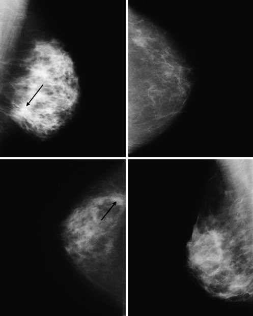
Four breast imaging radiologists read 86 mammograms (43 containing subtle cancerous masses and 43 normal) under low ( E = 1 lux) and elevated ( E = 50 lux) ambient lighting levels on a Digital Imaging and Communications in Medicine–calibrated, medical-grade LCD. Radiologists were asked to identify cancerous masses and to rate their detection confidence. Observer areas under the curve (AUCs) were calculated using a receiver-operating characteristic analysis of fully paired results. Additionally, average observer selection times under both ambient lighting levels were determined.
Results
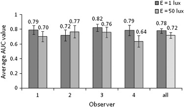
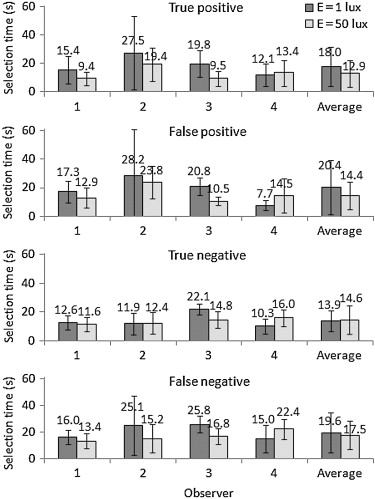
Average radiologist AUCs decreased with elevated ambient lighting (0.78 ± 0.03 to 0.72 ± 0.04). Observer performance differences, however, were of the same order of magnitude as interobserver variability and were not statistically significant. Average selection times under increased ambient lighting remained constant or decreased, with the greatest decrease occurring for false-positive (20.4 ± 18.9 to 14.4 ± 9.6 seconds) and true-positive (18.0 ± 13.8 to 12.9 ± 9.4 seconds) selections.
Conclusion
The results agree with those of previous studies in that observer performance differences under a controlled increase of ambient lighting are not statistically significant. On the basis of these findings and ACR guidelines, a moderate increase of ambient lighting in mammography reading rooms is still suggested, but further research with additional cases and observers should be considered.
The introduction of liquid crystal displays (LCDs) to diagnostic radiology has necessitated the re-evaluation of an optimum reading room environment. Although many reading room environmental conditions may affect observer performance, ambient lighting is known to be an especially important aspect of radiologist detection performance and comfort. In film-based mammography reading rooms, ambient lighting has typically been kept at a minimal level to maintain perceived image contrast . At this lighting level, however, a reader’s pupils will contract and dilate as the visual focus shifts between reading the high-luminance image ( L adp ) and the lower luminance surrounding background ( L s ). This pupillary action may lead to radiologist fatigue and result in the degradation of diagnostic performance . However, a moderate increase of ambient lighting, such that L s is close to L adp , might reduce pupillary action and potentially improve radiologist comfort and detection performance.
Modern calibration-capable, medical-grade LCDs, with intrinsically low diffuse reflection coefficients and high luminance ratios, may permit moderately elevated ambient lighting levels without a loss of image quality . Recent research has provided initial theoretical and physical evidence that a measured increase of reading room illuminance might lead to improved observer performance . In view of these findings, the American College of Radiology (ACR) has issued guidelines encouraging a moderate increase of ambient lighting levels in mammography reading rooms .
Get Radiology Tree app to read full this article<
Materials and methods
Image Data
Get Radiology Tree app to read full this article<
Get Radiology Tree app to read full this article<
Illuminance Levels
Get Radiology Tree app to read full this article<

Get Radiology Tree app to read full this article<
Display System
Get Radiology Tree app to read full this article<
Get Radiology Tree app to read full this article<
Observer Study
Get Radiology Tree app to read full this article<
Get Radiology Tree app to read full this article<
Performance Evaluation
Get Radiology Tree app to read full this article<
Results
Get Radiology Tree app to read full this article<
Get Radiology Tree app to read full this article<
Get Radiology Tree app to read full this article<
Get Radiology Tree app to read full this article<
Discussion
Get Radiology Tree app to read full this article<
Get Radiology Tree app to read full this article<
Get Radiology Tree app to read full this article<
Get Radiology Tree app to read full this article<
Get Radiology Tree app to read full this article<
Get Radiology Tree app to read full this article<
Get Radiology Tree app to read full this article<
Conclusion
Get Radiology Tree app to read full this article<
Acknowledgments
Get Radiology Tree app to read full this article<
Get Radiology Tree app to read full this article<
References
1. Mahesh M.: AAPM/RSNA physics tutorial for residents: digital mammography: an overview. RadioGraphics 2004; 24: pp. 1747-1760.
2. Fratt L. Redesigning the reading room. Available at: http://www.healthimaging.com/content/view/1389/68/ . Accessed April 2, 2007.
3. Reiner B.I., Siegel E.L., Rostenberg B.: Redesigning the PACS reading room: optimizing monitor and room lighting.Medical Imaging 1999: PACS design and evaluation: engineering and clinical issues.1999.SPIESan Diego, CA: 276–280
4. Kaiser CP. Report from SIIM: informatics specialist eyes visual problems in radiologists. Available at: http://www.diagnosticimaging.com/showNews.jhtml?articleID=199902386 . Accessed March 20, 2008.
5. Samei E., Badano A., Chakraborty D., et. al.: Assessment of display performance for medical imaging systems: executive summary of AAPM TG18 report. Med Phys 2005; 32: pp. 1205-1225.
6. Pollard B.J., Chawla A.S., Hashimoto N., Delong D., Samei E.: Object detectability at increased ambient lighting conditions. Med Phys 2008; 35: pp. 2204-2213.
7. Brennan P.C., McEntee M., Evanoff M., Phillips P., O’Connor W.T., Manning D.J.: Ambient lighting: effect of illumination on soft-copy viewing of radiographs of the wrist. AJR Am J Roentgenol 2007; 188: pp. W177-W180.
8. Chawla A.S., Samei E.: Ambient illumination revisited: a new adaptation-based approach for optimizing medical imaging reading environments. Med Phys 2007; 34: pp. 81-90.
9. Siegel E., Krupinski E., Samei E., et. al.: Digital mammography image quality: image display. J Am Coll Radiol 2006; 3: pp. 615-627.
10. Pisano ED, Seibert JA, Andriole KP, et al. Practice guideline for determinants of image quality in digital mammography. Available at: http://www.acr.org/SecondaryMainMenuCategories/quality_safety/guidelines/breast/image_quality_digital_mammo.aspx . Accessed March 20, 2008.
11. Heath M., Bowyer K.W., Kopans D., et. al.: Current status of digital database for screening mammography. Digit Mammogr 1998; pp. 457-460.
12. Saunders R., Ike R.: Mammo software.2007.Duke Advanced Imaging LaboratoriesDurham, NC
13. Samei E.: AAPM/RSNA physics tutorial for residents: technological and psychophysical considerations for digital mammographic displays. RadioGraphics 2005; 25: pp. 491-501.
14. National Electrical Manufacturers Association. Digital Imaging and Communications in Medicine (DICOM), part 14 grayscale standard display function PS 3.1-2006. Available at: http://medical.nema.org . Accessed March 20, 2008.
15. Metz C.: ROCKIT 0.9B.1998.University of ChicagoChicago
16. Chawla A.S., Samei E.: A method for reduction of eye fatigue by optimizing the ambient light conditions in radiology reading rooms.Medical Imaging 2006: PACS and imaging informatics.2006.SPIESan Diego, CA:pp. 614503-614512.