
Rationale and Objectives
To provide an updated evidence-based review of the literature on the role of fluorine-18-fluorodeoxyglucose positron emission tomography (FDG-PET) or PET/computed tomography (PET/CT) in patients with sarcoidosis.
Materials and Methods
A comprehensive literature search of PubMed/MEDLINE, Scopus, and Embase databases was conducted to find relevant published articles on FDG-PET or PET/CT in patients with sarcoidosis. Only those studies that satisfied all the following criteria were included: (1) FDG-PET or PET/CT performed in patients with histologically confirmed sarcoidosis, (2) sample size of at least 10 patients, and (3) whole-body PET studies performed. Information was collected concerning basic study, patient characteristics, and technical aspects. The main findings of the articles included have been described.
Results
Reviewing titles and abstracts, 21 articles were selected. The role of FDG-PET or PET/CT was analyzed in assessing disease extent and activity and in treatment response evaluation. Comparison with other tracers had also been investigated. Recent studies evaluated the influence of these methods in the clinical management and their role as predictive tools in patients with sarcoidosis.
Conclusions
FDG-PET and PET/CT seem to be useful in staging, evaluating disease activity, and monitoring treatment response in patients with sarcoidosis. PET appears to have higher diagnostic accuracy compared to Gallium-67-citrate scintigraphy. Conversely, there is not enough evidence about the use of other PET tracers in patients with sarcoidosis. FDG-PET and PET/CT seem to have a role as predictive tools and may influence the clinical management in patients with sarcoidosis, but more studies are needed in this regard.
Sarcoidosis is a granulomatous multisystem disease of unknown etiology, with different clinical course and one-third of incidence of chronic and progressive forms .
Clinical evaluation of patients with sarcoidosis needs many different diagnostic tools. Chest radiography (CXR) is often the first diagnostic imaging study to evaluate pulmonary involvement in sarcoidosis, but existing radiographic score (stages 0–IV) has limited value in predicting inflammatory activity . Computed tomography (CT) and high-resolution CT (HRCT) offer good anatomic details, but these methods are not able to investigate the functional deterioration caused by inflammatory disease. Serum inflammatory markers and respiratory function parameters have also a role in a correct evaluation of clinical status in patients with sarcoidosis .
Get Radiology Tree app to read full this article<
Get Radiology Tree app to read full this article<
Methods
Search Strategy
Get Radiology Tree app to read full this article<
Study Selection
Get Radiology Tree app to read full this article<
Get Radiology Tree app to read full this article<
Get Radiology Tree app to read full this article<
Get Radiology Tree app to read full this article<
Data Abstraction
Get Radiology Tree app to read full this article<
Results
Get Radiology Tree app to read full this article<
Table 1
Basic Characteristics of the Included Articles
Authors Year Country Type of Study Number of Patients with Sarcoidosis Performing PET Sex (% Male) Mean Age (years) Chest Radiographic Stage (Number of Patients) Yamada et al. 1998 Japan Prospective 31 61 NR I (18); II (13) Nishiyama et al. 2006 Japan Retrospective 18 33 59 0 (1); I (13); II (4) Kaira et al. 2007 Japan Prospective 24 37 43 I (11); II (13) Teirstein et al. 2007 USA Retrospective 137 52 50 0 (11), I (18); II (15); III (7); IV (22) Prager et al. 2008 Austria Retrospective 24 46 52 NR Braun et al. 2008 France Retrospective 20 55 51 I (2); II (3); III (1); IV (7) Keijsers et al. 2008 The Netherlands Retrospective 12 50 44 I (3); II (6); III (2); IV (1) Keijsers et al. 2009 The Netherlands Retrospective 36 44 39 0 (1), I (18); II (11); III (5); IV (1) Keijsers et al. 2010 The Netherlands Retrospective 77 58 39 0 (4), I (31); II (30); III (9); IV (3) Keijsers et al. 2011 The Netherlands Retrospective 43 63 43 0 (1), I (10), II/III (30); IV (2) Keijsers et al. 2011 The Netherlands Prospective 34 71 42 0 (3), I (9), II (18); III (2); IV (2) Mostard et al. 2011 The Netherlands Retrospective 89 56 46 0 (24); I (19); II (27), III (4); IV (15) Umeda et al. 2011 Japan NR 21 43 48 NR Milman et al. 2012 Denmark Prospective 10 20 47 I (2); II (6); III (1); IV (1) Mostard et al. 2012 The Netherlands Retrospective 95 58 45 0 (30); I (15); II (13), III (11); IV (26) Mostard et al. 2012 The Netherlands Retrospective 122 59 46 0 (36); I (26); II (22); III (11); IV (27) Sobic-Saranovic et al. 2012 Serbia Prospective 90 36 47 0 (10); I (42); II (25); III (8); IV (5) Sobic-Saranovic et al. 2013 Serbia NR 66 35 46 NR Mostard et al. 2013 The Netherlands Retrospective 95 58 46 0 (30); I (16); II (13); III (10); IV (26) Ambrosini et al. 2013 Italy Prospective 28 54 47 0 (1); I (6); II (17); III (2); IV (9) on 35 scans performed Imperiale et al. 2013 France Retrospective 13 62 44 NR
NR, not reported.
Table 2
Technical Characteristics of the Included Articles
Authors Year PET Device PET Tracers Other Tracers Mean Injected activity (MBq) Time Between Injection and Acquisition (minutes) PET Image Analysis Yamada et al. 1998 PET 18 F-FDG + 11 C-Met 67 Ga 148 60 Visual and semiquantitative Nishiyama et al. 2006 PET 18 F-FDG 67 Ga Range 185–200 60 Visual and semiquantitative Kaira et al. 2007 PET 18 F-FDG + 18 F-FMT — Range 4–6/kg 50 Visual and semiquantitative Teirstein et al. 2007 PET/CT 18 F-FDG — Range 370–740 NR Visual and semiquantitative Prager et al. 2008 PET 18 F-FDG 67 Ga Range 296–370 range 60-70 Visual and semiquantitative Braun et al. 2008 PET/CT 18 F-FDG 67 Ga 5.5/kg 60 Visual and semiquantitative Keijsers et al. 2008 PET 18 F-FDG 67 Ga Range 295–400 60 Visual and semiquantitative Keijsers et al. 2009 PET 18 F-FDG — Range 295–400 60 Visual and semiquantitative Keijsers et al. 2010 PET 18 F-FDG — Range 295–400 60 Visual and semiquantitative Keijsers et al. 2011 PET 18 F-FDG — Range 295–400 60 Visual and semiquantitative Keijsers et al. 2011 PET 18 F-FDG 67 Ga Range 295–400 60 Visual Mostard et al. 2011 PET/CT 18 F-FDG — 200 45 Visual and semiquantitative Umeda et al. 2011 PET 18 F-FDG 67 Ga 185 Dual time: 80–200 Visual and semiquantitative Milman et al. 2012 PET or PET/CT 18 F-FDG — 400 60 Visual and semiquantitative Mostard et al. 2012 PET/CT 18 F-FDG — 200 45 Visual and semiquantitative Mostard et al. 2012 PET/CT 18 F-FDG — 200 45 Visual Sobic-Saranovic et al. 2012 PET/CT 18 F-FDG — 5.5/kg 60 Visual and semiquantitative Sobic-Saranovic et al. 2013 PET/CT 18 F-FDG — 5.5/kg 60 Visual and semiquantitative Mostard et al. 2013 PET/CT 18 F-FDG — 200 45 Visual and semiquantitative Ambrosini et al. 2013 PET/CT 18 F-FDG — 370 60 Visual and semiquantitative Imperiale et al. 2013 PET/CT 18 F-FDG — 4.5/kg 60 Visual and semiquantitative
18 F-FDG, 18 F-fluorodeoxyglucose; 11 C-Met, 11 C-methionine; 18 F-FMT, 18 F-fluoromethyltyrosine, 67 Ga: Gallium-67-citrate; NR, not reported; PET/CT, positron emission tomography/computed tomography.
Get Radiology Tree app to read full this article<
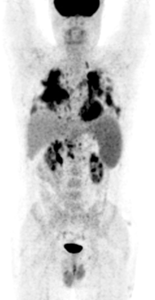
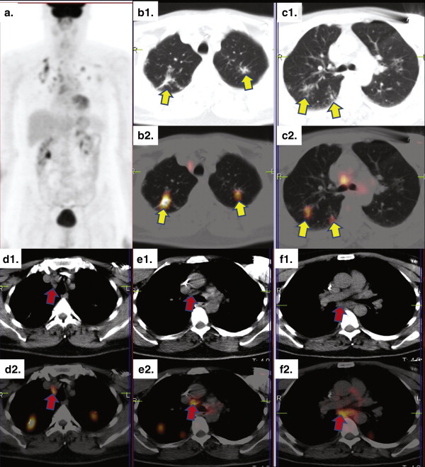
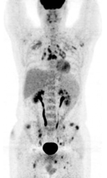
Role of FDG-PET and PET/CT in Evaluating Disease Extent in Patients with Sarcoidosis and Correlation with Conventional Imaging
Get Radiology Tree app to read full this article<
Get Radiology Tree app to read full this article<
Get Radiology Tree app to read full this article<
Get Radiology Tree app to read full this article<
Get Radiology Tree app to read full this article<
Get Radiology Tree app to read full this article<
Get Radiology Tree app to read full this article<
Get Radiology Tree app to read full this article<
Get Radiology Tree app to read full this article<
Get Radiology Tree app to read full this article<
Get Radiology Tree app to read full this article<
Get Radiology Tree app to read full this article<
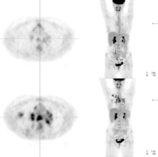
Role of FDG-PET and PET/CT as a Marker of Disease Activity in Patients with Sarcoidosis
Get Radiology Tree app to read full this article<
Get Radiology Tree app to read full this article<
Get Radiology Tree app to read full this article<
Get Radiology Tree app to read full this article<
Get Radiology Tree app to read full this article<
Get Radiology Tree app to read full this article<
Get Radiology Tree app to read full this article<
Role of FDG-PET and PET/CT in Treatment Evaluation of Patients with Sarcoidosis
Get Radiology Tree app to read full this article<
Get Radiology Tree app to read full this article<
Get Radiology Tree app to read full this article<
Get Radiology Tree app to read full this article<
Get Radiology Tree app to read full this article<
Get Radiology Tree app to read full this article<
Get Radiology Tree app to read full this article<
Get Radiology Tree app to read full this article<
Get Radiology Tree app to read full this article<
Comparison of FDG with Other PET Tracers
Get Radiology Tree app to read full this article<
Get Radiology Tree app to read full this article<
Get Radiology Tree app to read full this article<
Get Radiology Tree app to read full this article<
Comparison of FDG-PET and PET/CT with Gallium-67-citrate ( 67 Ga) Scintigraphy
Get Radiology Tree app to read full this article<
Get Radiology Tree app to read full this article<
Get Radiology Tree app to read full this article<
Get Radiology Tree app to read full this article<
Get Radiology Tree app to read full this article<
Get Radiology Tree app to read full this article<
Role of FDG-PET and PET/CT as Predictive Tools in Patients with Sarcoidosis
Get Radiology Tree app to read full this article<
Get Radiology Tree app to read full this article<
Get Radiology Tree app to read full this article<
Get Radiology Tree app to read full this article<
Get Radiology Tree app to read full this article<
Impact of FDG-PET/CT on Clinical Management of Patients with Sarcoidosis
Get Radiology Tree app to read full this article<
Get Radiology Tree app to read full this article<
Get Radiology Tree app to read full this article<
Role of Dual-time FDG-PET in Patients with Sarcoidosis
Get Radiology Tree app to read full this article<
Limitations of the Studies Included
Get Radiology Tree app to read full this article<
Pitfalls and Limitations of FDG-PET or PET/CT in Patients with Sarcoidosis
Get Radiology Tree app to read full this article<
Get Radiology Tree app to read full this article<
Recommendations for Use of FDG-PET or PET/CT in Patients with Sarcoidosis
Get Radiology Tree app to read full this article<
Get Radiology Tree app to read full this article<
Get Radiology Tree app to read full this article<
Conclusions
Get Radiology Tree app to read full this article<
Get Radiology Tree app to read full this article<
Get Radiology Tree app to read full this article<
Acknowledgements
Get Radiology Tree app to read full this article<
Get Radiology Tree app to read full this article<
References
1. Judson M.A.: Sarcoidosis: clinical presentation, diagnosis, and approach to treatment. Am J Med Sci 2008; 335: pp. 26-33.
2. Iannuzzi M.C., Rybicki B.A., Teirstein A.S.: Sarcoidosis. N Engl J Med 2007; 357: pp. 2153-2165.
3. Mostard R.L., Verschakelen J.A., van Kroonenburgh M.J., et. al.: Severity of pulmonary involvement and (18)F-FDG PET activity in sarcoidosis. Respir Med 2013; 107: pp. 439-447.
4. Treglia G., Cason E., Fagioli G.: Recent applications of nuclear medicine in diagnostics (first part). Ital J Med 2010; 4: pp. 84-91.
5. Treglia G., Cason E., Fagioli G.: Recent applications of nuclear medicine in diagnostics (second part). Ital J Med 2010; 4: pp. 159-166.
6. Treglia G., Taralli S., Giordano A.: Emerging role of whole-body 18F-fluorodeoxyglucose positron emission tomography as a marker of disease activity in patients with sarcoidosis: a systematic review. Sarcoidosis Vasc Diffuse Lung Dis 2011; 28: pp. 87-94.
7. Yamada Y., Uchida Y., Tatsumi K., et. al.: Fluorine-18-fluorodeoxyglucose and carbon-11-methionine evaluation of lymphadenopathy in sarcoidosis. J Nucl Med 1998; 39: pp. 1160-1166.
8. Nishiyama Y., Yamamoto Y., Fukunaga K., et. al.: Comparative evaluation of 18F-FDG-PET and 67Ga scintigraphy in patients with sarcoidosis. J Nucl Med 2006; 47: pp. 1571-1576.
9. Kaira K., Oriuchi N., Otani Y., et. al.: Diagnostic usefulness of fluorine-18-alpha-methyltyrosine positron emission tomography in combination with 18F-fluorodeoxyglucose in sarcoidosis patients. Chest 2007; 131: pp. 1019-1027.
10. Teirstein A.S., Machac J., Almeida O., et. al.: Results of 188 whole-body fluorodeoxyglucose positron emission tomography scans in 137 patients with sarcoidosis. Chest 2007; 132: pp. 1949-1953.
11. Prager E., Wehrschuetz M., Bisail B., et. al.: Comparison of 18F-FDG and 67Ga-citrate in sarcoidosis imaging. Nuklearmedizin 2008; 47: pp. 18-23.
12. Braun J.J., Kessler R., Constantinesco A., et. al.: 18F-FDGPET/CT in sarcoidosis management: review and report of 20 cases. Eur J Nucl Med Mol Imaging 2008; 35: pp. 1537-1543.
13. Keijsers R.G., Verzijlbergen J.F., van Diepen D.M., et. al.: 18F-FDG-PET in sarcoidosis: an observational study in 12 patients treated with infliximab. Sarcoidosis Vasc Diffuse Lung Dis 2008; 25: pp. 143-149.
14. Keijsers R.G., Verzijlbergen F.J., Oyen W.J., et. al.: 18F-FDG-PET, genotype-corrected ACE and sIL2R in newly diagnosed sarcoidosis. Eur J Nucl Med Mol Imaging 2009; 36: pp. 1131-1137.
15. Keijsers R.G., Grutters J.C., van Velzen-Blad H., et. al.: (18)F-FDG-PET patterns and BAL cell profiles in pulmonary sarcoidosis. Eur J Nucl Med Mol Imaging 2010; 37: pp. 1181-1188.
16. Keijsers R.G., Verzijlbergen F.G., van den Bosch J.M., et. al.: 18F-FDG PET as a predictor of pulmonary function in sarcoidosis. Sarcoidosis Vasc Diffuse Lung Dis 2011; 28: pp. 123-129.
17. Keijsers R.G., Grutters J.C., Thomeer M., et. al.: Imaging the inflammatory activity of sarcoidosis: sensitivity and inter observer agreement of (67)Ga imaging and (18)F-FDG PET. Q J Nucl Med Mol Imaging 2011; 55: pp. 66-71.
18. Mostard R.L., Vöö S., van Kroonenburgh M.J., et. al.: Inflammatory activity assessment by F18 FDG-PET/CT in persistent symptomatic sarcoidosis. Respir Med 2011; 105: pp. 1917-1924.
19. Umeda Y., Demura Y., Morikawa M., et. al.: Prognostic value of dual-time-point 18F-fluorodeoxyglucose positron emission tomography in patients with pulmonary sarcoidosis. Respirology 2011; 16: pp. 713-720.
20. Milman N., Graudal N., Loft A., et. al.: Effect of the TNF-α inhibitor adalimumab in patients with recalcitrant sarcoidosis: a prospective observational study using FDG-PET. Clin Respir J 2012; 6: pp. 238-247.
21. Mostard R.L., Van Kuijk S.M., Verschakelen J.A., et. al.: A predictive tool for an effective use of (18)F-FDG PET in assessing activity of sarcoidosis. BMC Pulm Med 2012; 12: pp. 57.
22. Mostard R.L., Prompers L., Weijers R.E., et. al.: F-18 FDG PET/CT for detecting bone and bone marrow involvement in sarcoidosis patients. Clin Nucl Med 2012; 37: pp. 21-25.
23. Sobic-Saranovic D., Grozdic I., Videnovic-Ivanov J., et. al.: The utility of 18F-FDG PET/CT for diagnosis and adjustment of therapy in patients with active chronic sarcoidosis. J Nucl Med 2012; 53: pp. 1543-1549.
24. Sobic-Saranovic D.P., Grozdic I.T., Videnovic-Ivanov J., et. al.: Responsiveness of FDG PET/CT to treatment of patients with active chronic sarcoidosis. Clin Nucl Med 2013; 38: pp. 516-521.
25. Ambrosini V., Zompatori M., Fasano L., et. al.: (18)F-FDG PET/CT for the assessment of disease extension and activity in patients with sarcoidosis: results of a preliminary prospective study. Clin Nucl Med 2013; 38: pp. e171-e177.
26. Imperiale A., Riehm S., Braun J.J.: Interest of [18F]FDG PET/CT for treatment efficacy assessment in aggressive phenotype of sarcoidosis with special emphasis on sinonasal involvement. Q J Nucl Med Mol Imaging 2013; 57: pp. 177-186.
27. Oberstein A., von Zitzewitz H., Schweden F., et. al.: Non invasive evaluation of the inflammatory activity in sarcoidosis with high-resolution computed tomography. Sarcoidosis Vasc Diffuse Lung Dis 1997; 14: pp. 65-72.
28. Sobic-Saranovic D., Artiko V., Obradovic V.: FDG PET imaging in sarcoidosis. Semin Nucl Med 2013; 43: pp. 404-411.