
Rationale and Objectives
The aim of this study was to evaluate the prognostic value of 18-fluoro-2-deoxyglucose positron emission tomography/computed tomography ( 18 F-FDG PET/CT) volume-metabolic combined parameters in patients with cervical carcinoma.
Materials and Methods
We retrospectively reviewed 91 consecutive patients’ whole-body FDG-PET/CT images, and further measured and calculated FDG PET/CT volume-metabolic parameters, including cervical metabolic tumor volume (CMTV), cervical total lesion glycolysis (CTLG), whole-body metabolic tumor volume (WB-MTV), whole-body total lesions glycolysis (WB-TLG). The prognostic value of these tumor volume-metabolic measures was assessed by Cox Proportional Hazard Regression Analysis.
Results
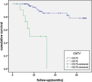
The overall survival (OS) was 88.8% for patients with low CMTV (≤53.75 mL) and 45.5% for those with high CMTV (>53.75 mL), respectively ( P < 0.01, 95% confidence interval). Univariate analysis showed that CMTV and CTLG were significant prognostic factors for OS, in addition to International Federation of Gynecology and Obstetrics (FIGO) stage, age, lymphadenopathy, and maximum standardized uptake value (SUVmax) ( P < 0.05 for all). On multivariate analysis, CMTV remained significant for OS, in addition to FIGO stage ( P < 0.05 for all). CMTV remains as prognostic factor for OS regardless of patients’ FIGO stages ( P < 0.05). In patients in the metastatic diseases group, univariate and multivariate analyses demonstrated that CMTV, WB-MTV, and WB-TLG were independent prognostic factors for OS ( P < 0.05 for all).
Conclusion
CMTV, WB-MTV, and WB-TLG are reliable prognostic factors for patients with cervical carcinoma and should be included in FDG-PET/CT reports to guide referral clinicians for risk-adapted therapies.
Introduction
Cervical carcinoma is the third most common cancer in women worldwide , with 85% of cases occurring in developing countries, where it is the second most frequent cause of cancer death in the female population . 18-Fluoro-2-deoxyglucose positron emission tomography/computed tomography ( 18 F-FDG PET/CT) has been shown to be useful in staging, monitoring therapy response , and predicting prognostication of primary cervical cancer. Recent studies have shown that the volume-based parameter of 18 F-FDG PET, three-dimensional measurements of total tumor metabolic activity, is a more useful measure than the concentration-based standardized uptake value (SUV) in many cancers . Various quantitative and qualitative 18 F-FDG PET/CT parameters have been found to be useful predictors of survival in primary cervical cancer . Among those, maximum standardized uptake value (SUVmax) and metabolic tumor volume (MTV) have been reported to be independent prognostic factors in patients with primary cervical cancer . In this retrospective study, we intended to identify the prognostic value of volume-based PET/CT metabolic parameters (whole-body metabolic tumor volume [WB-MTV], whole-body total lesions glycolysis [WB-TLG], cervical metabolic tumor volume [CMTV], and cervical total lesion glycolysis [CTLG]) in patients with cervical carcinoma.
Materials and Methods
Patients
The institutional review board approved this retrospective study, and the requirement to obtain informed consent was waived. We retrospectively reviewed consecutive patients (from April 2007 and September 2013) with International Federation of Gynecology and Obstetrics (FIGO) stage I B1 through FIGO stage IV B cervical cancer who underwent 18 F-FDG PET/CT. A total of 91 eligible patients were identified for this analysis. The characteristics of the 91 eligible patients with cervical cancer are listed in Table 1 . The median of OS duration was 25 months with a range of 11–69 months. The 1- and 5-year OS were 85% and 71%, respectively.
TABLE 1
Patient Characteristics
Characteristic N(%) Age (months) Mean ± SD 52.02 ± 11.21 Range 21–83 FIGO stage I 24 (26%) II 44 (48%) III 16 (18%) IV 7 (8%) Pathologic subtype Adenocarcinoma 11 (12%) Squamous 80 (88%) Parametrial involvement Yes 23 (25%) No 68 (75%) Lymph node metastasis Yes 26 (29%) No 65 (71%) Treatment method Sur 14 (16%) Sur + CR 21 (23%) Sur + ChT 13 (14%) CR 43 (47%)
ChT, chemothrapy; CR, concurrent chemoradiation therapy; SD, standard deviation; Sur, surgery.
Get Radiology Tree app to read full this article<
PET/CT Acquisition
Get Radiology Tree app to read full this article<
Visual Image Inspection
Get Radiology Tree app to read full this article<
Measurement of PET Quantitative Parameters
Get Radiology Tree app to read full this article<
Get Radiology Tree app to read full this article<
Statistical Analysis
Get Radiology Tree app to read full this article<
Get Radiology Tree app to read full this article<
Results
Image Quantification
Get Radiology Tree app to read full this article<
Kaplan-Meier Survival Analysis
Get Radiology Tree app to read full this article<
TABLE 2
Cox Proportional Hazard Model Analysis of Potential Prognostic Factors Influencing OS
Variable Univariate Analysis Multivariate Analysis Hazard Ratio (95% CI)P Value Hazard Ratio (95% CI)P Value Age 1.044 (1.001, 1.089) 0.045 — 0.840 FIGO stage 7.835 (2.137, 28.716) 0.002 3.040 (1.401, 6.593) 0.005 Pathologic subtype — 0.162 — — Parametrial involvement — 0.128 — — Treatment — <0.01 — 0.670 Lymphadenopathy 4.161 (1.426, 12.142) 0.009 — 0.576 CMTV 1.027 (1.014, 1.041) <0.01 1.019 (1.006, 1.033) 0.005 CTLG 1.002 (1.001, 1.003) <0.01 — 0.091 SUVmax 1.035 (1.009, 1.063) 0.009 — 0.316
CI, confidence interval; CMTV, cervical metabolic tumor volume; CTLG, cervical tumor lesions glycolysis; FIGO, International Federation of Gynecology and Obstetrics; OS, overall survival; SUVmax, maximum standardized uptake values.
TABLE 3
Cox Proportional Hazard Model Analysis of Potential Prognostic Factors Influencing OS of Patients with Metastatic Diseases Group
Variable Univariate Analysis Multivariate Analysis Hazard Ratio (95% CI)P Value Hazard Ratio (95% CI)P Value Age — 0.119 — — FIGO stage 3.665 (1.176, 11.421) 0.025 — 0.057 Pathologic subtype — 0.660 — — Parametrial involvement — 0.815 — — Treatment — 0.238 — — Lymphadenopathy — 0.268 — — CMTV 1.022 (1.006, 1.039) <0.01 1.022 (1.002, 1.042) 0.028 CTLG 1.002 (1.000, 1.003) <0.01 — 0.747 WB-MTV 1.008 (1.002, 1.014) <0.01 1.008 (1.002, 1.014) 0.007 WB-TLG 1.002 (1.001, 1.003) <0.01 1.031 (1.001, 1.072) 0.016 SUVmax 1.065 (1.017, 1.116) <0.01 — 0.590
CI, confidence interval; CMTV, cervical metabolic tumor volume; CTLG, cervical tumor lesions glycolysis; FIGO, International Federation of Gynecology and Obstetrics; OS, overall survival; SUVmax, maximum standardized uptake values; WB-MTV, whole-body metabolic tumor volume; WB-TLG, whole-body tumor lesions glycolysis.
Get Radiology Tree app to read full this article<
Get Radiology Tree app to read full this article<
Get Radiology Tree app to read full this article<
Discussion
Get Radiology Tree app to read full this article<
Get Radiology Tree app to read full this article<
Get Radiology Tree app to read full this article<
Conclusions
Get Radiology Tree app to read full this article<
References
1. Parkin D.M., Bray F., Ferlay J., et. al.: Global cancer statistics, 2002. CA Cancer J Clin 2005; 55: pp. 74-108.
2. Kamangar F., Dores G.M., Anderson W.F.: Patterns of cancer incidence, mortality, and prevalence across five continents: defining priorities to reduce cancer disparities in different geographic regions of the world. J Clin Oncol 2006; 24: pp. 2137-2150.
3. Jemal A., Bray F., Center M.M., et. al.: Global cancer statistics. CA Cancer J Clin 2011; 61: pp. 69-90.
4. Barwick T.D., Taylor A., Rockall A.: Functional imaging to predict tumor response in locally advanced cervical cancer. Curr Oncol Rep 2013; 15: pp. 549-558.
5. Zhang H., Wroblewski K., Liao S., et. al.: Prognostic value of metabolic tumor burden from (18)F-FDG PET in surgical patients with non-small-cell lung cancer. Acad Radiol 2013; 20: pp. 32-40.
6. Chung H.H., Cheon G.J., Kang K.W., et. al.: Preoperative PET/CT FDG standardized uptake value of pelvic lymph nodes as a significant prognostic factor in patients with uterine cervical cancer. Eur J Nucl Med Mol Imaging 2014; 41: pp. 674-681.
7. Abgral R., Keromnes N., Robin P., et. al.: Prognostic value of volumetric parameters measured by (18)F-FDG PET/CT in patients with head and neck squamous cell carcinoma. Eur J Nucl Med Mol Imaging 2014; 41: pp. 659-667.
8. Hyun S.H., Choi J.Y., Shim Y.M., et. al.: Prognostic value of metabolic tumor volume measured by 18F-fluorodeoxyglucose positron emission tomography in patients with esophageal carcinoma. Ann Surg Oncol 2010; 17: pp. 115-122.
9. Lee H.Y., Hyun S.H., Lee K.S., et. al.: Volume-based parameter of (18) F-FDG PET/CT in malignant pleural mesothelioma: prediction of therapeutic response and prognostic implications. Ann Surg Oncol 2010; 17: pp. 2787-2794.
10. Chung M.K., Jeong H.S., Park S.G., et. al.: Metabolic tumor volume of [18F]-fluorodeoxyglucose positron emission tomography/computed tomography predicts short-term outcome to radiotherapy with or without chemotherapy in pharyngeal cancer. Clin Cancer Res 2009; 15: pp. 5861-5868.
11. Chen S.W., Hsieh T.C., Yen K.Y., et. al.: Pretreatment (18)F-FDG PET/CT in whole-body total lesion glycolysis to predict survival in patients with pharyngeal cancer treated with definitive radiotherapy. Clin Nucl Med 2014; 39: pp. e296-e300.
12. Pan L., Cheng J., Zhou M., et. al.: The SUVmax (maximum standardized uptake value for F-18 fluorodeoxyglucose) and serum squamous cell carcinoma antigen (SCC-ag) function as prognostic biomarkers in patients with primary cervical cancer. J Cancer Res Clin Oncol 2012; 138: pp. 239-246.
13. Chung H.H., Kim J.W., Han K.H., et. al.: Prognostic value of metabolic tumor volume measured by FDG-PET/CT in patients with cervical cancer. Gynecol Oncol 2011; 120: pp. 270-274.
14. Miller T.R., Grigsby P.W.: Measurement of tumor volume by PET to evaluate prognosis in patients with advanced cervical cancer treated by radiation therapy. Int J Radiat Oncol Biol Phys 2002; 53: pp. 353-359.
15. Kidd E.A., Siegel B.A., Dehdashti F., et. al.: Pelvic lymph node F-18 fluorodeoxyglucose uptake as a prognostic biomarker in newly diagnosed patients with locally advanced cervical cancer. Cancer 2010; 116: pp. 1469-1475.
16. Fonti R., Larobina M., Del Vecchio S., et. al.: Metabolic tumor volume assessed by 18F-FDG PET/CT for the prediction of outcome in patients with multiple myeloma. J Nucl Med 2012; 53: pp. 1829-1835.
17. Ryu I.S., Kim J.S., Roh J.L., et. al.: Prognostic value of preoperative metabolic tumor volume and total lesion glycolysis measured by 18F-FDG PET/CT in salivary gland carcinomas. J Nucl Med 2013; 54: pp. 1032-1038.
18. Chung H.W., Lee K.Y., Kim H.J., et. al.: FDG PET/CT metabolic tumor volume and total lesion glycolysis predict prognosis in patients with advanced lung adenocarcinoma. J Cancer Res Clin Oncol 2014; 140: pp. 89-98.
19. Kidd E.A., Thomas M., Siegel B.A., et. al.: Changes in cervical cancer FDG uptake during chemoradiation and association with response. Int J Radiat Oncol Biol Phys 2013; 85: pp. 116-122.
20. Showalter T.N., Miller T.R., Huettner P., et. al.: 18 F-fluorodeoxyglucose-positron emission tomography and pathologic tumor size in early-stage invasive cervical cancer. Int J Gynecol Cancer 2009; 19: pp. 1412-1414.
21. Chung H.H., Nam B.H., Kim J.W., et. al.: Preoperative [18F]FDG PET/CT maximum standardized uptake value predicts recurrence of uterine cervical cancer. Eur J Nucl Med Mol Imaging 2010; 37: pp. 1467-1473.
22. Larson S.M., Erdi Y., Akhurst T., et. al.: Tumor treatment response based on visual and quantitative changes in global tumor glycolysis using PET-FDG imaging. The visual response score and the change in total lesion glycolysis. Clin Positron Imaging 1999; 2: pp. 159-171.
23. Chen H.H., Chiu N.T., Su W.C., et. al.: Prognostic value of whole-body total lesion glycolysis at pretreatment FDG PET/CT in non-small cell lung cancer. Radiology 2012; 264: pp. 559-566.
24. Liu F.Y., Chao A., Lai C.H., et. al.: Metabolic tumor volume by (18)F-FDG PET/CT is prognostic for stage IVB endometrial carcinoma. Gynecol Oncol 2012; 125: pp. 566-571.
25. Kim K., Kim S.J., Kim I.J., et. al.: Prognostic value of volumetric parameters measured by F-18 FDG PET/CT in surgically resected non-small-cell lung cancer. Nucl Med Commun 2012; 33: pp. 613-620.
26. Chung H.H., Kwon H.W., Kang K.W., et. al.: Prognostic value of preoperative metabolic tumor volume and total lesion glycolysis in patients with epithelial ovarian cancer. Ann Surg Oncol 2012; 19: pp. 1966-1972.
27. Hatt M., Visvikis D., Albarghach N.M., et. al.: Prognostic value of 18F-FDG PET image-based parameters in oesophageal cancer and impact of tumour delineation methodology. Eur J Nucl Med Mol Imaging 2011; 38: pp. 1191-1202.