
Rationale and Objectives
The purpose of this study was to compare the efficacy of the color scale with regard to focal detection with computed tomography in acute ischemic stroke.
Materials and Methods
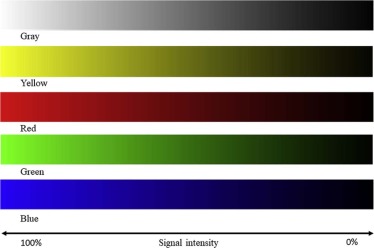
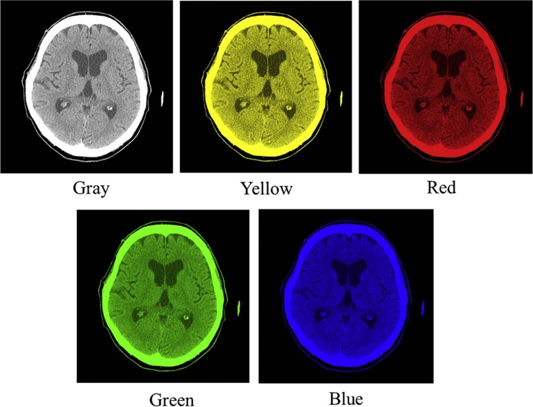
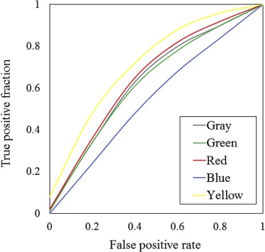
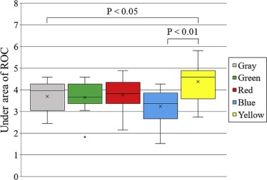
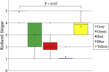
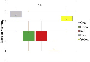
Computed tomography images of the brain of 19 patients diagnosed with acute stroke, based on magnetic resonance diffusion-weighted images obtained within an onset of 24 hours, and the images of five normal patients were displayed in each color look-up table on a monitor. The detection of acute stroke was compared among 15 radiologists. The images were compared in the gray, green, yellow, red, and blue scales of the look-up tables. The observers recorded acute ischemic stroke as “present” or “absent.” They also located the position of the stroke lesion and described the degree of their conviction as to whether a lesion existed. Detection was evaluated by receiver operating characteristic analysis. The area under the receiver operating characteristic curves was compared. In addition, reduced fatigue and the ease in image observation were compared.
Results
Compared to the other scales, the yellow scale had a significantly higher area under the receiver operating characteristic curve, which indicated that this scale allowed better detection of acute ischemic stroke. The gray scale produced the least fatigue in image observation.
Conclusions
The detection of acute ischemic stroke is improved by changing the display monitor from the gray scale to the yellow scale. From the perspective of color psychology, yellow is associated with higher arousal, cheerfulness, confidence, creativity, and excitement. Therefore, the yellow scale may be suitable for a medical imaging display.
Introduction
Stroke is the second leading cause of death worldwide, and many researchers have analyzed the developmental mechanism and the defense against stroke . If cerebral infarction is detected early and a patient receives appropriate treatment such as thrombolytic therapy, then the survival rate and convalescence are improved. In particular, adaptation to ischemic cerebrovascular disorder of recombinant tissue-type plasminogen activator is an epoch-making therapy . Patients can expect a good outcome after stroke onset if these treatments are administered early (ie, within 4.5 hours) . Computed tomography (CT) of the brain is generally performed as an emergency study when a stroke is suspected. CT of the brain provides cerebral infarction views such as early CT sign without bleeding, and the aforementioned treatments can be applied. Therefore, early diagnosis is necessary. However, interpreting cerebral infarction with few contrast changes is difficult, even for expert radiology physicians.
In recent years, an image monitor has often been used for the interpretation of a radiograph image. On an image monitor, the gray scale display (which was initially used for backlight and film diagnosis) is commonly used, although a color display can be used to determine a monitor diagnosis. However, in one report , the detectability of a low-contrast image was improved more by a color scale than by the gray scale. Changing the color scale of the monitor may improve the detectability of an acute cerebral infarction. In this study, we investigated whether the detectability of an acute cerebral infarction would be improved by using a color scale on the imaging monitor.
Materials and Methods
Image Evaluation
Get Radiology Tree app to read full this article<
Get Radiology Tree app to read full this article<
Observation Evaluation
Get Radiology Tree app to read full this article<
Analysis of the Evaluation
Get Radiology Tree app to read full this article<
Results
Get Radiology Tree app to read full this article<
Get Radiology Tree app to read full this article<
Get Radiology Tree app to read full this article<
Get Radiology Tree app to read full this article<
Discussion
Get Radiology Tree app to read full this article<
Get Radiology Tree app to read full this article<
Get Radiology Tree app to read full this article<
Conclusions
Get Radiology Tree app to read full this article<
Acknowledgments
Get Radiology Tree app to read full this article<
References
1. Krishnamuthi R.V., Feigin V.L., Forouzanfar M.H., et. al.: Global and regional burden of first-ever ischaemic and haemorrhagic stroke during 1990–2010: findings from the Global Burden of Disease Study 2010. Lancet Glob Health 2013; 1: pp. e259-e281.
2. Feigin V.L., Forouzanfar M.H., Krishnamuthi R.V., et. al.: Global and regional burden of stroke during 1990–2010: findings from the Global Burden of Disease Study 2010. Lancet 2014; 383: pp. 245-255.
3. Adams H.P., Bendixen B.H., Kappelle L.J., et. al.: Classification of subtype of acute ischemic stroke. Definitions for use in a multicenter clinical trial. TOAST. Trial of Org 10172 in Acute Stroke Treatment. Stroke 1993; 24: pp. 35-41.
4. Yew K.S., Cheng E.M.: Diagnosis of acute stroke. Am Fam Physician 2015; 91: pp. 528-536.
5. Nor A.M., Davis J., Sen B., et. al.: The recognition of stroke in the emergency room (ROSIER) scale: development and validation of a stroke recognition instrument. Lancet Neurol 2005; 4: pp. 727-734.
6. Brott T., Adams H.P., Olinger C.P., et. al.: Measurements of acute cerebral infarction: a clinical examination scale. Stroke 1989; 20: pp. 864-870.
7. Ovbiagele B.: The emergency department: first line of defense in preventing secondary stroke. Acad Emerg Med 2006; 13: pp. 215-222.
8. National Institute of Neurological Disorders and Stroke rt-PA Stroke Study Group: Tissue plasminogen activator for acute ischemic stroke. N Engl J Med 1995; 333: pp. 1581-1587.
9. Chiu D., Krieger D., Villar-Cordova C., et. al.: Intravenous tissue plasminogen activator for acute ischemic stroke: feasibility, safety, and efficacy in the first year of clinical practice. Stroke 1998; 29: pp. 18-22.
10. Clark W.M., Wissman S., Albers G.W., et. al.: Recombinant tissue-type plasminogen activator (Alteplase) for ischemic stroke 3 to 5 hours after symptom onset. The ATLANTIS study: a randomized controlled trial. Alteplase Thrombolysis for Acute Noninterventional Therapy in Ischemic Stroke. JAMA 1999; 282: pp. 2019-2026.
11. Graham G.D.: Tissue plasminogen activator for acute ischemic stroke in clinical practice: a meta-analysis of safety data. Stroke 2003; 34: pp. 2847-2850.
12. Wardlaw J.M., Murray V., Berge E., et. al.: Recombinant tissue plasminogen activator for acute ischemic stroke: an updated systematic review and meta-analysis. Lancet 2012; 379: pp. 2364-2372.
13. Hacke W., Donnan G., Fieschi C., et. al.: Association of outcome with early stroke treatment: pooled analysis of ATLANTIS, ECASS, and NINDOS rt-PA stroke trials. Lancet 2004; 363: pp. 768-774.
14. Ogura A., Kamakura A., Kaneko Y., et. al.: Comparison of grayscale and color-scale renderings of digital medical images for diagnostic interpretation. Radiol Phys Technol 2017; 10: pp. 359-363. First online 27 March 2017
15. Rose A.: The sensitivity performance of the human eye on an absolute scale. J Opt Soc Am 1948; 38: pp. 196-208.
16. Meeteren A.V., Vos J.J.: Resolution and contrast sensitivity at low luminances. Vision Res 1972; 12: pp. 825-833.
17. Barten P.G.J.: Contrast sensitivity of the human eye and its effects on image quality.1999.SPIE PressBellingham, WApp. 149-170.
18. Lovegrove W.J., Bowling A., Badclock D., et. al.: Specific reading disability: differences in contrast sensitivity as a function of spatial frequency. Science 1980; 210: pp. 439-440.
19. Mullen K.T.: The contrast sensitivity of human colour vision to red-green and blue-yellow chromatic gratings. J Physiol 1985; 359: pp. 381-400.
20. Green D.G.: The contrast sensitivity of the colour mechanisms of the human eye. J Physiol 1968; 196: pp. 415-429.
21. Kolenda N.: Color psychology: the complete guide for marketers. Available at http://nickkolenda.com/color-psychology/
22. Hill R.A., Barton R.A.: Psychology: red enhances human performance in contests. Nature 2005; 435: pp. 293.
23. Moutoussis K.: The physiology and psychophysics of the color-form relationship: a review. Front Psychol 2015; 6: pp. 1407.
24. Bartels A., Zeki S.: The architecture of the colour centre in the human visual brain: new results and a review. Eur J Neurosci 2000; 12: pp. 172-193.
25. Ikeda M., Ashizawa S.: Equivalent lightness of colored of equal Munsell chroma and equal Munsell value at various illuminances. Color Res Appl 1991; 16: pp. 72-80.