
Rationale and Objectives
The aim of this study was to compare high-resolution magnetic resonance (MR) arthrography and multislice computed tomographic (MSCT) arthrography in the evaluation of the entire knee cartilage obtained from cadavers.
Materials and Methods
MR arthrography and MSCT arthrography were performed on 16 cadaver knees, and their findings were compared to those found during macroscopic assessment. The sensitivity and specificity of MR arthrography and MSCT arthrography for detecting cartilage lesions of grade ≥ 2 and Spearman’s correlation coefficients between arthrographic and macroscopic grades were determined. In addition, cartilage surface conspicuity of the two techniques was measured using a subjective ranking system.
Results
The sensitivity and specificity, respectively, for the detection of cartilage disorder (grade ≥ 2) were 87% and 97% on MR arthrography and 84% and 99% on MSCT arthrography. There was no statistically significant difference between the two techniques in sensitivity ( P = 1.000) or specificity ( P = .625). Spearman’s correlation coefficients between MR arthrography or MSCT arthrography and macroscopic grading were 0.783 and 0.800, respectively, with no statistically difference ( P = .492). Both MR arthrography and MSCT arthrography enabled the accurate depiction of cartilage surface.
Conclusions
High-resolution MR arthrography and MSCT arthrography were comparably accurate for the assessment of cartilage lesions of the entire knee.
Articular cartilage lesions are commonly seen in athletes and the elderly, but imaging the cartilage remains one of the most difficult and controversial areas in musculoskeletal imaging. Although conventional magnetic resonance (MR) has been regarded as the imaging modality of choice for the noninvasive assessment of articular cartilage, its efficiency had a strong dependence on the selected sequences, and limited performance in depicting cartilage lesions caused by relatively low resolution has been reported . Traditional computed tomographic (CT) arthrography and MR arthrography enable the relatively accurate depiction of cartilage lesions , but most of the reported trials studied the patellar cartilage , not the hyaline cartilage of the entire joint.
With the advent of multislice CT (MSCT) imaging, there have been remarkable advances in musculoskeletal imaging . MSCT imaging can be used to acquire isotropic or near isotropic data sets from which high–spatial resolution multiplanar reformatted images can be generated, so it is possible to evaluate the entire cartilage in a single MSCT arthrographic examination with three-dimensional high resolution. MSCT arthrography had been used in the evaluation of the entire hyaline cartilage in cadaver knees as well as the measurement of cartilage thickness in cadaver ankles . The findings of these previous studies showed that MSCT arthrography depicted cartilage more accurately than MR imaging.
Get Radiology Tree app to read full this article<
Materials and methods
Specimen Preparation
Get Radiology Tree app to read full this article<
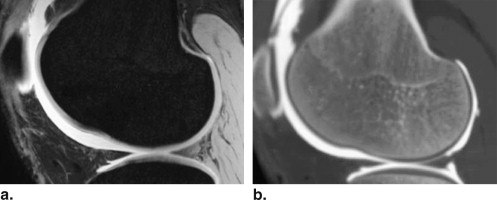
High-Resolution Three-Dimensional MR Arthrography
Get Radiology Tree app to read full this article<
Get Radiology Tree app to read full this article<
MSCT Arthrography
Get Radiology Tree app to read full this article<
Macroscopic Analysis
Get Radiology Tree app to read full this article<
Table 1
International Cartilage Repair Society Classification of Cartilage Injuries
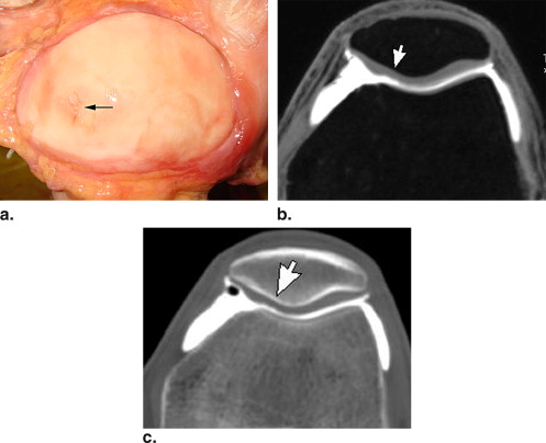
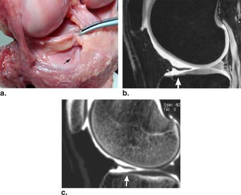
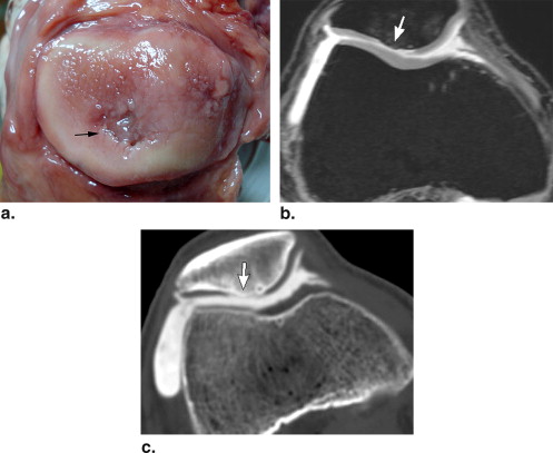
Grade Macroscopic Findings 0 Normal 1 Superficial lesions, fissures, cracks, indentations 2 Cartilage defects extending down to <50% of cartilage depth 3 Cartilage defects extending down to >50% of cartilage depth 4 Complete loss of cartilage thickness, bone only
Get Radiology Tree app to read full this article<
Image Analysis
Get Radiology Tree app to read full this article<
Get Radiology Tree app to read full this article<
Table 2
Grading System for Interpretation of Magnetic Resonance Arthrographic or Multislice Computed Tomographic Arthrographic Images
Grade Findings 0 Smooth articular surface 1 Loss of smooth contour without contrast within cartilage 2 Penetration of contrast in cartilage, <50% of cartilage depth 3 Penetration of contrast material, >50% of cartilage depth 4 Penetration of contrast material, down to subchondral bone
Get Radiology Tree app to read full this article<
Get Radiology Tree app to read full this article<
Statistical Analysis
Get Radiology Tree app to read full this article<
Get Radiology Tree app to read full this article<
Get Radiology Tree app to read full this article<
Get Radiology Tree app to read full this article<
Results
Macroscopic Analysis
Get Radiology Tree app to read full this article<
Get Radiology Tree app to read full this article<
Detection of Cartilage Lesions of Grade ≥ 2
Get Radiology Tree app to read full this article<
Get Radiology Tree app to read full this article<
Grading of Articular Cartilage
Get Radiology Tree app to read full this article<
Get Radiology Tree app to read full this article<
Cartilage Surface Conspicuity
Get Radiology Tree app to read full this article<
Table 3
Rating Scores for Cartilage Surface Conspicuity
MR Arthrography MSCT Arthrography_P_ Femoropatellar joint 5.00 ± 0.00 5.00 ± 0.00 1.000 Femorotibial joint 4.50 ± 0.52 4.44 ± 0.81 .763 Posterior surface of femoral condyles 4.63 ± 0.50 4.94 ± 0.25 .025 ∗
MR, magnetic resonance; MSCT, multislice computed tomographic.
Data are expressed as mean ± standard deviation. P values are based on nonparametric paired-data Wilcoxon’s signed rank test.
Get Radiology Tree app to read full this article<
Get Radiology Tree app to read full this article<
Discussion
Get Radiology Tree app to read full this article<
Get Radiology Tree app to read full this article<
Get Radiology Tree app to read full this article<
Get Radiology Tree app to read full this article<
Get Radiology Tree app to read full this article<
Get Radiology Tree app to read full this article<
Get Radiology Tree app to read full this article<
Get Radiology Tree app to read full this article<
Get Radiology Tree app to read full this article<
References
1. Recht M.P., Goodwin D.W., Winalski C.S., et. al.: MRI of articular cartilage: revisiting current status and future directions. AJR Am J Roentgenol 2005; 185: pp. 899-914.
2. McCauley T.R., Disler D.G.: MR imaging of articular cartilage. Radiology 1998; 209: pp. 629-640.
3. Kneeland J.B.: Magnetic resonance imaging of articular cartilage. Semin Roentgenol 2000; 35: pp. 249-255.
4. Gold G.E., McCauley T.R., Gray M.L., et. al.: What’s new in cartilage?. Radiographics 2003; 23: pp. 1227-1242.
5. Gagliardi J.A., Chung E.M., Chandnani V.P., et. al.: Detection and staging of chondromalacia patellae: relative efficacies of conventional MR imaging, MR arthrography, and CT arthrography. AJR Am J Roentgenol 1994; 163: pp. 629-636.
6. Daenen B.R., Ferrara M.A., Marcelis S., et. al.: Evaluation of patellar cartilage surface lesions: comparison of CT arthrography and fat-suppressed FLASH 3D MR imaging. Eur Radiol 1998; 8: pp. 981-985.
7. Rand T., Brossmann J., Pedowitz R., et. al.: Analysis of patellar cartilage. Comparison of conventional MR imaging and MR and CT arthrography in cadavers. Acta Radiol 2000; 41: pp. 492-497.
8. Buckwalter K.A., Rydberg J., Kopecky K.K., et. al.: Musculoskeletal imaging with multislice CT. AJR Am J Roentgenol 2001; 176: pp. 979-986.
9. Vande Berg B.C., Lecouvet F.E., Poilvache P., et. al.: Assessment of knee cartilage in cadavers with dual-detector spiral CT arthrography and MR imaging. Radiology 2002; 222: pp. 430-436.
10. El-Khoury G.Y., Alliman K.J., Lundberg H.J., et. al.: Cartilage thickness in cadaveric ankles: measurement with double-contrast multi-detector row CT arthrography versus MR imaging. Radiology 2004; 233: pp. 768-773.
11. Masi J.N., Sell C.A., Phan C., et. al.: Cartilage MR imaging at 3.0 versus that at 1.5 T: preliminary results in a porcine model. Radiology 2005; 236: pp. 140-150.
12. Kleemann R.U., Krocker D., Cedraro A., et. al.: Altered cartilage mechanics and histology in knee osteoarthritis: relation to clinical assessment (ICRS grade). Osteoarthritis Cartilage 2005; 13: pp. 958-963.
13. Dwyer A.J.: Matchmaking and McNemar in the comparison of diagnostic modalities. Radiology 1991; 178: pp. 328-330.
14. Hauger O., Dumont E., Chateil J.F., et. al.: Water excitation as an alternative to fat saturation in MR imaging: preliminary results in musculoskeletal imaging. Radiology 2002; 224: pp. 657-663.
15. Waldt S., Bruegel M., Ganter K., et. al.: Comparison of multislice CT arthrography and MR arthrography for the detection of articular cartilage lesions of the elbow. Eur Radiol 2005; 15: pp. 784-791.
16. Schmid M.R., Pfirrmann C.W., Hodler J., et. al.: Cartilage lesions in the ankle joint: comparison of MR arthrography and CT arthrography. Skeletal Radiol 2003; 32: pp. 259-265.
17. Wyler A., Bousson V., Bergot C., et. al.: Comparison of MR-arthrography and CT-arthrography in hyaline cartilage-thickness measurement in radiographically normal cadaver hips with anatomy as gold standard. Osteoarthritis Cartilage 2009; 17: pp. 19-25.
18. Kataoka M., Ueda H., Koyama T., et. al.: Contrast-enhanced volumetric interpolated breath-hold examination compared with spin-echo T1-weighted imaging of head and neck tumors. AJR Am J Roentgenol 2005; 184: pp. 313-319.
19. Wyler A., Bousson V., Bergot C., et. al.: Hyaline cartilage thickness in radiographically normal cadaveric hips: comparison of spiral CT arthrographic and macroscopic measurements. Radiology 2007; 242: pp. 441-449.
20. Nishii T., Tanaka H., Nakanishi K., et. al.: Fat-suppressed 3D spoiled gradient-echo MRI and MDCT arthrography of articular cartilage in patients with hip dysplasia. AJR Am J Roentgenol 2005; 185: pp. 379-385.