
Rationale and Objectives
Ultrasound image quality can be improved by imaging an object (here: the female breast) from different viewing angles in one image plane. With this technique, which is commonly referred to as spatial compounding, a more isotropic resolution is achieved while speckle noise and further artifacts are reduced. We present results obtained from a combination of spatial compounding with contrast-enhanced ultrasound imaging in three dimensions to reduce contrast specific artifacts (depth dependency, shadowing, speckle) and reconstruct vascular structures.
Materials and Methods
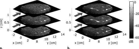
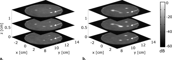
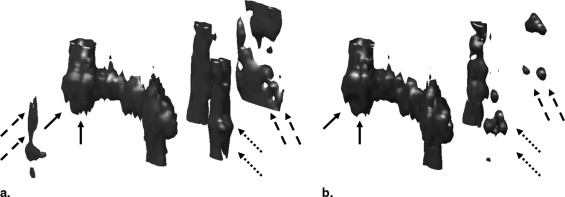
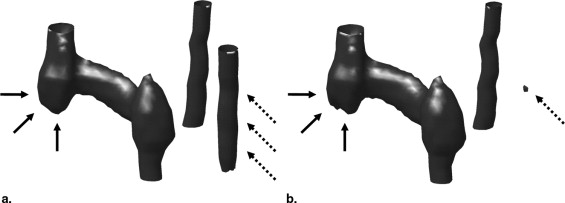
We used a conventional ultrasound scanner and a custom made mechanical system to rotate an ultrasound curved array probe around an object (360°, 36 transducer positions). For 10 parallel image planes, ultrasound compound images were generated of a flow-mimicking phantom consecutively supplied with water and contrast agent. These compound images were combined to form a volume dataset and postprocessed to obtain a sonographic subtraction angiography.
Results
Image quality was significantly improved by spatial compounding for the native (ie, without contrast agent), and, in particular, for the contrast-enhanced case. After subtracting the native images from the contrast-enhanced ones, only structures supplied with contrast agent remain. This technique yields much better results for compound images than for conventional ultrasound images because speckle noise and an anisotropic resolution affect the latter.
Conclusions
With the presented approach contrast specific artifacts can be eliminated efficiently, and a subtraction angiography can be computed. A speckle reduced three-dimensional reconstruction of submillimeter vessel structures was achieved for the first time. In the future, this technique can be applied in vivo to image the vascularity of cancer in the female breast.
The early detection and staging of breast masses is one of the most challenging diagnostic problems in medical imaging. Because magnetic resonance imaging is too expensive to be applied routinely for breast screening, mammography is considered the gold standard. However, because of radiation exposure, this technique is generally not unobjectionable and limited (eg, in pregnant patients). Furthermore, the specificity in mammography screening is rather low, featuring high false-positive rates ( ). In particular, for dense breast tissue or diffuse lesions, mammography is inconclusive. As a second modality before biopsies or surgical inventions, sonographic breast imaging is routinely used for differential diagnosis. Primarily, high-frequency ultrasound of center frequencies above 10 MHz is capable to assess the size, texture, and morphology of the tumor with high spatial resolution. In the case that the tumor is sonographically visible, these features can be helpful to differentiate between benign and malignant lesions ( ). Moreover, many different kinds of tumors induce intense angiogenesis in the affected tissue. Because of a nonphysiologic and rapid genesis of tumor vascularity, structural abnormalities are typical, such as irregular and variable vessel diameters, elongated and coiled vessels, anarchic vascular networks with tortuous vessel courses, rings, sinusoids, arteriovenous shunts, vessel loops, and incomplete vascular walls ( ). Thus, vascularity and tissue perfusion are highly significant parameters for the assessment of breast cancer. Ultrasound Doppler techniques (eg, color Doppler, power Doppler, spectral Doppler) are capable of imaging blood flow in vessels and provide direct, absolute measurements of flow velocity. Much work has been published on Doppler two-dimensional (2D) and three-dimensional (3D) imaging of breast masses ( ). However, these techniques fail for low flow rates in small vessels, because tissue motion creates higher velocities than the flow, and the two phenomena cannot be differentiated ( ). Contrast-enhanced ultrasound imaging assesses both the macrovascular blood supply and the microvascular perfusion of tissue ( ). Ultrasound contrast agents (CA) consist of gas-filled microbubbles stabilized by a shell. By enhancing the weak echo signals from blood by strong linear and nonlinear scattering, they can not only be used to enhance Doppler signals but also directly for a contrast specific imaging. For this, modalities have been developed to detect CA using their nonlinear response to ultrasound ( ). In particular, different implementations of harmonic imaging, such as phase inversion technique ( ) or contrast pulse sequences ( ), are widely used. Unlike methods known from computed tomography or magnetic resonance imaging, the imaging process itself (ie, ultrasound insonification) can cause changes in the microbubble concentration (eg, ultrasound induced destruction of CA). Thus, ultrasound transmit power is usually kept low to achieve a nonlinear response of the CA and, at the same time, avoid bubble rupture. Having a diameter of less then 10 μm, the geometric dimension of the microbubbles is too small to be resolved by ultrasound with clinically used frequencies. Considering typical CA concentrations in the blood pool, multiple microbubbles contribute to the intensity of a pixel, and image intensity is a measure of CA concentration in a resolution cell. Furthermore, it depends on the type of vascularity whether vessel structures can be visualized: if a vessel is big enough to be spatially resolved with ultrasound or if it is too small but spatially separated from others, its structure and course can be seen from contrast enhancement. Here, a quasi-angiographic view of the macrovascular network can be realized ( ). If the vessel, however, is too small to be resolved (<1–3 mm), flow velocities are slow (<1–5 cm/s) and other adjacent vessels are too close, the vascular network cannot be identified. In such cases, the intensity of the imaged tissue increases with CA concentration and tissue types can be differentiated by their microvascular perfusion. Here, the perfusion can either be analyzed qualitatively from the image intensity of a single image or semiquantitatively by evaluating perfusion kinetics in an image series using, for example, the bolus, depletion, or replenishment method ( ). Contrast-enhanced ultrasound imaging has already been reported to provide potential for the detection of breast cancer ( ).
Although the application of ultrasound is generally promising in the context of breast imaging, there are restrictions due to the underlying physical principles of the imaging modality.
Get Radiology Tree app to read full this article<
Get Radiology Tree app to read full this article<
Get Radiology Tree app to read full this article<
Get Radiology Tree app to read full this article<
Get Radiology Tree app to read full this article<
Get Radiology Tree app to read full this article<
Get Radiology Tree app to read full this article<
 Open full size image
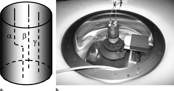
Figure 1
Experimental setup: (a) principle of spatial compounding. (b) Mechanical applicator installed in a bed, ultrasound scanner, and PC.
Get Radiology Tree app to read full this article<
Materials and methods
Experimental Setup
Get Radiology Tree app to read full this article<
Get Radiology Tree app to read full this article<
Get Radiology Tree app to read full this article<
Get Radiology Tree app to read full this article<
Flow Mimicking Phantom
Get Radiology Tree app to read full this article<
Get Radiology Tree app to read full this article<
Get Radiology Tree app to read full this article<
Data Processing
Get Radiology Tree app to read full this article<
Get Radiology Tree app to read full this article<
Get Radiology Tree app to read full this article<
Results
Get Radiology Tree app to read full this article<
Get Radiology Tree app to read full this article<
Discussion
Get Radiology Tree app to read full this article<
Get Radiology Tree app to read full this article<
Get Radiology Tree app to read full this article<
Get Radiology Tree app to read full this article<
Get Radiology Tree app to read full this article<
Get Radiology Tree app to read full this article<
Acknowledgment
Get Radiology Tree app to read full this article<
Get Radiology Tree app to read full this article<
Get Radiology Tree app to read full this article<
Get Radiology Tree app to read full this article<
References
1. Madjar H.: Contrast ultrasound in breast tumor characterization: present situation and future tracks. Eur Radiol 2001; 11: pp. E41-E46.
2. Leucht D., Madjar H.: Teaching atlas of breast ultrasound.2nd ed.1995.ThiemeStuttgart
3. Kolb T.M., Lichy J., Newhouse J.H.: Occult cancer in women with dense breasts: detection with screening US—diagnostic yield and tumor characteristics. Radiology 1998; 207: pp. 191-199.
4. Schroeder R.J., Bostanjoglo M., Rademaker J., et. al.: Role of power Doppler techniques and ultrasound contrast enhancement in the diagnosis of focal breast lesions. Eur Radiol 2003; 13: pp. 68-79.
5. Cosgrove D.O., Kedar R.P., Bamber J.C., et. al.: Breast disease: color Doppler US in differential diagnosis. Radiology 1993; 189: pp. 99-104.
6. Callida F., Raieli G., Sala G., et. al.: Doppler color-echo in the echographic evaluation of solid neoplasms of the breast: 5 years of experience. Radiol Med 1994; 87: pp. 28-35.
7. Carson P.L., Fowlkes J.B., Roubidoux M.A., et. al.: 3-D color Doppler image quantification of breast masses. Ultrasound Med Biol 1997; 23: pp. 837-849.
8. Jensen J.: Estimation of blood velocities using ultrasound: a signal processing approach.1996.University PressCambridge
9. Goldberg B.B., Raichlen J.S., Forsberg F.: Ultrasound contrast agents.2nd ed.2001.Martin Dunitz LtdLondon
10. Lindner J.R.: Microbubbles in medical imaging: current applications and future directions. Nat Rev Drug Discovery 2004; 3: pp. 527-532.
11. Postema M., Schmitz G.: Bubble dynamics involved in ultrasonic imaging. Expert Rev Mol Diagn 2006; 6: pp. 493-502.
12. Chapman C., Lazenby J.: Ultrasound imaging system employing phase inversion subtraction to enhance the image. US Patent No. 5, 632,27771997.
13. Hope Simpson D., Chin C., Burns P.: Pulse inversion Doppler: a new method for detecting nonlinear echoes from microbubble contrast agents. IEEE Trans Ultrason Ferroelect Freq Contr 1999; 46: pp. 372-382.
14. Phillips P.: Contrast pulse sequences (CPS): imaging non-linear microbubbles. Proc IEEE Intern Ultrason Symp 2001; 2: pp. 1739-1745.
15. Holscher T., Wilkening W., Lyden P.D., et. al.: Transcranial ultrasound angiography (T USA): a new approach for contrast specific imaging of intracranial arteries. Ultrasound Med Biol 2005; 31: pp. 1001-1006.
16. Eyding J., Krogias C., Wilkening W., et. al.: Parameters of cerebral perfusion in phase-inversion harmonic imaging (PIHI) ultrasound examinations. Ultrasound Med Biol 2003; 29: pp. 1379-1385.
17. Engelhardt M., Hansen C., Eyding J., et. al.: Feasibility of contrast-enhanced sonography during resection of cerebral tumours—initial results of a prospective study. Ultrasound Med Biol 2007; 33: pp. 571-575.
18. Wei K., Jayaweera A., Firoozan S., et. al.: Quantification of myocardial blood flow with ultrasound-induced destruction of microbubbles administered as a constant venous infusion. Circulation 1998; 97: pp. 473-483.
19. Arditi M., Frinking P.J.A., Zhou X., Rognin N.G.: A new formalism for the quantification of tissue perfusion by the destruction-replenishment method in contrast ultrasound imaging. IEEE Trans Ultrason Ferroelect Freq Contr 2006; 53: pp. 1118-1129.
20. Hansen C., Hüttebräuker N., Wilkening W., et. al.: A fast method for data acquisition in contrast replenishment analyses. Proc IEEE Intern Ultrason Symp 2007; pp. 2187-2190.
21. Eyding J., Wilkening W., Krogias , et. al.: Validation of the depletion kinetic in semiquantitative ultrasonographic cerebral perfusion imaging using 2 different techniques of data acquisition. Ch J Ultrasound Med 2004; 23: pp. 1035-1040.
22. Rizzato G., Martegani A., Chersevani R., et. al.: Importance of staging of breast cancer and role of contrast ultrasound. Eur Radiol 2001; 11: pp. E47-E52.
23. Jung E.M., Jungius K.P., Rupp N., et. al.: Contrast enhanced harmonic ultrasound for differentiating breast tumors—first results. Clin Hemorheol Microcirc 2005; 33: pp. 109-120.
24. Riccci P., Cantisani V., Ballesio L., et. al.: Benign and malignant breast lesions: efficacy of real time contrast-enhanced ultrasound vs. magnetic resonance imaging. Ultraschall Med 2007; 28: pp. 57-62.
25. Burkhardt C.: Speckle in ultrasound B-mode scans. IEEE Trans Sonics Ultrason 1978; 25: pp. 1-6.
26. Robinson D., Knight P.: Computer reconstruction techniques in compound scan pulse-echo imaging. Ultrasonic Imag 1981; 3: pp. 217-234.
27. Hiller D., Ermert H.: System analysis of ultrasound reflection mode computerized tomography. IEEE Trans Sonics Ultrason 1984; 31: pp. 240-250.
28. Röhrlein G., Ermert H.: Limited angle reflection mode computerized tomography.Berkhout A.J.Ridder J.van der Wal L.F.1985.Plenum PressNew York:pp. 413-424.
29. Trahey G., Smith S., von Ramm O.: Speckle pattern correlation with lateral aperture translation: experimental results and implications for spatial compounding. IEEE Trans Ultrason Ferroelect Freq Contr 1986; 33: pp. 257-264.
30. Pesavento A., Ermert H., Broll-Zeitvogel E., et. al.: Ultrasonic reflection tomography of post-disomic scarring. Acoust Imag 1997; 23: pp. 635-640.
31. Huber S., Wagner M., Medl M., et. al.: Real-time spatial compound imaging in breast ultrasound. Ultras Med Biol 2002; 28: pp. 155-163.
32. Hansen C., Hüttebräuker N., Wilkening W., et. al.: Full angle spatial compounding for improved replenishment analyses in contrast perfusion imaging: in vitro studies. IEEE Trans Ultrason Ferroelect Freq Contr 2008; 55: pp. 819-831.
33. Hansen C., Hüttebräuker N., Ashfaq M., et. al.: Contrast enhanced perfusion imaging by means of spatial compounding. Proc IEEE Intern Ultrason Symp 2006; pp. 424-427.
34. Eilebrecht B., Hansen C., Hüttebräuker N., et. al.: Ultrasonographic spatial compounding and contrast perfusion imaging using depletion method. Proc Biomed Tech (Berlin) 2007; 51: pp. v78.
35. Hansen C., Hüttebräuker N., Wilkening W., et. al.: Ultrasonographic contrast agent imaging of sub-millimeter vessel structures with spatial compounding: in vitro analyses. Biomed Tech (Berlin) 2007; 33: pp. 274-283.
36. Hansen Ch, Hüttebräuker N., Wilkening W., et. al.: 3D reconstruction of sub-millimeter vessels by means of ultrasound contrast enhanced spatial compounding: in vitro analyses. Int J Comput Assist Radiol Surg 2007; 2: pp. S35-S37.
37. Brunke S., Insana M., Dahl J., et. al.: An ultrasound research interface for a clinical system. IEEE Trans Ultrason Ferroelect Freq Contr 2007; 54: pp. 198-210.
38. van der Meer S., Verslius M., Lohse D.: The resonance frequency of SonoVue as observed by high-speed optical imaging. Proc IEEE Intern Ultrason Symp 2004; pp. 343-345.
39. Jago J.: An automated method for determining the centre of rotation of a mechanically scanned reflection UCT System. Phys Med Biol 1994; 39: pp. 2367-2371.
40. Chen C.-T., Chen L.-S., Millero F.: Speed of sound in NaCl, MgCl 2 , Na 2 SO 4 and MgSO 4 aqueous solutions as functions of concentration, temperature and pressure. J Acoust Soc Am 1978; 63: pp. 1795-1800.
41. Chen C.-T., Millero F.: Reevaluation of Wilson’s sound-speed measurements for pure water. J Acoust Soc Am 1976; 60: pp. 1270-1273.
42. Schreiman J.S., Gisvold J.J., Greenleaf J.F., et. al.: Ultrasound transmission computed tomography of the breast. Radiology 1984; 150: pp. 523-530.
43. Robinson D.E., Ophir J.O., Wilson L.S., et. al.: Pulse-echo ultrasound speed measurements: progress and prospects. Ultrasound Med Biol 1991; 17: pp. 633-646.
44. Moskalik A., Carson P.L., Meyer C.R., et. al.: Registration of three-dimensional compound ultrasound scans of the breast for refraction and motion correction. Ultrasound Med Biol 1995; 21: pp. 769-778.
45. Krücker J.F., Fowlkes J.B., Carson P.L.: Sound speed estimation using automatic ultrasound image registration. IEEE Trans Ultrason Ferroelect Freq Contr 2004; 51: pp. 1095-1106.
46. Ashfaq M., Hansen C., Hüttebräuker N., et. al.: A quantitative study of the time delay correction for full angle spatial compounding. Proc IEEE Intern Ultrason Symp 2006; pp. 2156-2159.
47. Hansen C., Schasse A., Hüttebräuker N., et. al.: Reconstruction of speed of sound for a correction of full angle spatial compounding. Proc IEEE Intern Ultrason Symp 2007; pp. 785-788.