
Rationale and Objectives
Correlation imaging (CI) is a form of multiprojection imaging in which multiple images of a patient are acquired from slightly different angles. Information from these images is combined to make the final diagnosis. A critical factor affecting the performance of CI is its data acquisition scheme, because nonoptimized acquisition may distort pathologic indicators. The authors describe a computer-aided detection (CADe) methodology to optimize the acquisition scheme of CI for superior diagnostic accuracy.
Materials and Methods
Images from 106 subjects were used. For each subject, 25 angular projections of a single breast were acquired. Projection images were supplemented with a simulated 3-mm three-dimensional lesion. Each projection was then processed using a traditional CADe algorithm at high sensitivity, followed by the reduction of false-positives by combining the geometric correlation information available from the multiple images. The performance of the CI system was determined in terms of free-response receiver-operating characteristic curves and the areas under receiver-operating characteristic curves. For optimization, the components of acquisition, such as the number of projections and their angular span, were systematically changed to investigate which of the many possible combinations maximized the obtainable CADe sensitivity and specificity.
Results
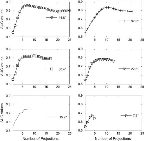
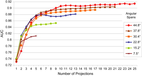
The performance of the CI system was improved by increasing the angular span. Increasing the number of angular projections beyond a certain number did not improve performance. Maximum performance was obtained between 7 and 10 projections spanning a maximum angular arc of 45°.
Conclusion
The findings suggest the existence of an optimum acquisition scheme for CI of the breast. CADe results confirmed earlier predictions on the basis of observer models. An optimized CI system may be an important diagnostic tool for improved breast cancer detection.
The field of diagnostic imaging is fast adopting the use of multiple images of the same patient for clinical workup. These images may be acquired by one or a combination of different imaging modalities. The information from these images is combined by either a clinician or a computer algorithm to extract knowledge about the presence as well as the morphology of a potential pathology within the patient.
In the context of digital radiographic imaging, the multi-image scheme takes the form of multiprojection imaging in which different projection images of a patient may be obtained by a single modality from different positions around the patient along a limited angular arc. This imaging scheme can take the form of tomosynthesis , stereoscopic imaging , or correlation imaging (CI) . In CI, projection images are directly analyzed, thereby avoiding reconstruction artifacts inherent to tomosynthesis. CI thus builds on the advantages of standard projection techniques and combines them with the proven benefits of fusing information from multiple images to potentially improve the accuracy of cancer detection . In practice, CI can take different forms, including scrolling images manually or in cine mode, the stereoscopic display of projections images, and computer-aided analysis of multiple images, and it may also be used as an adjunct application to tomosynthesis.
Get Radiology Tree app to read full this article<
Get Radiology Tree app to read full this article<
Materials and methods
Image Database
Get Radiology Tree app to read full this article<
Get Radiology Tree app to read full this article<
Get Radiology Tree app to read full this article<
CADe Processor
Get Radiology Tree app to read full this article<
Get Radiology Tree app to read full this article<
Get Radiology Tree app to read full this article<
Optimization of Data Acquisition
Get Radiology Tree app to read full this article<
Get Radiology Tree app to read full this article<
Get Radiology Tree app to read full this article<
Results
Get Radiology Tree app to read full this article<
Get Radiology Tree app to read full this article<
Get Radiology Tree app to read full this article<
![Figure 3, Average positive predictive index (true-positives/[true-positives + false-positives]) as a function of the number of projections spanning different angular ranges in a multiprojection correlation imaging setup.](https://storage.googleapis.com/dl.dentistrykey.com/clinical/TowardsOptimizedAcquisitionSchemeforMultiprojectionCorrelationImagingofBreastCancer/2_1s20S1076633208005710.jpg)
Get Radiology Tree app to read full this article<
Get Radiology Tree app to read full this article<
Get Radiology Tree app to read full this article<
Get Radiology Tree app to read full this article<
Get Radiology Tree app to read full this article<
Discussion
Get Radiology Tree app to read full this article<
Get Radiology Tree app to read full this article<
Get Radiology Tree app to read full this article<
Get Radiology Tree app to read full this article<
Get Radiology Tree app to read full this article<
Conclusions
Get Radiology Tree app to read full this article<
Acknowledgment
Get Radiology Tree app to read full this article<
Get Radiology Tree app to read full this article<
References
1. Dobbins J.T., Godfrey D.J.: Digital x-ray tomosynthesis: current state of the art and clinical potential. Phys Med Biol 2003; 48: pp. R65-R106.
2. Maidment A.D.A., Bakic P.R., Albert M.: Effects of quantum noise and binocular summation on dose requirements in stereoradiography. Med Phys 2003; 30: pp. 3061-3071.
3. Samei E., Stebbins S.A., Dobbins J.T., Lo J.Y.: Multiprojection correlation imaging for improved detection of pulmonary nodules. AJR Am J Roentgenol 2007; 188: pp. 1239-1245.
4. Chawla A., Samei E., Abbey C.: A mathematical model approach toward combining information from multiple image projections of the same patient. Proc SPIE 2007; 6510: pp. 1-11.
5. Chawla A., Samei E., Saunders R., Lo J., Baker J.: A mathematical model platform for optimizing a multi-projection breast imaging system. Med Phys 2008; 35: pp. 1337-1345.
6. Saunders R.S., Samei E., Majdi-Nasab N., Lo J.Y.: Initial human subject results for breast bi-plane correlation imaging technique. Proc SPIE 2007; 6514: pp. 1-7.
7. Singh S., Tourassi G.D., Chawla A.S., Saunders R.S., Samei E., Lo J.Y.: Computer-aided detection of breast masses in tomosynthesis reconstructed volumes using information-theoretic similarity measures. Proc SPIE 2008; 6915: pp. 1-8.
8. Bissonnette M., Hansroul M., Masson E., et. al.: Digital breast tomosynthesis using an amorphous selenium flat panel detector. Proc SPIE 2005; 5745: pp. 529-540.
9. Saunders R.S., Samei E., Hoeschen C.: Impact of resolution and noise characteristics of digital radiographic detectors on the detectability of lung nodules. Med Phys 2004; 31: pp. 1603-1613.
10. Catarious D.M., Baydush A.H., Floyd C.E.: Characterization of difference of Gaussian filters in the detection of mammographic regions. Med Phys 2006; 33: pp. 4104-4114.
11. Jesneck J.L., Nolte L.W., Baker J.A., Floyd C.E., Lo J.Y.: Optimized approach to decision fusion of heterogeneous data for breast cancer diagnosis. Med Phys 2006; 33: pp. 2945-2954.
12. Wu T., Moore R.H., Kopans D.B.: Voting strategy for artifact reduction in digital breast tomosynthesis. Med Phys 2006; 33: pp. 2461-2471.
13. Reiser I., Nishikawa R.M., Giger M.L., et. al.: Computerized mass detection for digital breast tomosynthesis directly from the projection images. Med Phys 2006; 33: pp. 482-491.
14. Chan H.-P., Wei J., Zhang Y., et. al.: Computer-aided detection of masses in digital tomosynthesis mammography: combination of 3D and 2D detection information. Proc SPIE 2007; 6514: pp. 1-6.
15. Chawla A., Samei E., Lo J.Y., Mertelmeier T.: Multi-projection correlation imaging as a new diagnostic tool for improved breast cancer detection. Proc Int Workshop Digital Mammogr 2008; 9: pp. 635-642.