
Rationale and Objectives
The aim of this study was to develop a more reliable ultrasonic elastographic diagnostic method than a five-point scoring system by analyzing the difference in stiffness between benign and malignant breast lesions.
Materials and Methods
From January 2008 to April 2009, 559 solid lesions (415 benign, 144 malignant) in 437 consecutive patients (age range, 12–77 years) were examined using ultrasound elastography (UE). Final diagnosis was made on the basis of histopathologic findings. The strain ratios of the lesions were calculated. The area under the curve and cutoff point, both of which were obtained using receiver-operating characteristic curve analysis, were used to assess diagnostic performance. Diagnostic performance was further compared to that generated using a five-point scoring system with the z test. The sensitivity, specificity, and accuracy of these two evaluation systems were compared using McNemar’s test.
Results
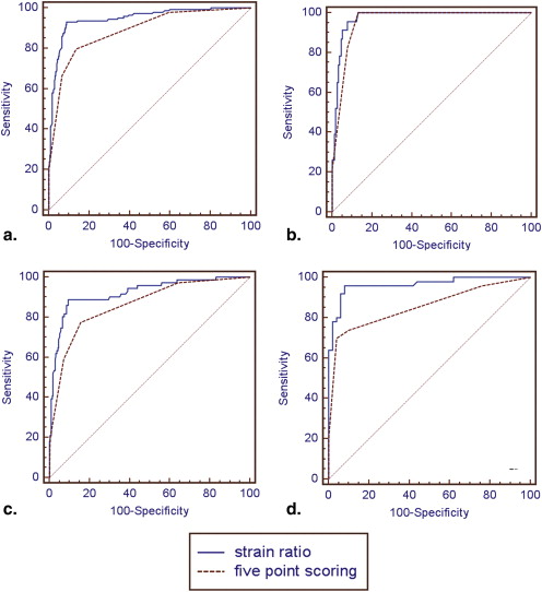
The strain ratios of benign lesions (mean, 1.83 ± 1.22) and malignant lesions (mean, 8.38 ± 7.65) were significantly different ( P < .00001). When a cutoff point of 3.05 was introduced, UE had 92.4% sensitivity, 91.1% specificity, and 91.4% accuracy. The area under the curve for strain ratio–based elastographic analysis was 0.944, and the area under the curve for the five-point scoring system was 0.885. The diagnostic performance of strain ratio–based elastographic analysis was better than that of the five-point scoring system with UE ( P < .05).
Conclusions
Strain ratio–based elastographic analysis can provide a new, more reliable diagnostic tool in comparison to a five-point scoring system for UE.
Biologic tissue possesses a specific inherent elasticity that may be altered by pathophysiologic processes, such as tumor development . This property serves as the basis for the physical examination method for detecting breast cancers, or palpation. Unfortunately, palpation is a highly empirical method and is dependent on the size and location of lesions and the skill of practitioners. The inaccuracy in elasticity caused by these factors resulted in the development of more accurate elasticity measurements, which began in the 1980s .
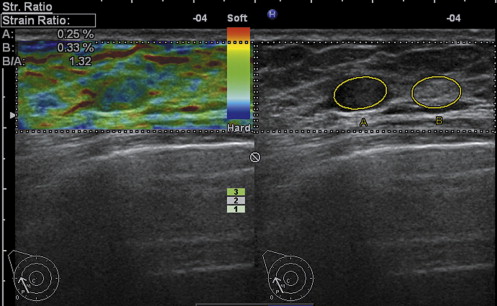
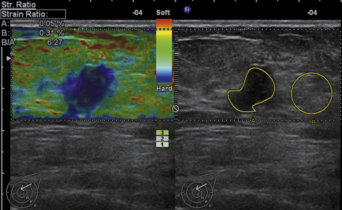
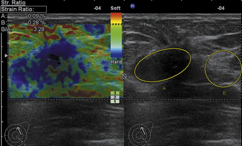
Young’s modulus, a measure of the stiffness of an elastic material, is too difficult to be calculated in vivo. Elastography is a new technology that can be applied to ultrasound for the reconstruction of tissue elasticity . The principle of elastography is that tissue compression results in deformation within the tissue; the displacement is smaller in harder tissue than in softer tissue. Real-time ultrasound elastography (UE) allows the calculation of tissue elasticity in real time and superimposes the information in color on B-mode images, with each color representing a certain level of elasticity. Although UE has not become a routine clinical examination, it has been shown to be useful in the diagnosis of breast cancer , thyroid cancer , prostate cancer , and liver cirrhosis .
Get Radiology Tree app to read full this article<
Get Radiology Tree app to read full this article<
Get Radiology Tree app to read full this article<
Materials and methods
Get Radiology Tree app to read full this article<
Patients
Get Radiology Tree app to read full this article<
Methods
Get Radiology Tree app to read full this article<
Get Radiology Tree app to read full this article<
Get Radiology Tree app to read full this article<
Statistical Analysis
Get Radiology Tree app to read full this article<
Get Radiology Tree app to read full this article<
Get Radiology Tree app to read full this article<
Get Radiology Tree app to read full this article<
Results
Pathologic Diagnosis
Get Radiology Tree app to read full this article<
Table 1
Histologic Diagnoses in 428 Patients with Benign or Malignant Breast Lesions
Benign Lesions ( n = 415) Malignant Lesions ( n = 144) Diagnosis_n_ Diagnosis_n_ Fibroadenoma 243 Invasive ductal carcinoma 130 Fibrocystic mastopathy 124 Papillocarcinoma 3 Papilloma 21 Invasive lobular carcinoma 3 Lipoma 7 Mucinous carcinoma 2 Chronic inflammation 5 Lymphoma 1 Hyperplasia 4 Ductal carcinoma in situ 1 Phyllodes tumor 2 Paget’s disease 2 Adenomyoepithelioma 2 Neuroendocrine carcinoma 1 Adenoleiomyoma 2 Invasive papillary carcinoma 1 Foreign body reaction 2 Tubular adenoma 1 Sclerosing adenosis 1 Hamartoma 1
Get Radiology Tree app to read full this article<
Strain Ratio of Breast Lesions
Get Radiology Tree app to read full this article<
Get Radiology Tree app to read full this article<
Get Radiology Tree app to read full this article<
Get Radiology Tree app to read full this article<
Get Radiology Tree app to read full this article<
Comparing Diagnostic Performance Between Strain Ratio Analysis and Five-point Scoring System
Get Radiology Tree app to read full this article<
Get Radiology Tree app to read full this article<
Get Radiology Tree app to read full this article<
Get Radiology Tree app to read full this article<
Get Radiology Tree app to read full this article<
Table 2
Comparison of Sensitivity, Specificity, PPV, and NPV Obtained Using Strain Ratio Measurement Method and Five-point Scoring System in the Differentiation of Benign From Malignant Breast Lesions
Lesions Method Sensitivity (%) Specificity (%) Accuracy (%) PPV (%) NPV (%) Total lesions Strain ratio 92.4 (133/144) ∗ 91.1 (378/415) 91.4 (511/559) 78.2 (133/170) 97.2 (378/389) Five-point scoring system 70.1 (101/144) 93.0 (386/415) 87.1 (487/559) 77.7 (101/130) 90.0 (386/429) Small lesions (≤10 mm) Strain ratio 95.7 (22/23) 91.6 (141/154) 92.1 (163/177) 62.9 (22/35) 99.3 (141/142) Five-point scoring system 82.6 (19/23) 92.2 (142/154) 91.0 (161/177) 61.3 (19/31) 97.3 (142/146) Median lesions (>10–20 mm) Strain ratio 88.7 (63/71) ∗ 91.0 (191/210) 90.4 (254/281) 76.8 (63/82) 96.0 (191/199) Five-point scoring system 64.8 (46/71) 93.3 (196/210) 86.1 (242/281) 76.7 (46/60) 88.7 (196/221) Large lesions (≥20 mm) Strain ratio 96 (48/50) ∗ 92.2 (47/51) 94.1 (95/101) ∗ 92.3 (48/52) 95.9 (47/49) Five-point scoring system 70 (35/50) 96.1 (49/51) 83.2 (84/101) 94.6 (35/37) 76.6 (49/64)
NPV, negative predictive value; PPV, positive predictive value.
Get Radiology Tree app to read full this article<
Get Radiology Tree app to read full this article<
False-negative and False-positive Diagnoses with the Strain Ratio Assessment Method
Get Radiology Tree app to read full this article<
Table 3
Analysis of False-negative and False-positive Diagnosis by Strain Ratio Assessment Method
False-negative ( n = 11) False-positive ( n = 37) Diagnosis_n_ Diagnosis_n_ Invasive ductal carcinoma 10 Fibroadenoma 16 Paget’s disease 1 Fibrocystic mastopathy 10 Papilloma 8 Lipoma 2 Chronic inflammation 1
Get Radiology Tree app to read full this article<
Discussion
Get Radiology Tree app to read full this article<
Get Radiology Tree app to read full this article<
Get Radiology Tree app to read full this article<
Get Radiology Tree app to read full this article<
Get Radiology Tree app to read full this article<
Get Radiology Tree app to read full this article<
Conclusions
Get Radiology Tree app to read full this article<
References
1. Bogomoletz W.V.: Elastosis in breast cancer. Pathol Annu 1986; 21: pp. 347-366.
2. Ophir J., Barra B., Kallel F., et. al.: Elastographic imaging. Ultrasound Med Biol 2000; 26: pp. S23-S29.
3. Ophir J., Céspedes I., Ponnekanti H., et. al.: Elastography: a quantitative method for imaging the elasticity of biological tissues. Ultrason Imaging 1991; 13: pp. 111-134.
4. O’Donnell M., Skovoroda A., Shapo B., et. al.: Internal displacement and strain imaging using ultrasonic speckle tracking. IEEE Trans Ultrason Ferroel Freq Cont 1994; 41: pp. 314-325.
5. Zhi H., Ou B., Luo B.M., et. al.: Comparison of ultrasound elastography, mammography, and sonography in the diagnosis of solid breast lesions. J Ultrasound Med 2007; 26: pp. 807-815.
6. Itoh A., Ueno E., Tohno E., et. al.: Breast disease: clinical application of US elastography for diagnosis. Radiology 2006; 239: pp. 341-350.
7. Lyshchik A., Higashi T., Asato R., et. al.: Thyroid gland tumor diagnosis at US elastography. Radiology 2005; 237: pp. 202-211.
8. Asteria C., Giovanardi A., Pizzocaro A., et. al.: US-elastography in the differential diagnosis of benign and malignant thyroid nodules. Thyroid 2008; 18: pp. 523-531.
9. Kamoi K., Okihara K., Ochiai A., et. al.: The utility of transrectal real-time elastography in the diagnosis of prostate cancer. Ultrasound Med Biol 2008; 34: pp. 1025-1032.
10. Nelson E.D., Slotoroff C.B., Gomella L.G., et. al.: Targeted biopsy of the prostate: the impact of color Doppler imaging and elastography on prostate cancer detection and Gleason score. Urology 2007; 70: pp. 1136-1140.
11. Tatsumi C., Kudo M., Ueshima K., et. al.: Noninvasive evaluation of hepatic fibrosis using serum fibrotic markers, transient elastography (FibroScan) and real-time tissue elastography. Intervirology 2008; 51: pp. 27-33.
12. Waki K, Murayama N, Matsumura T, et al. Investigation ofstrain ratio using ultrasound elastography technique. In: Proceedings of ISICE: The First International Symposium on Information and Computer Elements, Kitakyushu, Japan, September 12–14, 2007:449–452.
13. Zhi H., Xiao X.Y., Yang H.Y., et. al.: Semi-quantitating stiffness of breast solid lesions in ultrasonic elastography. Acad Radiol 2008; 15: pp. 1347-1353.
14. Levy L., Suissa M., Chiche J.F., Teman G., Martin B.: BIRADS ultrasonograhy. Eur J Radiol 2007; 61: pp. 202-211.
15. Thomas A., Fischer T., Frey H., et. al.: Real-time elastography—an advanced method of ultrasound: first results in 108 patients with breast lesions. Ultrasound Obstet Gynecol 2006; 28: pp. 335-340.
16. Samani A., Zubovits J., Plewes D.: Elastic moduli of normal and pathological human breast tissues: an inversion-technique-based investigation of 169 samples. Phys Med Biol 2007; 52: pp. 1565-1576.
17. Giannoula A., Cobbold R.S.: Propagation of shear waves generated by a modulated finite amplitude radiation force in a viscoelastic medium. IEEE Trans Ultrason Ferroelectr Freq Control 2009; 56: pp. 575-588.
18. Lupsor M., Badea R., Stefanescu H., et. al.: Performance of a new elastographic method (ARFI technology) compared to unidimensional transient elastography in the noninvasive assessment of chronic hepatitis C. Preliminary results. J Gastrointestin Liver Dis 2009; 18: pp. 303-310.
19. Sivesgaard K., Christensen S.D., Nygaard H., et. al.: Speckle tracking ultrasound is independent of insonation angle and gain: an in vitro investigation of agreement with sonomicrometry. J Am Soc Echocardiogr 2009; 22: pp. 852-858.
20. Shiina T., Doyley M.M., Bamber J.C.: Strain imaging using combined RF and envelope autocorrelation processing. Proc IEEE Ultrason Symp 1996; 2: pp. 1331-1336.
21. Thomas A., Degenhardt F., Farrokh A., et. al.: Significant differentiation of focal breast lesions calculation of strain ratio in breast sonoelastography. Acad Radiol 2010; 17: pp. 558-563.
22. Bodian C.A., Perzin K.H., Lattes R., et. al.: Prognostic significance of benign proliferative breast disease. Cancer 1993; 71: pp. 3896-3907.
23. Hiltawsky K.M., Kruger M., Starke C., et. al.: Freehand ultrasound elastography of breast lesions: clinical results. Ultrasound Med Biol 2001; 27: pp. 1461-1469.