
Rationale and Objectives
The aim of this study was to investigate whether ultrasound-mediated microbubble destruction enhances the transduction efficiency of recombinant adeno-associated virus (rAAV)–mediated enhanced green fluorescent protein (EGFP) gene into retinal ganglion cells (RGCs) of rats and whether it causes relevant adverse effects.
Materials and Methods
Thirty-two adult Sprague-Dawley rats were divided into four groups with different ultrasound powers, and retinal flat mounts and hematoxylin and eosin staining sections were made for optimization of parameters. A further 70 adult Sprague-Dawley rats were divided into four groups randomly. The first group (group A) was used as a normal control with 10 rats, and the remaining rats were evenly divided into groups B, C, and D. Each group included 20 rats. Groups B and C received rAAV-encoding EGFP gene (rAAV 2 -EGFP) in phosphate-buffered saline without and with ultrasound to the retina, respectively. Group D received microbubbles and rAAV 2 -EGFP mixture and ultrasound to the retina. The injection approach was intravitreal injection for all eyes. After 21 days, RGCs were labeled retrogradely with Fluoro-Gold. After 28 days, retinal flat mounts, frozen sections, and pathologic sections were assessed in each group. Expression of EGFP reporter gene was observed on laser confocal microscopy and evaluated according to average optical density and transfected RGC rate. To evaluate adverse effects with retinal flat mounts, labeled RGCs were counted, and retinal pathologic sections were examined.
Results
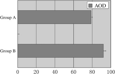
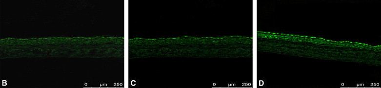
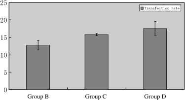
When ultrasound parameters (frequency, 0.3 MHz; power, 0.5 W/cm 2 ; total time, 60 seconds [irradiation time, 5 seconds; interval time, 10 seconds; four times]) were selected, EGFP expression was stronger, and retinas were not damaged. In the second part of the experiment, RGCs were labeled with Fluoro-Gold successfully. Green fluorescence can be observed in labeled RGCs in groups B to D. While average optical density and transfected RGC rate in group D were the highest compared to the other groups, no significant reduction in RGC number was detected with retrograde labeling. No obvious damage was observed with pathologic sections.
Conclusions
Ultrasound-mediated microbubble destruction can effectively and safely enhance rAAV delivery to RGCs in rats, and it may represent a novel gene delivery method in gene therapy for glaucomatous optic neuroprotection.
Retinal ganglion cell (RGC) apoptosis results in optic nerve damage in glaucoma. It has been proven that RGC apoptosis can be adjusted with apoptosis-related genes , and the progress of apoptosis can be prevented. Thus, gene therapy will probably become a potential treatment for glaucomatous optic neuroprotection . However, how to make foreign gene expression safe and efficient in target cells has been the key issue of gene therapy. Recombinant adeno-associated virus (rAAV) vectors have become promising gene delivery tools for optic neuroprotection treatment. These vectors possess a number of features that render them suited for this purpose, including a lack of pathogenicity and the ability to transfect retinal cells in a stable and long-term manner. But the application of viral vectors has been limited because of difficult production in sufficient quantities with viral particles, so that gene transfection efficiency is not easy to enhance . Unger et al showed that ultrasound contrast agent microbubbles can be a targeted gene therapy vector by using ultrasound cavitation erosion. As the cavitation nuclei in the cavitation progress, the gene released after microbubble destruction can enter into the vessel wall or even the tissue, depending on the energy of the ultrasound radiation . Microbubbles, as a novel gene vector, could enhance target gene transfection and expression safely and efficiently . In this study, we investigated whether ultrasound-mediated microbubble destruction enhances rAAV delivery of enhanced green fluorescent protein (EGFP) reporter gene into RGCs of rats and whether it causes relevant adverse effects.
Materials and methods
rAAV and Microbubble Mixture Preparation
According to a previously described method , lipid microbubbles were prepared at the Institute of Ultrasound Imaging at Chongqing University of Medical Sciences. The lipid shells of the microbubbles were composed of 90 mol% 1,2-distearoyl-sn-glycero-3-phosphocholine, 5 mol% 1,2-distearoyl-sn-glycero-3-phosphoethanolamine, and 1 mol% 1,2-distearoyl-sn-glycero-phosphoacid (Avanti Polar Lipids, Inc, Alabaster, AL). The lipids were mixed in chloroform and dried for 20 minutes under flowing nitrogen gas. To further dry the lipids, they were heated under vacuum for 45 minutes at −50°C. Glycerol (Sigma-Aldrich Corporation, St Louis, MO) and phosphate-buffered saline (PBS) (pH 7.0; Sigma-Aldrich Corporation) were added to the lipids. The solution was placed into a sonicating bath for 45 minutes and then aliquoted into gas-tight vials into which perfluoropropane gas (SynQuest Labs, Alachua, FL) was added. Microbubbles were formed by the shaking method using a VialMix mixing machine (DuPont Pharmaceuticals, Billerica, MA) for 45 seconds.
Get Radiology Tree app to read full this article<
Part 1 of the Experiment
Selection of Ultrasound Parameters
Get Radiology Tree app to read full this article<
Retinal Flat-mount Preparation
Get Radiology Tree app to read full this article<
Retinal Pathologic Section Preparation
Get Radiology Tree app to read full this article<
Part 2 of the Experiment: Comparison of Gene Transfection Efficiency
Intravitreal Injection and Gene Transfer
Get Radiology Tree app to read full this article<
Get Radiology Tree app to read full this article<
Get Radiology Tree app to read full this article<
Retrograde Labeling of RGCs
Get Radiology Tree app to read full this article<
Retinal Flat-mount preparation
Get Radiology Tree app to read full this article<
Retinal Frozen Section Preparation
Get Radiology Tree app to read full this article<
Retinal Pathologic Section Preparation
Get Radiology Tree app to read full this article<
EGFP Expression in RGCs and RGC Counting
Get Radiology Tree app to read full this article<
Get Radiology Tree app to read full this article<
Statistical Methods
Get Radiology Tree app to read full this article<
Results
Ultrasound Parameter Selection
Get Radiology Tree app to read full this article<
Get Radiology Tree app to read full this article<
EGFP Expression in Retina
Get Radiology Tree app to read full this article<
Get Radiology Tree app to read full this article<
Get Radiology Tree app to read full this article<
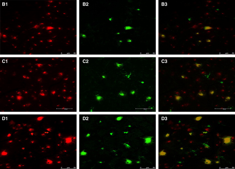
Table 1
AODs for EGFP Expression in Retinal Ganglion Cells on Retinal Flat Mounts
Group AOD 1 (%) AOD 2 (%) AOD 3 (%) AOD 4 (%) AOD 5 (%) Group B (rAAV 2 -EGFP) 45.55 53.67 49.20 64.97 49.08 Group C (rAAV 2 -EGFP plus ultrasound irradiation) 73.72 87.84 58.68 57.53 65.48 Group D (ultrasound irradiation plus rAAV 2 -EGFP plus microbubbles) 104.92 88.93 95.23 98.81 87.23
AOD, average optical density; EGFP, enhanced green fluorescent protein; rAAV, recombinant adeno-associated virus.
P < .05, group D versus groups B and C.
Get Radiology Tree app to read full this article<
RGC Counting
Get Radiology Tree app to read full this article<
Histologic Analyses of the Retinas
Get Radiology Tree app to read full this article<
Discussion
Get Radiology Tree app to read full this article<
Get Radiology Tree app to read full this article<
Get Radiology Tree app to read full this article<
Get Radiology Tree app to read full this article<
Get Radiology Tree app to read full this article<
Get Radiology Tree app to read full this article<
Get Radiology Tree app to read full this article<
Get Radiology Tree app to read full this article<
Get Radiology Tree app to read full this article<
References
1. Nickells R.W., Semaan S.J., Schlamp C.L.: Involvement of the Bcl2 gene family in the signaling and control of retinal ganglion cell death. Prog Brain Res 2008; 173: pp. 423-435.
2. Levin L.A., Schlamp C.L., Spieldoch R.L., et. al.: Identification of the bcl-2 family of genes in the rat retina. Invest Ophthalmol Vis Sci 1997; 38: pp. 2545-2553.
3. Mazzoni F., Novelli E., Strettoi E.: Retinal ganglion cells survive and maintain normal dendritic morphology in a mouse model of inherited photoreceptor degeneration. J Neurosci 2008; 28: pp. 14282-14292.
4. Yamamoto T.: The dawn of neuroprotective therapy for glaucomatous optic neuropathy. Nippon Ganka Gakkai Zasshi 2001; 105: pp. 866-883.
5. Martin K.R., Klein R.L., Quigley H.A.: Gene delivery to the eye using adeno-associated viral vectors. Methods 2002; 28: pp. 267-275.
6. Li W., Kong F., Li X., et. al.: Gene therapy following subretinal AAV5 vector delivery is not affected by a previous intravitreal AAV5 vector administration in the partner eye. Mol Vis 2009; 15: pp. 267-275.
7. Stieger K., Schroeder J., Provost N., et. al.: Detection of intact rAAV particles up to 6 years after successful gene transfer in the retina of dogs and primates. Mol Ther 2009; 17: pp. 516-523.
8. Negrete A., Kotin R.M.: Strategies for manufacturing recombinant adeno-associated virus vectors for gene therapy applications exploiting baculovirus technology. Brief Funct Genomic Proteomic 2008; 7: pp. 303-311.
9. Dinculescu A., Glushakova L., Min S.H., et. al.: Adeno-associated virus-vectored gene therapy for retinal disease. Hum Gene Ther 2005; 16: pp. 649-663.
10. Unger E.C.H.E., Vannan M., Matsunaga T.O., et. al.: Local drug and gene delivery through microbubbles. progress in cardiovascular diseases 2001; 44: pp. 45-54.
11. Skyba D.M., Price R.J., Linka A.Z., et. al.: Direct in vivo visualization of intravascular destruction of microbubbles by ultrasound and its local effects on tissue. Circulation 1998; 98: pp. 290-293.
12. Taniyama Y., Tachibana K., Hiraoka K., et. al.: Development of safe and efficient novel nonviral gene transfer using ultrasound: enhancement of transfection efficiency of naked plasmid DNA in skeletal muscle. Gene Ther 2002; 9: pp. 372-380.
13. Liu S., Wang Z.G.: Ultrasound-mediated microbubble destruction enhances green fluorescent protein gene expression in retinal ganglion cells in vitro. Zhonghua Chaosheng Yingxiangxue Zazhi 2007; 16: pp. 353-356.
14. Xu Y., Zhou X.Y., Wang Z.G., et. al.: Experimental study on transferring EGFP gene into the retina of rat mediated by microbubbles. Chin J Med Imaging Technol 2007; 23: pp. 188-190.
15. Ren J., Xu C., Zhou Z., et. al.: A novel ultrasound microbubble carrying gene and tat peptide: preparation and characterization. Acad Radiol 2009; 16: pp. 1457-1466.
16. Su H., Liu S., Wang Z.G., et. al.: In vivo transfection of enhanced green fluorescent protein in rat retinal ganglion cells mediated by ultrasound-induced microbubbles. Neural regeneration research 2009; 4: pp. 413-417.
17. Yin X.L., Ye J., Chen C.L.: Investigation of retinal ganglion cells and axons of normal rats using FluoroGold retrograde labeling. Label Immunoassays Clin Med 2006; 13: pp. 218-220.
18. Nakazawa T., Tamai M., Mori N.: Brain-derived neurotrophic factor prevents axotomized retinal ganglion cell death through MAPK and PI3K signaling pathways. Invest Ophthalmol Vis Sci 2002; 43: pp. 3319-3326.
19. Zalewska R., Reszec J., Mariak Z., et. al.: The expression of Bcl-2, Bcl-xl, Bak and Bax proteins in axons of the optic nerve in closed-angle glaucoma. Klin Oczna 2004; 106: pp. 155-157.
20. Ran H.T., Ren H., Wang Z.G., et. al.: Effects of ultrasonic cavitation on cultured cell membrane in vitro. Chin J Ultrasonogr 2003; 12: pp. 499-501.
21. Li W., Liu S., Ren J., et. al.: Gene transfection to retinal ganglion cells mediated by ultrasound microbubbles in vitro. Acad Radiol 2009; 16: pp. 1086-1094.
22. Muller O.J., Schinkel S., Kleinschmidt J.A., et. al.: Augmentation of AAV-mediated cardiac gene transfer after systemic administration in adult rats. Gene Ther 2008; 15: pp. 1558-1565.
23. Xia J.T., Xu B., Wang S.C., et. al.: Adeno-associated virus mediated tumor necrosis factor related apoptosis inducing ligand gene transfected into hepatocellular carcinoma cells by ultrasound microbubble intensifier: an experimental study. Zhonghua Yi Xue Za Zhi 2008; 88: pp. 1846-1850.
24. Schmued L.C., Fallon J.H.: Fluoro-Gold: a new fluorescent retrograde axonal tracer with numerous unique properties. Brain Res 1986; 377: pp. 147-154.
25. Buning H., Ried M.U., Perabo L., et. al.: Receptor targeting of adeno-associated virus vectors. Gene Ther 2003; 10: pp. 1142-1151.
26. Taniyama Y., Tachibana K., Hiraoka K., et. al.: Local delivery of plasmid DNA into rat carotid artery using ultrasound. Circulation 2002; 105: pp. 1233-1239.
27. Krasovitski B., Kimmel E.: Gas bubble pulsation in a semiconfined space subjected to ultrasound. J Acoust Soc Am 2001; 109: pp. 891-898.
28. Harvey A.R., Kamphuis W., Eggers R., et. al.: Intravitreal injection of adeno-associated viral vectors results in the transduction of different types of retinal neurons in neonatal and adult rats: a comparison with lentiviral vectors. Mol Cell Neurosci 2002; 21: pp. 141-157.
29. Dreyer E.B., Vorwerk C.K., Zurakowski D., et. al.: Infection with adeno-associated virus may protect against excitotoxicity. Neuroreport 1999; 10: pp. 2887-2890.