
Rationale and Objectives
Three-dimensional (3D) manufacturing is shaping personalized medicine, in which radiologists can play a significant role, be it as consultants to surgeons for surgical planning or by creating powerful visual aids for communicating with patients, physicians, and trainees. This report illustrates the steps in development of custom 3D models that enhance the understanding of complex anatomy.
Materials and Methods
We graphically designed 3D meshes or modified imported data from cross-sectional imaging to develop physical models targeted specifically for teaching complex segmental and branch anatomy. The 3D printing itself is easily accessible through online commercial services, and the models are made of polyamide or gypsum.
Results
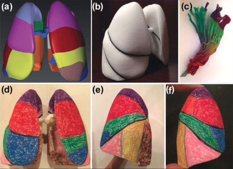
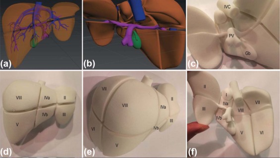
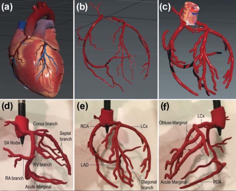
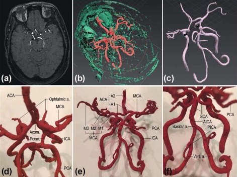
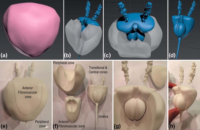
Anatomic models of the liver, lungs, prostate, coronary arteries, and the Circle of Willis were created. These models have advantages that include customizable detail, relative low cost, full control of design focusing on subsegments, color-coding potential, and the utilization of cross-sectional imaging combined with graphic design.
Conclusions
Radiologists have an opportunity to serve as leaders in medical education and clinical care with 3D printed models that provide beneficial interaction with patients, clinicians, and trainees across all specialties by proactively taking on the educator’s role. Complex models can be developed to show normal anatomy or common pathology for medical educational purposes. There is a need for randomized trials, which radiologists can design, to demonstrate the utility and effectiveness of 3D printed models for teaching simple and complex anatomy, simulating interventions, measuring patient satisfaction, and improving clinical care.
Introduction
Three-dimensional (3D) printing is contributing to the revolution of personalized and precision medicine . The use of 3D products in medicine is burgeoning : planning surgical procedures for hepatic and renal cancer removal; innovative cardiac and vascular device testing for pediatric and adult populations; visualization of complex head and neck anatomy for neurosurgeons and neurologists; practicing spine and lung procedures ex vivo for residents and medical students; personalized drug tablets and delivery with potential for nanotechnology; training models for image guided spinal pain management; and educating medical providers and the patients.
Patient-customized 3D products are available because of advanced 3D printing in conjunction with the evolution of cross-sectional imaging , which includes high-resolution multidetector computed tomography, computed tomography angiography, steady-state free precession and fast spoiled gradient-echo, magnetic resonance imaging (MRI), and magnetic resonance angiogram (MRA). Similar to an inkjet printer that reproduces a digital image with ink and paper, a 3D printer takes virtual data that are derived from cross-sectional imaging. The anatomic data are then processed by 3D reconstruction software into a virtual 3D mesh. Various materials are then loaded onto 3D printers to fabricate solid forms in a layer-by-layer fashion. The exquisite and customizable detail can be produced in a reasonable time frame and at a relatively low cost. Innovations in 3D printing have the potential to afford the medical team direct production capabilities and advance the practice of medicine through unique and customizable educational tools.
Get Radiology Tree app to read full this article<
Get Radiology Tree app to read full this article<
Get Radiology Tree app to read full this article<
Materials and Methods
Get Radiology Tree app to read full this article<
Table 1
Combination of Methods for Creating Final Digital 3D File with Relative Time and Cost Involved, Where the Simplest Model, the Lung, Is Used as a Reference
Anatomy Initial 3D Mesh Method of Obtaining Modification Time Cost Lung Graphically designed Free online library Radiologist − − Liver Graphically designed Commercially customized Graphic artist +++ +++ Coronary Graphically designed Commercially available Radiologist + ++ COW 3D Reconstructed (MRA) Performed with free software Radiologist ++ + Prostate 3D Reconstructed (MRI) Freely available online Radiologist +++ ++
COW, Circle of Willis, MRA, magnetic resonance angiography; MRI, magnetic resonance imaging.
Get Radiology Tree app to read full this article<
Get Radiology Tree app to read full this article<
Get Radiology Tree app to read full this article<
Get Radiology Tree app to read full this article<
Get Radiology Tree app to read full this article<
Get Radiology Tree app to read full this article<
Get Radiology Tree app to read full this article<
Get Radiology Tree app to read full this article<
Get Radiology Tree app to read full this article<
Get Radiology Tree app to read full this article<
Get Radiology Tree app to read full this article<
Get Radiology Tree app to read full this article<
Results
Get Radiology Tree app to read full this article<
Scaletarget=PricetargetPricecurrent−−−−−−−√3 Scal
e
target
=
Pric
e
target
Pric
e
current
3
Get Radiology Tree app to read full this article<
Get Radiology Tree app to read full this article<
Get Radiology Tree app to read full this article<
Get Radiology Tree app to read full this article<
Discussion
Get Radiology Tree app to read full this article<
Get Radiology Tree app to read full this article<
Get Radiology Tree app to read full this article<
Get Radiology Tree app to read full this article<
Get Radiology Tree app to read full this article<
Get Radiology Tree app to read full this article<
Get Radiology Tree app to read full this article<
Get Radiology Tree app to read full this article<
Financial Disclosure
Get Radiology Tree app to read full this article<
References
1. Ventola C.L.: Medical applications for 3D printing: current and projected uses. P T 2014; 39: pp. 704-711.
2. Mitsouras D., Liacouras P., Imanzadeh A., et. al.: Medical 3D printing for the radiologist. Radiographics 2015; 35: pp. 1965-1988.
3. Wilson C.A., Arthurs O.J., Black A.E., et. al.: Printed three-dimensional airway model assists planning of single-lung ventilation in a small child. Br J Anaesth 2015; 115: pp. 616-620.
4. Rehder R., Abd-El-Barr M., Hooten K., et. al.: The role of simulation in neurosurgery. Childs Nerv Syst 2015; 32: pp. 43-54.
5. Jones D.B., Sung R., Weinberg C., et. al.: Three-dimensional modeling may improve surgical education and clinical practice. Surg Innov 2015; 23: pp. 189-195.
6. Matsumoto J.S., Morris J.M., Foley T.A., et. al.: Three-dimensional physical modeling: applications and experience at Mayo clinic. Radiographics 2015; 35: pp. 1989-2006.
7. Bernhard J.C., Isotani S., Matsugasumi T., et. al.: Personalized 3D printed model of kidney and tumor anatomy: a useful tool for patient education. World J Urol 2015; 34: pp. 337-345.
8. Valverde I., Gomez G., Coserria J.F., et. al.: 3D printed models for planning endovascular stenting in transverse aortic arch hypoplasia. Catheter Cardiovasc Interv 2015; 85: pp. 1006-1012.
9. Javan R., Bansal M., Tangestanipoor A.: A prototype hybrid gypsum-based 3D printed training model for CT-guided spinal pain management. J Comput Assist Tomogr 2016; in press
10. Gross B.C., Erkal J.L., Lockwood S.Y., et. al.: Evaluation of 3D printing and its potential impact on biotechnology and the chemical sciences. Anal Chem 2014; 86: pp. 3240-3253.
11. Murphy S.V., Atala A.: 3D bioprinting of tissues and organs. Nat Biotechnol 2014; 32: pp. 773-785.
12. Brodland G.W.: How computational models can help unlock biological systems. Semin Cell Dev Biol 2015; 47-48: pp. 62-73.
13. Price compare—3D printers; Available at: http://www.i.Materialise.com Accessed March 15, 2016
14. Tutorials; Available at: http://www.3ders.org/pricecompare/3dprinters/ Accessed March 15, 2016
15. Friedman T., Michalski M., Goodman T.R., et. al.: 3D printing from diagnostic images: a radiologist’s primer with an emphasis on musculoskeletal imaging—putting the 3D printing of pathology into the hands of every physician. Skeletal Radiol 2016; 45: pp. 307-321.
16. Kong X., Nie L., Zhang H., et. al.: Do three-dimensional visualization and three-dimensional printing improve hepatic segment anatomy teaching? A randomized controlled study. J Surg Educ 2016; 73: pp. 264-269.
17. Zheng Y.X., Yu D.F., Zhao J.G., et. al.: 3D printout models vs. 3D-rendered images: which is better for preoperative planning?. J Surg Educ 2016; 73: pp. 518-523.
18. Crossingham J.L., Jenkinson J., Woolridge N., et. al.: Interpreting three-dimensional structures from two-dimensional images: a web-based interactive 3D teaching model of surgical liver anatomy. HPB (Oxford) 2009; 11: pp. 523-528.
19. Preece D., Williams S.B., Lam R., et. al.: “Let’s get physical”: advantages of a physical model over 3D computer models and textbooks in learning imaging anatomy. Anat Sci Educ 2013; 6: pp. 216-224.
20. Cheng G.Z., Folch E., Brik R., et. al.: Three-dimensional modeled T-tube design and insertion in a patient with tracheal dehiscence. Chest 2015; 148: pp. e106-e108.
21. Schmauss D., Haeberle S., Hagl C., et. al.: Three-dimensional printing in cardiac surgery and interventional cardiology: a single-centre experience. Eur J Cardiothorac Surg 2015; 47: pp. 1044-1052.
22. Perry D.J., Kwan S.W., Bhargava P.: Patient-centered clinical training in radiology. J Am Coll Radiol 2015; 12: pp. 724-727.
23. Knechtges P.M., Carlos R.C.: The evolving role of the radiologist within the health care system. J Am Coll Radiol 2007; 4: pp. 626-635.
24. Itri J.N.: Patient-centered radiology. Radiographics 2015; 35: pp. 1835-1846.
25. Itri J.N., Ballard D.H., Kantartzis S., et. al.: Entrepreneurship in the academic radiology environment. Acad Radiol 2015; 22: pp. 14-24.
26. Kansagra A.P., Shute T.S.: Space: the final frontier for IR. J Vasc Interv Radiol 2015; 26: pp. 825-828.
27. Ferrara S.: Interventional radiology in the austere environment. Semin Intervent Radiol 2010; 27: pp. 3-13.
28. Charalel R.A., Hentel K.D., Min R.J., et. al.: Adding value to health care: where radiologists may contribute. AJNR Am J Neuroradiol 2014; 35: pp. 1883-1884.
29. Silberstein J.L., Maddox M.M., Dorsey P., et. al.: Physical models of renal malignancies using standard cross-sectional imaging and 3-dimensional printers: a pilot study. Urology 2014; 84: pp. 268-272.
30. Ollivier L., Apiou F., Leclère J., et. al.: Patient experiences and preferences: development of practice guidelines in a cancer imaging department. Cancer Imaging 2009; 9: pp. S92-S97.