
Rationale and Objectives
The aim of this study was to evaluate the usefulness of a novel computerized method for lung nodule detection on digital chest radiographs using temporal subtraction images.
Materials and Methods
To significantly reduce the number of false-positive results while maintaining high sensitivity, temporal subtraction images, which can enhance interval changes on sequential chest radiographs, were used. Fifty-one cases with lung nodules <3 cm and 51 cases without lung nodules were selected for an observer performance test. Twelve radiologists participated in this observer performance test. The radiologists’ performance was evaluated using receiver-operating characteristic analysis, on a continuous rating scale. To estimate the numbers of cases affected beneficially and those affected detrimentally using this computerized method, the computer output was assumed to have an effect on an observer’s diagnosis when there was a difference in rating score of ≥30% between the first and second ratings.
Results
The average area under the curve for all radiologists increased significantly from 0.849 to 0.950 with the computerized method ( P < .001). The mean number of cases affected beneficially was significantly higher than that of cases affected detrimentally (8.92 vs 1.25, P < .001).
Conclusions
The novel computerized method using temporal subtraction images would be useful in detecting lung nodules on digital chest radiographs.
Although chest radiography is the most prevalent screening procedure for detecting lung lesions, because it is economical and easy to use, radiologists are sometimes unaware of lung nodules that are perceptible on chest radiographs in retrospect , and it is even more difficult to detect small lung nodules . Computer-aided diagnosis (CAD) is now considered an approach that might improve the efficacy of radiologic image interpretation. For the detection of subtle lung nodules, however, CAD of a single chest radiograph suffers from interference by anatomic noise, leading to low specificity for an acceptable level of sensitivity . The temporal subtraction technique is a CAD method in which a previous chest radiograph is subtracted from a current radiograph so that interval changes are enhanced. Several studies have found that this technique improves the diagnostic accuracy of newly formed lung nodule . However, misregistration artifacts of this system, due to mismatching of normal anatomic structures in current and previous images, cause false-positive findings and sometimes lead to misinterpretation. In addition, both previous and current images are required for this CAD system, which may limit its clinical use.
We are developing a novel computerized method to assist radiologists in the detection of lung nodules by using temporal subtraction images. Our purpose in this study was to evaluate the usefulness of this computerized method on radiologists’ performance for lung nodule detection.
Materials and methods
Scanning Protocol
Get Radiology Tree app to read full this article<
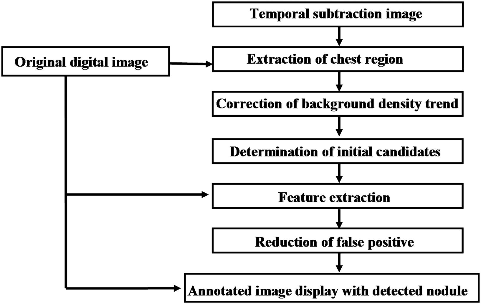
Computerized Scheme
Get Radiology Tree app to read full this article<
Get Radiology Tree app to read full this article<
Performance of the CAD System
Get Radiology Tree app to read full this article<
Observer Performance Study
Get Radiology Tree app to read full this article<
Get Radiology Tree app to read full this article<
Get Radiology Tree app to read full this article<
Statistical Analysis
Get Radiology Tree app to read full this article<
Results
Get Radiology Tree app to read full this article<
Table 1
Comparison of AUC Values without and with CAD
Observer AUC_P_ Without CAD With CAD Residents A 0.814 0.961 B 0.826 0.959 C 0.768 0.927 D 0.759 0.874 Average 0.792 0.930 <.001 Fellows E 0.853 0.951 F 0.895 0.952 G 0.799 0.937 H 0.812 0.934 Average 0.840 0.944 <.001 Attending radiologists I 0.931 0.982 J 0.903 0.970 K 0.933 0.972 L 0.900 0.979 Average 0.917 0.976 <.001 Average total 0.849 0.950 <.001
AUC, area under the curve; CAD, computer-aided diagnosis.
![Figure 3, Comparison of average receiver-operating characteristic curves of all radiologists for lung nodule detection without and with the computer-aided diagnosis (CAD) system. The average area under the curve (Az) for all radiologists increased significantly from 0.849 to 0.950 with the CAD system ( P < .001, Dorfman-Berbaum-Metz method [19] ).](https://storage.googleapis.com/dl.dentistrykey.com/clinical/UsefulnessofComputerizedMethodforLungNoduleDetectioninDigitalChestRadiographsUsingTemporalSubtractionImages/2_1s20S1076633211002078.jpg)
Get Radiology Tree app to read full this article<
Get Radiology Tree app to read full this article<
Get Radiology Tree app to read full this article<
Discussion
Get Radiology Tree app to read full this article<
Get Radiology Tree app to read full this article<
Get Radiology Tree app to read full this article<
Get Radiology Tree app to read full this article<
Get Radiology Tree app to read full this article<
Get Radiology Tree app to read full this article<
Get Radiology Tree app to read full this article<
References
1. Austin J.H., Romney B.M., Goldmith L.S.: Missed bronchogenic carcinoma: radiographic findings in 27 patients with a potentially resectable lesion evident in retrospect. Radiology 1992; 182: pp. 115-122.
2. Muhm J.R., Miller W.E., Fontana R.S., et. al.: Lung cancer detected during a screening program using 4-month chest radiographs. Radiology 1983; 148: pp. 609-615.
3. Heelan R.T., Flehinger B.J., Melamed M.R., et. al.: Non-small-cell lung cancer: results of the New York Screening Program. Radiology 1984; 151: pp. 289-293.
4. Samei E., Flynn M.J., Eyler W.R.: Detection of subtle lung nodules: relative influence of quantum and anatomical noise on chest radiographs. Radiology 1999; 213: pp. 727-734.
5. Uozumi T., Nakamura K., Watanabe H., et. al.: ROC analysis of detection of metastatic pulmonary nodules on digital chest radiographs with temporal subtraction. Acad Radiol 2001; 8: pp. 871-878.
6. Johkoh T., Kozuka T., Tomiyama N., et. al.: Temporal subtraction for detection of solitary pulmonary nodules on chest radiographs: evaluation of a commercially available computer-aided diagnosis system. Radiology 2002; 223: pp. 806-811.
7. Kakeda S., Nakamura K., Kamada K., et. al.: Improved detection of lung nodules by using a temporal subtraction technique. Radiology 2002; 224: pp. 145-151.
8. Oda N., Kido S., Shono H., et. al.: Development of computerized system for detection of pulmonary nodules on digital chest radiographs using subtraction images. Jpn J IEICE Trans 2004; 87D: pp. 208-218.
9. Kano A., Doi K., MacMahon H., et. al.: Digital image subtraction of temporally sequential chest images for detection of interval change. Med Phys 1994; 21: pp. 453-461.
10. Ishida T., Katsuragawa S., Nakamura K., et. al.: Iterative image warping technique for temporal subtraction of sequential chest radiographs to detect interval change. Med Phys 1999; 26: pp. 1320-1329.
11. Xu X.W., Doi K.: Image feature analysis for computer-aided diagnosis: accurate determination of ribcage boundary in chest radiographs. Med Phys 1995; 22: pp. 617-626.
12. Katsuragawa S., Doi K., MacMahon H.: Image feature analysis and computer-aided diagnosis in digital radiography: detection and characterization of interstitial lung disease in digital chest radiographs. Med Phys 1988; 15: pp. 311-319.
13. Xu X.W., Doi K., Kobayashi T., et. al.: Development of an improved CAD scheme for automated detection of lung nodules in digital chest images. Med Phys 1997; 24: pp. 1395-1403.
14. Duda R.O., Hart R.E., Stork D.G.: Pattern Classification.2nd ed.2001.John WileyNew York
15. Jain A.K., Duin P.W., Mao J.: Statistical pattern recognition; a review. IEEE Trans Pattern Anal Mach Intell 2000; 22: pp. 17-24.
16. Shiraishi J., Katsuragawa S., Ikezoe J., et. al.: Development of a digital image database for chest radiographs with and without a lung nodule: receiver operating characteristic analysis of radiologists’ detection of pulmonary nodules. AJR Am J Roentgenol 2000; 174: pp. 71-74.
17. Kobayashi T., Xu X.W., MacMahon H., et. al.: Effect of a computer-aided diagnosis scheme on radiologists’ performance in detection of lung nodules on radiographs. Radiology 1996; 199: pp. 843-848.
18. Metz C.E., Herman B.A., Shen J.H.: Maximum-likelihood estimation of receiver operating characteristic (ROC) curves from continuously-distributed data. Stat Med 1998; 17: pp. 1033-1053.
19. Dorfman D.D., Berbaum K.S., Metz C.E.: ROC rating analysis: generalization to the population of readers and cases with the jackknife method. Invest Radiol 1992; 27: pp. 723-731.
20. Freedman M.T., Osicka T., Lo S.C.B., et. al.: Methods for identifying changes in radiologists’ behavioral operating point of sensitivity-specificity tradeoffs within an ROC study of the use of computeraided detection of lung cancer. Prop SPIE 2001; 4324: pp. 184-194.
21. Henschke C.I., Naidich D.P., Yankelevitz D.F., et. al.: Early lung cancer action project: initial findings on repeat screenings. Cancer 2001; 92: pp. 153-159.
22. Patz E.F., Black W.C., Goodman P.C.: CT screening for lung cancer: not ready for routine practice. Radiology 2001; 221: pp. 587-591.