
Rationale and Objectives
We evaluated the usefulness of temporal subtraction images obtained from two successive whole-body bone scans, in terms of improvement in radiologists’ diagnostic accuracy in detecting interval changes and of a reduction in reading time, by use of a jackknife free-response receiver operating characteristic (JAFROC) analysis method.
Materials and Methods
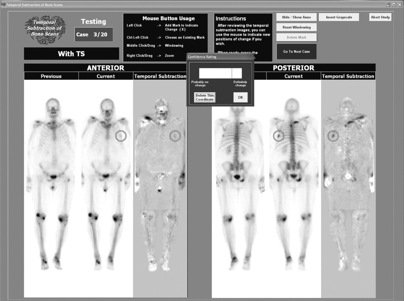
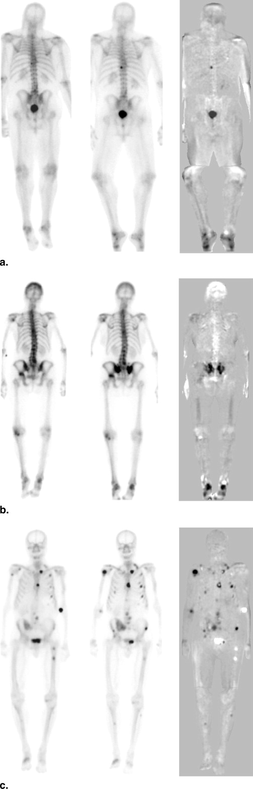
Twenty pairs of successive whole-body bone scans (72 consented interval changes) and their temporal subtraction images were used for an observer performance study. Our institutional review board approved the use of this database and the participation of radiologists in this study. In the first session of the observer study, without temporal subtraction images, the previous and current images were shown to five radiologists independently for their marking of the locations on current images and confidence ratings on potential interval changes from previous images. In the second session, temporal subtraction images were shown together with the modified previous and current images. JAFROC analysis was used for assessing the statistical significance of differences between radiologists’ performance without and with temporal subtraction images.
Results
The average sensitivity for detecting interval changes was improved from 58.6% to 73.2% at a false-positive rate of two per case by use of temporal subtraction images, and the difference was statistically significant by use of JAFROC analysis ( P = .035). In addition, the mean reading time per case was reduced considerably from 134 seconds to 91 seconds ( P < .01).
Conclusions
Temporal subtraction imaging for successive whole-body bone scans has the potential greatly to assist radiologists by increasing both their accuracy and productivity.
Bone scintigraphy is one of the most frequent examinations among various diagnostic nuclear medicine procedures. For example, based on the results of a comprehensive survey of radiology practice worldwide, the United Nations Scientific Committee on the Effects of Atomic Radiation ( ) reported that bone scintigraphy accounted for 24.0% of all diagnostic nuclear medicine procedures for the period 1991–1996 in the specific group of countries of health care level I, in which a country has more than one physician per 1,000 population. The whole-body bone scan examination with use of the radioisotope of technetium-99m methylene diphosphonate hydroxymethane diphosphonate is commonly employed for imaging of new bone formation and for demonstrating all-inclusively increased or decreased gamma-ray emissions localized at the site of bone abnormalities. These bone abnormalities may occur because of the presence of almost any skeletal pathology such as skeletal metastases, osteosarcoma, osteomyelitis, or nondisplaced fractures. Although the sensitivity of bone scan examinations for the detection of bone abnormalities has been considered to be relatively high, it is time-consuming to identify multiple lesions such as bone metastases of prostate or breast cancers. In addition, because of variations in patient conditions, the accumulation of radioisotopes during each examination, and the image quality of gamma cameras, it is difficult to detect subtle changes between two successive abnormal bone scans.
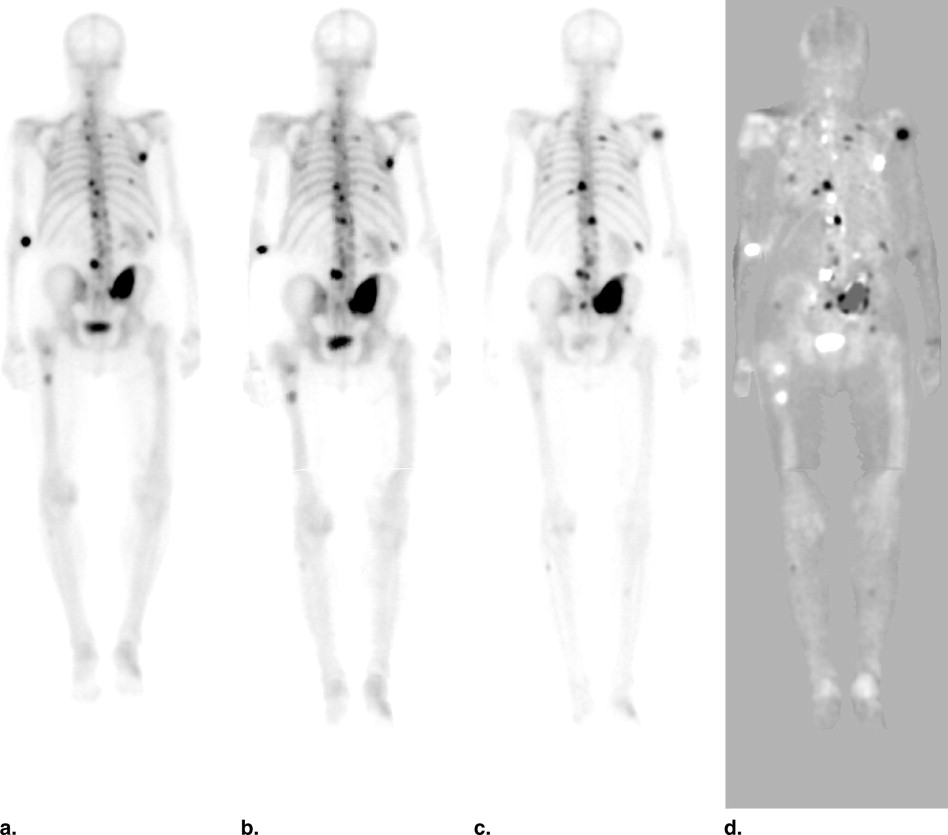
To assist radiologists in interpreting whole-body bone scans, we developed a computerized temporal subtraction technique that can highlight interval changes between successive whole-body bone scans ( ). A temporal subtraction technique for detecting interval changes between two successive chest radiographs has been described previously ( ). To our knowledge, however, there has been no application, in bone scans, of a temporal subtraction scheme by use of a nonlinear image-warping technique, and thus no observer performance study was reported on the effect of the temporal subtraction scheme on the detection of interval changes in successive whole-body bone scans.
Get Radiology Tree app to read full this article<
Materials and methods
Get Radiology Tree app to read full this article<
Get Radiology Tree app to read full this article<
Get Radiology Tree app to read full this article<
Get Radiology Tree app to read full this article<
Get Radiology Tree app to read full this article<
Get Radiology Tree app to read full this article<
Get Radiology Tree app to read full this article<
Get Radiology Tree app to read full this article<
Get Radiology Tree app to read full this article<
Get Radiology Tree app to read full this article<
Get Radiology Tree app to read full this article<
Get Radiology Tree app to read full this article<
Get Radiology Tree app to read full this article<
Get Radiology Tree app to read full this article<
Get Radiology Tree app to read full this article<
Results
Get Radiology Tree app to read full this article<
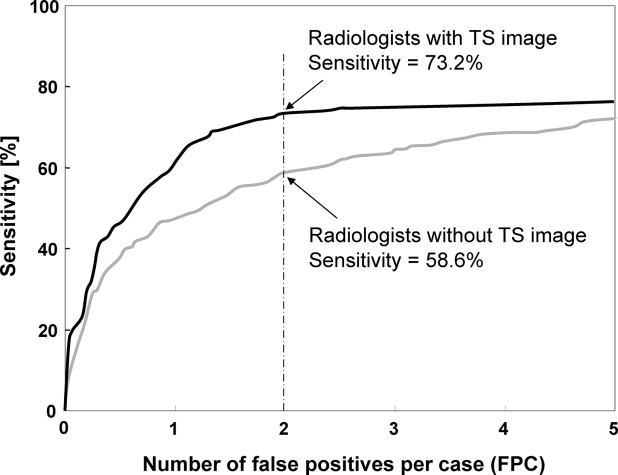
Table 1
Figure-of-Merit (FOM) Values Obtained From Jackknife Free-Response Receiver Operating Characteristic Analysis (Method 1) and Mean Reading Time per Case Without and With Temporal Subtraction (TS) Images for the Five Radiologists
FOM Mean Reading Time per Case [seconds] Reader Without TS With TS Without TS With TS_P_ Value A 0.587 0.695 164.7 98.3 (59.7%) <.0001 B 0.532 0.657 94.8 73.0 (77.0%) <.001 C 0.452 0.478 125.5 72.3 (57.6%) <.0001 D 0.506 0.690 188.7 149.4 (79.1%) .164 E 0.461 0.545 98.0 60.8 (62.0%) <.001 Mean 0.508 0.613 134.3 90.7 (67.5%)
Get Radiology Tree app to read full this article<
Get Radiology Tree app to read full this article<
Discussion
Get Radiology Tree app to read full this article<
Get Radiology Tree app to read full this article<
Get Radiology Tree app to read full this article<
Get Radiology Tree app to read full this article<
Get Radiology Tree app to read full this article<
Get Radiology Tree app to read full this article<
Get Radiology Tree app to read full this article<
Acknowledgments
Get Radiology Tree app to read full this article<
Get Radiology Tree app to read full this article<
References
1. UNSCEAR: 2000.United Nations Scientific Committee on the Effects of Atomic RadiationNew York
2. Shiraishi J., Appelbaum D., Pu Y., et. al.: Development of a computer-aided diagnostic scheme for detection of interval changes in successive whole-body bone scans. Med Phys 2006; 34: pp. 25-36.
3. Ishida T., Katsuragawa S., Nakamura K., et. al.: Iterative image warping technique for temporal subtraction of sequential chest radiographs to detect interval change. Med Phys 1999; 26: pp. 1320-1329.
4. Ishida T., Ashizawa K., Engelmann R., et. al.: Application of temporal subtraction for detection of interval changes on chest radiographs: improvement of subtraction images using automated initial image matching. J Digit Imaging 1999; 12: pp. 77-86.
5. Kano A., Doi K., MacMahon H., et. al.: Digital image subtraction of temporally sequential chest images for detection of interval change. Med Phys 1994; 21: pp. 453-461.
6. Chakraborty D.P., Berbaum K.S.: Observer studies involving detection and localization: modeling, analysis, and validation. Med Phys 2004; 31: pp. 2313-2330.
7. Li Q., Katsuragawa S., Doi K.: Improved contralateral subtraction images by use of elastic matching technique. Med Phys 2000; 27: pp. 1934-1942.
8. Zheng B., Chakraborty D.P., Rockette H.E., et. al.: A comparison of two data analyses from two observer performance studies using jackknife ROC and JAFROC. Med Phys 2005; 32: pp. 1031-1034.
9. Chakraborty D.P., Winter L.H.: Free-response methodology: alternate analysis and a new observer-performance experiment. Radiology 1990; 174: pp. 873-881.
10. Dorfman D.D., Berbaum K.S., Metz C.E.: Receiver operating characteristic rating analysis. Invest Radiol 1992; 27: pp. 723-731.
11. Sakai S., Soeda H., Furuya A., et. al.: Evaluation of the image quality of temporal subtraction images produced automatically in a PACS environment. J Digit Imaging 2006; 19: pp. 383-390.
12. Kakeda S., Nakamura K., Kamada K., et. al.: Improved detection of lung nodules by using a temporal subtraction technique. Radiology 2002; 224: pp. 145-151.
13. Uozumi T., Nakamura K., Watanabe H., et. al.: ROC analysis of detection of metastatic pulmonary nodules on digital chest radiographs with temporal subtraction. Acad Radiol 2001; 8: pp. 871-878.
14. Difazio M.C., MacMahon H., Xu X.W., et. al.: Digital chest radiography: effect of temporal subtraction images on detection accuracy. Radiology 1997; 202: pp. 447-452.
15. Johkoh T., Kozuka T., Tomiyama N., et. al.: Temporal subtraction for detection of solitary pulmonary nodules on chest radiographs: evaluation of a commercially available computer-aided diagnosis system. Radiology 2002; 223: pp. 806-811.
16. Abe H., Ishida T., Shiraishi J., et. al.: Effect of temporal subtraction images on radiologists’ detection of lung cancer on CT: results of the observer performance study with use of film computed tomography images. Acad Radiol 2004; 11: pp. 1337-1343.