
Rationale and Objectives
Anatomy is an essential component of medical education as it is critical for the accurate diagnosis in organs and human systems. The mental representation of the shape and organization of different anatomical structures is a crucial step in the learning process. The purpose of this pilot study is to demonstrate the feasibility and benefits of developing innovative teaching modules for anatomy education of first-year medical students based on three-dimensional (3D) reconstructions from actual patient data.
Materials and Methods
A total of 196 models of anatomical structures from 16 anonymized computed tomography datasets were generated using the 3D Slicer open-source software platform. The models focused on three anatomical areas: the mediastinum, the upper abdomen, and the pelvis. Online optional quizzes were offered to first-year medical students to assess their comprehension in the areas of interest. Specific tasks were designed for students to complete using the 3D models.
Results
Scores of the quizzes confirmed a lack of understanding of 3D spatial relationships of anatomical structures despite standard instruction including dissection. Written task material and qualitative review by students suggested that interaction with 3D models led to a better understanding of the shape and spatial relationships among structures, and helped illustrate anatomical variations from one body to another.
Conclusions
The study demonstrates the feasibility of one possible approach to the generation of 3D models of the anatomy from actual patient data. The educational materials developed have the potential to supplement the teaching of complex anatomical regions and help demonstrate the anatomical variation among patients.
Introduction
The teaching of anatomy is in crisis, as the discipline is disappearing from many academic institutions, being subsumed by disciplines such as cell biology . However, anatomy remains a vital element of any medical educational curriculum. Accurate diagnosis of alterations in organs or human systems requires a deep knowledge of anatomy. Radiology can play a critical role in helping medical students to recognize gross anatomical structures and their relationships to one another. The rapid development and application of imaging in medicine over the past 20 years has led to a better assessment and understanding of organ function in health and disease. In parallel, the emergence of increasingly sophisticated mathematical models, image processing, and visualization tools in the field of biomedical imaging research has enabled sophisticated three-dimensional (3D) representation of anatomical structures . The Visible Human Project (VHP) of the National Library of Medicine pioneered the development of digital image libraries of volumetric data to serve as a common reference point for the study of human anatomy. The VHP was initiated to attempt to provide rich datasets from computed tomography (CT), magnetic resonance imaging (MRI), and cryosection of representative male and female cadavers at an average of 1-mm intervals . Detailed animations and interactive 3D models of the human body, such as the Visible Human 3D Anatomical Structure Viewer, have been developed using the VHP data to facilitate learning of anatomy, radiological, and surgical procedures . By offering the possibility to add or remove anatomical structures and to observe an organ from different angles, 3D computer-aided visualization can enhance teaching of complex anatomical areas. A wealth of innovative anatomy education resources have been developed including tools such as Anatomy.tv and the Visible Body , as well as digital atlases generated from cadaver and clinical imaging data such as the Visible Ear , the Digital Anatomist Project , and RadStax . Although these resources provide a very rich set of tools for radiological anatomy education, one of the most essential aspects of anatomy education is for students to realize the often dramatic degree of variation from one body to another. With the advent and wide availability of multi-detector CT scanners, it is now a routine to obtain data in living patients that can be reconstructed at 1 mm or smaller increments. We propose to use this volume of data to demonstrate the degree of anatomical variation between patients for medical education purpose. This paper presents the results of our pilot study to assess the feasibility of the development of teaching modules for anatomy education of first-year medical students based on 3D reconstructions from actual patient data. We chose three of the most complicated regions of the body for this project, and recruited fourth-year medical students to participate in building models. In this way, we used senior students in a manner analogous to a traditional approach, where senior students could serve as preceptors for junior students, sometimes preparing prosections for anatomical study. Our students prepared “digital prosections”, learning the anatomy of these regions themselves in the process. Our goal was to determine the feasibility of this method of teaching, and to elicit feedback from first-year anatomy students on whether they felt that the use of 3D models helped them gain a deeper understanding of the shape and spatial organization of anatomical structures, as well as an appreciation of anatomical variation among patients that is so important in actual clinical practice.
Needs Assessment
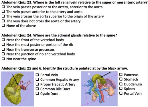
Medical school institutional review board approval was obtained for use of anonymous student feedback and anonymous optional test results. Two anonymous optional online quizzes consisting of 13 questions each were offered to students in a first-year medical anatomy course. Students were informed that quiz results would have no affect on their course grade and that quiz material would not be used in the final exam for the course. The first quiz covered structures in the upper abdomen and the second quiz covered structures in the pelvis. A quiz for mediastinal structures was also developed but was not administered because of technical difficulties. Questions in the quizzes were designed to assess both student ability to recognize structures in 3D representations in various orientations and demonstrate their understanding of spatial relationships between structures. Figure 1 shows a set of representative quiz questions for the upper abdomen.
Get Radiology Tree app to read full this article<
3D Modeling of Anatomy
Get Radiology Tree app to read full this article<
Get Radiology Tree app to read full this article<
Table 1
Anatomical Models of the Mediastinum
Organs Vessels Bones Heart Arteries Thoracic spine Left atrium Ascending aorta Ribs Left ventricle Aortic Arch Sternum Right atrium Descending aorta Right ventricle Pulmonary artery Esophagus Veins Thymus Superior vena cava Inferior vena cava Azygos vein
Table 2
Anatomical Models of the Upper Abdomen
Organs Vessels Muscles Adrenal glands Arteries External Obliques Duodenum Common Iliac Internal Obliques Esophagus Internal Iliac Rectus abdominis Gall bladder External Iliac Iliacus Liver Veins Psoas Kidneys Common Iliac Pancreas Internal Iliac Spleen External Iliac Stomach
Table 3
Anatomical Models of the Pelvis
Organs Vessels Bones Muscles Urinary Arteries Pelvis Pelvic Bladder Common Iliac Sacrum Coccygeus Urethra Internal Iliac Femur External anal sphincter Gastrointestinal External Iliac Levator ani Sigmoid colon Veins Thigh Anus Common Iliac Quadratus femoris Genital (male) Internal Iliac Obturator externus Internal genital External Iliac Obturator internus Prostate Piriformis Vas deferens Pectineus Testes Adductor brevis External Genital Cura of penis Bulb of penis Genital (female) Internal Genital Uterus Cervix Vagina Ovaries External Genital Crura of clitoris Bulb of vestibule Bartholin’s glands
Get Radiology Tree app to read full this article<
Computer-assisted Teaching of Radiological Anatomy
Get Radiology Tree app to read full this article<
Get Radiology Tree app to read full this article<
Get Radiology Tree app to read full this article<
Get Radiology Tree app to read full this article<
Get Radiology Tree app to read full this article<
Results
Generation of 3D Models
Get Radiology Tree app to read full this article<
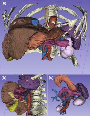
Table 4
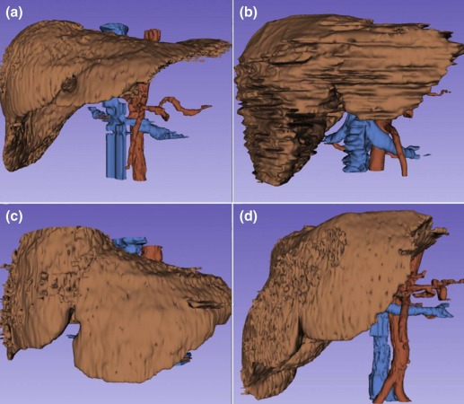
Dimensions and Volume of the Liver in Three Anatomical Directions
Case A Case B Case C Case D I-S (mm) 159 165 130 185 R-L (mm) 251 234 323 207 A-P (mm) 199 225 190 226 Volume (mL) 1302 2322 1380 2184
A-P, anterior-posterior; I-S, inferior-superior; R-L, right-left.
Get Radiology Tree app to read full this article<
Get Radiology Tree app to read full this article<
Results of Upper Abdomen and Pelvis Quiz
Get Radiology Tree app to read full this article<
3D Anatomy Workshops
Get Radiology Tree app to read full this article<
Task Forms
Get Radiology Tree app to read full this article<
Get Radiology Tree app to read full this article<
Get Radiology Tree app to read full this article<
Qualitative Feedback
Get Radiology Tree app to read full this article<
Get Radiology Tree app to read full this article<
Get Radiology Tree app to read full this article<
Get Radiology Tree app to read full this article<
“The 3D imaging was really helpful in visualizing areas of the body that we could not see clearly in lab. Because the spatial relationships and surface anatomy are so important for clinical practice, the 3D imaging is such a useful learning tool, and I hope that all of the areas of the body can be studied this way (especially the pelvis!).” “Bravo! I think this should replace cadaver study.” “I found the comparison of the model with CT images to be the most helpful aspect of this program.” “Visualizing 3D objects in 2D images (such as in CT, MR, and US) is really difficult, and I think Slicer is an invaluable tool to help us to do that!” “It would be nice to be able to easily compare images on Slicer from different cases, in particular to view various anatomical variations or to compare a variation to the normal structure.” “I think it would be a useful tool in radiology lab to use alongside the current online modules.” “This session was fantastic! I gained a new appreciation of being to see the body in 3D terms. Initially I got a little frustrated trying to figure the program out—but, in time I was able to use it sufficiently well to learn from the program. I only wish that Slicer could be installed on all of the computers in the other skills areas so more students could use it.” “This is something that is very useful if you have a hard time seeing things in dissection, plus it really helped my visualize CT scans. I also worked in a small group which I also thought was very beneficial.” “As long as it’s complete, I think it will be a fantastic learning tool.”
Get Radiology Tree app to read full this article<
Get Radiology Tree app to read full this article<
Conclusion
Get Radiology Tree app to read full this article<
Get Radiology Tree app to read full this article<
Get Radiology Tree app to read full this article<
Get Radiology Tree app to read full this article<
Get Radiology Tree app to read full this article<
Get Radiology Tree app to read full this article<
Get Radiology Tree app to read full this article<
Get Radiology Tree app to read full this article<
Get Radiology Tree app to read full this article<
Get Radiology Tree app to read full this article<
Get Radiology Tree app to read full this article<
Get Radiology Tree app to read full this article<
Get Radiology Tree app to read full this article<
Get Radiology Tree app to read full this article<
Acknowledgments
Get Radiology Tree app to read full this article<
Appendix
Supplementary material
Get Radiology Tree app to read full this article<
Figure S1
Get Radiology Tree app to read full this article<
Get Radiology Tree app to read full this article<
Get Radiology Tree app to read full this article<
Figure S2
Get Radiology Tree app to read full this article<
Get Radiology Tree app to read full this article<
Get Radiology Tree app to read full this article<
Figure S3
Get Radiology Tree app to read full this article<
Get Radiology Tree app to read full this article<
Get Radiology Tree app to read full this article<
Table S1
Get Radiology Tree app to read full this article<
Get Radiology Tree app to read full this article<
Get Radiology Tree app to read full this article<
References
1. Vazquez R., Riesco J.M., Carretero J.: Reflections and challenges in the teaching of human anatomy at the beginning of the 21st century. Eur J Anat 2005; 9: pp. 111-115.
2. Duncan J.S., Ayache N.: Medical image analysis: progress over two decades and the challenges ahead. IEEE Trans PAMI 2000; 22: pp. 85-105.
3. Spitzer V.M., Ackerman M.J., Scherzinger A.L., et. al.: The Visible Human Male: a technical report. J Am Med Inform Assoc 1996; 3: pp. 118-130.
4. Pommert A., Hohne K.H., Plesser B., et. al.: Creating a high-resolution spatial/symbolic model of the inner organs based on the Visible Human. Med Image Anal 2001; 5: pp. 221-228.
5. Gehrmann S., Hohne K.H., Linhart W., et. al.: A detailed 3D model of the human hand. J Vis 2004; 7: pp. 221.
6. Burmester E., Leineweber T., Hacker S., et. al.: EUS Meets Voxel-Man: three-dimensional anatomical animation of linear-array endoscopic ultrasound images. Endoscopy 2004; 36: pp. 726-730.
7. Pommert A., Hohne K.H., Burmester E., et. al.: Computer-based anatomy: a prerequisite for computer-assisted radiology and surgery. Acad Radiol 2006; 13: pp. 104-112.
8. Visible Body : Argosy Publishing Inc., Newton, MA; Available at: http://www.visiblebody.com Accessed August 26, 2015
9. Anatomy.tv : Primal Pictures Ltd., London, UK; Available at: http://www.anatomy.tv Accessed August 26, 2015
10. Wang H., Merchant S.N., Sorensen M.S.: A downloadable three-dimensional virtual model of the Visible Ear. ORL J Otorhinolaryngol Relat Spec 2007; 69: pp. 63-67. Available at http://research.meei.harvard.edu/Otopathology/3dmodels/visible_ear.html Accessed August 26, 2015
11. Brinkley J.F., Bradley S.W., Sundsten J.W., et. al.: The digital anatomist information system and its use in the generation and delivery of web-based anatomy atlases. Comput Biomed Res 1997; 30: pp. 472-503. Available at http://www9.biostr.washington.edu/da.html Accessed August 26, 2015
12. Colucci P.G., Kostandy P., Shrauner W.R., et. al.: Development and utilization of a web-based application as a robust radiology teaching tool (RadStax) for medical student anatomy teaching. Acad Radiol 2015; 22: pp. 247-255.
13. Rosset A., Spadola L., Ratib O.: OsiriX: an open-source software for navigating in multidimensional DICOM images. J Digit Imaging 2004; 17: pp. 205-216.
14. Pieper S., Halle M., Kikinis R.: 3D Slicer. Proceedings of the IEEE Symposium on Biomedical Imaging. Arlington, Virginia, USA; April 15–182004.pp. 632-635.
15. Lorensen W.E., Cline H.E.: Marching Cubes: a high resolution 3D surface construction algorithm. Proceedings of the 14th annual conference on Computer Graphics and Interactive Techniques, SIGGRAPH’87; Anaheim, California, USA; July 27–311987.pp. 163-169.