
Rationale and Objectives
Chest radiographs are recommended for prevention and detection of pneumoconiosis. In 2011, the International Labour Office (ILO) released a revision of the International Classification of Radiographs of Pneumoconioses that included a digitized standard images set. The present study compared results of classifications of digital chest images performed using the new ILO 2011 digitized standard images to classification approaches used in the past.
Materials and Methods
Underground coal miners ( N = 172) were examined using both digital and film-screen radiography (FSR) on the same day. Seven National Institute for Occupational Safety and Health-certified B Readers independently classified all 172 digital radiographs, once using the ILO 2011 digitized standard images (DR ILO2011-D ) and once using digitized standard images used in the previous research (DR RES ). The same seven B Readers classified all the miners’ chest films using the ILO film-based standards.
Results
Agreement between classifications of FSR and digital radiography was identical, using a standard image set (either DR ILO2011-D or DR RES ). The overall weighted κ value was 0.58. Some specific differences in the results were seen and noted. However, intrareader variability in this study was similar to the published values and did not appear to be affected by the use of the new ILO 2011 digitized standard images.
Conclusions
These findings validate the use of the ILO digitized standard images for classification of small pneumoconiotic opacities. When digital chest radiographs are obtained and displayed appropriately, results of pneumoconiosis classifications using the 2011 ILO digitized standards are comparable to film-based ILO classifications and to classifications using earlier research standards.
Chest radiographs are recommended for the detection and prevention of pneumoconiosis in workers involved in dusty trades, such as underground mining . In clinical practice and public health surveillance, digital chest radiographs (DR) presented on medical-grade monitors have largely replaced the conventional film-screen radiograph (FSR) technology. The International Labour Office (ILO) Guidelines for the Classification of the Pneumoconioses has been an invaluable tool for standardization of interpretations of chest radiographs for epidemiologic studies of the pneumoconioses . To enhance accuracy and precision in applying the ILO classification scoring system, readers are required to perform a side-by-side comparison of each individual worker’s radiograph to one or more prototypical chest images, which illustrate a variety of types and severity of radiographic abnormalities induced by dust inhalation. The ILO classification system includes a standard set of chest images for comparison purposes. Until recently, the ILO classification system only provided a set of standard images in the film-screen radiograph (FSR) format. However, in 2011, the ILO revised its guidelines to “extend the applicability of the Classification to digital radiographic images of the chest” . In the 2011 revision of the classification, the ILO includes a set of electronic image files (ILO Standard Digital Images [2011-D]) that was digitized from the film-based standards included in the 2000 revision of the classification.
Prior to the adoption of the ILO Standard Digital Images (2011-D), a series of research studies was undertaken to assess any potential impact of image modality on the outcomes of ILO classifications of chest radiographs (comparing FSR to DR displayed on medical-grade diagnostic monitors) . These investigations obtained both FSR and DR chest radiographs from study participants on the same day. FSR chest radiographs were interpreted using the ILO 2000 version of the classification system with the traditional film-based standard images. To enable classification of the DR chest radiographs displayed as soft copies on a medical-grade computer monitor, an existing set of the ILO standard films was scanned and digitized (see Franzblau et al. for methods). The resulting image files (“research” digitized standards) were used as the ILO standards for classifying digital images in a number of previous investigations . These “research” digitized standards appear quite similar to the current set of standard films that are included in the ILO 2000 classification, but were digitized from a prior version of the ILO standard films that had been issued with a black label rather than the current white label. The methods used in these modality studies required that at least two National Institute for Occupational Safety and Health (NIOSH)-certified B Readers 1
Get Radiology Tree app to read full this article<
interpret each participant’s DR chest radiograph presented as a soft copy image side-by-side with the digitized ILO standard radiographs, using two identical medical-grade monitors. In brief, these studies concluded that, with appropriate attention to image acquisition and soft copy display, both of the widely available digital radiography systems 2
Get Radiology Tree app to read full this article<
can be equivalent to FSR in the visualization and classification of small interstitial lung opacities.
Get Radiology Tree app to read full this article<
Get Radiology Tree app to read full this article<
Materials and methods
Get Radiology Tree app to read full this article<
Results
Image Quality
Get Radiology Tree app to read full this article<
Table 1
Image Quality Ratings for 3612 Classifications by Image Modality and Standard Set
Quality FSR [ n (%)] DR RES [ n (%)] DR ILO2011-D [ n (%)] Good 536 (44.5) 792 (65.8) 827 (68.7) Acceptable, no defects 410 (34.1) 332 (27.6) 337 (28.0) Acceptable, some defects 228 (18.9) 74 (6.1) 39 (3.2) Unacceptable 15 (1.2) 0 (0.0) 1 (0.1) Missing/no indication 15 (1.2) 6 (0.5) 0 (0.0)
DR ILO2011-D , digital radiograph interpreted using digitized ILO reference standards (2011-D); DR RES , digital radiograph interpreted using the “research” digitized standards; FSR, film-screen radiograph interpreted using 2000 ILO film-based reference standard; ILO, International Labour Organization.
Each chest radiograph from 172 miners was classified by seven National Institute for Occupational Safety and Health (NIOSH) B Readers for a total of 1204 classifications.
Get Radiology Tree app to read full this article<
Profusion of Small Opacities
Get Radiology Tree app to read full this article<
Get Radiology Tree app to read full this article<
Get Radiology Tree app to read full this article<
Table 2
Small Opacity Profusion by Image Type and Standard Set
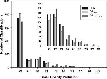
Profusion FSR [ n (%)] DR RES [ n (%)] DR ILO2011-D [ n (%)] 0/0 743 (62.8) 817 (69.0) 691 (58.4) 0/1 133 (11.2) 109 (9.2) 133 (11.2) 1/0 136 (11.5) 121 (10.2) 158 (13.3) 1/1 93 (7.9) 61 (5.2) 85 (7.2) 1/2 28 (2.4) 26 (2.2) 46 (3.9) 2/1 24 (2.0) 19 (1.6) 33 (2.8) 2/2 19 (1.6) 16 (1.4) 21 (1.8) 2/3 6 (0.5) 10 (0.8) 10 (0.8) 3/2 1 (0.1) 5 (0.4) 6 (0.5) 3/3 1 (0.1) 0 1 (0.1)
DR ILO2011-D , digital radiograph interpreted using digitized ILO reference standards (2011-D), DR RES , digital radiograph interpreted using the “research” digitized standards; FSR, film-screen radiograph interpreted using 2000 ILO film-based reference standard; ILO, International Labour Organization.
One hundred seventy-two images were classified by seven B readers, for a total of 1204 possible classifications for each method. Small opacity profusion categories are ILO designations
Get Radiology Tree app to read full this article<
Get Radiology Tree app to read full this article<
Table 3
Intrareader Weighted Kappa Values and 95% Confidence Intervals (CIs) Comparing ILO Small Opacity Profusion
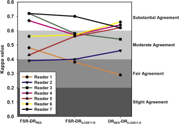
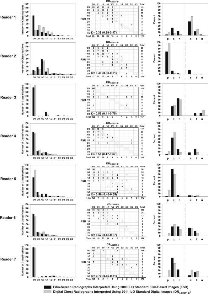
Reader 1 Reader 2 Reader 3 Reader 4 Reader 5 Reader 6 Reader 7 Overall FSR-DR RES ∗ 0.48 (0.37–0.59) 0.39 (0.29–0.49) 0.72 (0.56–0.87) 0.67 (0.59–0.74) 0.43 (0.31–0.54) 0.56 (0.45–0.67) 0.72 (0.58–0.86) 0.58 (0.54–0.62) FSR-DR ILO2011-D 0.38 (0.29–0.47) 0.40 (0.30–0.51) 0.58 (0.41–0.75) 0.57 (0.47–0.67) 0.56 (0.46–0.65) 0.57 (0.48–0.67) 0.70 (0.60–0.81) 0.58 (0.54–0.61) DR RES -DR ILO2011-D 0.29 (0.20–0.39) 0.46 (0.37–0.55) 0.54 (0.35–0.73) 0.64 (0.54–0.75) 0.62 (0.49–0.74) 0.66 (0.58–0.75) 0.62 (0.49–0.76) 0.58 (0.54–0.62)
DR ILO2011-D , digital radiograph interpreted using digitized ILO reference standards (2011-D), DR RES , digital radiograph interpreted using the “research” digitized standards; FSR, film-screen radiograph interpreted using 2000 ILO film-based reference standard; ILO, International Labour Organization.
Date represented as κ Value (95% CI).
Get Radiology Tree app to read full this article<
Get Radiology Tree app to read full this article<
Small Opacity Shape and Size
Get Radiology Tree app to read full this article<
Table 4
Small Opacity Shape and Size by Image Modality and Standard Set
Shape and Size FSR [ n (%)] DR RES [ n (%)] DR ILO2011-D [ n (%)] Rounded 328 (74.4) 245 (66.4) 338 (68.6) p 108 (24.5) 107 (29.0) 187 (37.9) q 160 (36.3) 102 (27.6) 121 (24.5) r 60 (13.6) 36 (9.8) 30 (6.1) Irregular 113 (25.6) 124 (33.6) 155 (31.4) s 72 (16.3) 88 (23.9) 108 (21.9) t 36 (8.2) 34 (9.2) 44 (8.9) u 5 (1.1) 2 (0.5) 3 (0.6)
DR ILO2011-D , digital radiograph interpreted using digitized ILO reference standards (2011-D), DR RES , digital radiograph interpreted using the “research” digitized standards; FSR, film-screen radiograph interpreted using 2000 ILO film-based reference standard; ILO, International Labour Organization.
One hundred seventy-two images were classified by seven B readers, for a total of 3612 possible classifications for each method. Primary shape and size designations are standard ILO designations. Readers did not record shape and size for images with 0/0 profusion.
Get Radiology Tree app to read full this article<
Get Radiology Tree app to read full this article<
Table 5
Simple κ Values and 95% Confidence Intervals (CIs) Comparing Primary Shape and Size Designation by Image Modality and Standard Set
κ Value 95% CI FSR-DR RES ∗ 0.51 (0.44–0.58) FSR-DR ILO2011-D 0.47 (0.41–0.54) DR RES -DR ILO2011-D 0.57 (0.50–0.63)
DR ILO2011-D , digital radiograph interpreted using digitized ILO reference standards (2011-D), DR RES , digital radiograph interpreted using the “research” digitized standards; FSR, film-screen radiograph interpreted using 2000 ILO film-based reference standard; ILO, International Labour Organization.
Get Radiology Tree app to read full this article<
Get Radiology Tree app to read full this article<
Within-reader Variability
Get Radiology Tree app to read full this article<
Discussion
Get Radiology Tree app to read full this article<
Get Radiology Tree app to read full this article<
Get Radiology Tree app to read full this article<
Get Radiology Tree app to read full this article<
Get Radiology Tree app to read full this article<
Acknowledgments
Get Radiology Tree app to read full this article<
Get Radiology Tree app to read full this article<
References
1. Wagner G.: Screening and surveillance of workers exposed to mineral dust.1996.World Health OrganizationGeneva
2. International Labour Office: Guidelines for the use of the ILO international classification of radiographs of pneumoconioses.2011.International Labour OfficeGeneva
3. Franzblau A., Kazerooni E.A., Sen A., et. al.: Comparison of digital radiographs with film radiographs for the classification of pneumoconiosis. Acad Radiol 2009; 16: pp. 669-677.
4. Laney A.S., Petsonk E.L., Wolfe A.L., et. al.: Comparison of storage phosphor computed radiography with conventional film-screen radiography in the recognition of pneumoconiosis. Eur Respir J 2010; 36: pp. 122-127.
5. Sen A., Lee S.Y., Gillespie B.W., et. al.: Comparing film and digital radiographs for reliability of pneumoconiosis classifications: a modeling approach. Acad Radiol 2010; 17: pp. 511-519.
6. Laney A.S., Petsonk E.L., Attfield M.D.: Intramodality and intermodality comparisons of storage phosphor computed radiography and conventional film-screen radiography in the recognition of small pneumoconiotic opacities. Chest 2011; 140: pp. 1574-1580.
7. Viera A.J., Garrett J.M.: Understanding interobserver agreement: the kappa statistic. Fam Med 2005; 37: pp. 360-363.