
Rationale and Objectives
We sought to compare coronary artery calcium (CAC) scores, the variability and radiation doses on 64- and 16-slice computed tomography (CT) scanners by both prospective electrocardiographically (ECG)-triggered and retrospective ECG-gated scans.
Materials and Methods
Coronary artery models ( n = 3) with different plaque CT densities (∼240 Hounsfield units [HU], ∼600 HU, and ∼1000 HU) of four sizes (1, 3, 5, and 10 mm in length) on a cardiac phantom were scanned three times in five heart rate sequences. The tube current-time products were set to almost the same on all four protocols (32.7 mAs for 64-slice prospective and retrospective scans, 33.3 mAs for 16-slice prospective and retrospective scans). Slice thickness was set to 2.5 mm to keep the radiation dose low. Overlapping reconstruction with a 1.25-mm increment was applied on the retrospective ECG-gated scan.
Results
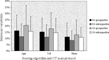
The CAC scores were not different between the four protocols (one-factor analysis of variance: Agatston, P = .32; volume, P = .19; and mass, P = .09). Two-factor factorial analysis of variance test revealed that the interscan variability was different between protocols ( P < .01) and scoring algorithms ( P < .01). The average variability of Agatston/volume/mass scoring and effective doses were as follows: 64-slice prospective scan: 16%/15%/11% and 0.5 mSv; 64-slice retrospective scan: 11%/11%/8% and 3.7 mSv; 16-slice prospective scan: 20%/18%/13% and 0.6 mSv; and 16-slice retrospective scan: 16%/15%/11% and 2.9 to 3.5 mSv (depending on the pitch).
Conclusion
Retrospective ECG-gated 64-slice CT showed the lowest variability. Prospective ECG-triggered 64-slice CT, with low radiation dose, shows low variability on CAC scoring comparable to retrospective ECG-gated 16-slice CT.
Coronary artery calcium (CAC) scoring is performed to evaluate the presence of coronary atherosclerosis or to assess the progression and regression of coronary atherosclerosis ( ). Therefore, low variability and low radiation exposure are both key requirements on CAC scoring. Interscan variability of Agatston score ( ) on electron beam computed tomography (CT), however, yielding 20% to 37% ( ), is high, considering that normal progression of CAC scores per year is 14% to 27% (average 24%) ( ) and is accelerated up to 33% to 48% with significant coronary disease ( ). To reduce the variability, the volumetric approach ( ) and the calcium mass ( ) were devised as alternative CAC scoring algorithms. Also, on multidetector CT (MDCT), CAC scoring using the conventional Agatston method on nonoverlapping reconstruction yields high interscan variability: 23% to 43% ( ) on 4-slice spiral CT and 22% ( ) on 16-slice CT. Through retrospective electrocardiographically (ECG)-gated overlapping scan, a considerable reduction in interscan variability of Agatston scores can be achieved: 23% to 12% ( ) and 22% to 13% ( ), but at the expense of increased radiation exposure compared with ECG-triggered scan. Thin-slice images (1.25 or 1.5 mm) are shown to also reduce variability of CAC in both electron beam CT ( ) and 64-slice CT ( ). It does, however, require an increased radiation dose to maintain required image quality. In these circumstances, CAC scoring is preferably performed with a standard image thickness (2.5 or 3 mm), offering the best balance of low scoring variability and low radiation dose. The purpose of this study is, using a pulsating cardiac phantom, to assess the variability of CAC scoring on 64- and 16-slice CT scanners by both prospective ECG-triggered and retrospective ECG-gated scans.
Materials and methods
Cardiac Phantom
A prototype cardiac phantom is commercially available (ALPHA 2; Fuyo Corp., Tokyo, Japan). The phantom consists of five components: driver, control, support, rubber balloon, and electrocardiograph. A controller with an electrocardiographic synchronizer drives the balloon. The main characteristics of this phantom are programmable variable heart rate sequences and mimicking of natural heart movements. Details of the phantom are described elsewhere ( ).
Get Radiology Tree app to read full this article<

Get Radiology Tree app to read full this article<
Coronary Artery Calcium Models
Get Radiology Tree app to read full this article<
![Figure 2, Cardiac balloon phantom. A pulsating phantom is shown with three coronary artery models, indicated with arrows ( a ). The coronary artery models with different computed tomographic densities were attached to a balloon filled with a mixture of water and contrast medium (45 Hounsfield units [HU]) to simulate noncontrast blood. The balloon was submerged in corn oil (−112 HU), simulating epicardial and pericardial fat ( b ). The drawing shows four coronary artery calcium models (1, 3, 5, and 10 mm in length), resulting in 75% area stenosis, inserted into a coronary artery model with a diameter of 4 mm ( c ).](https://storage.googleapis.com/dl.dentistrykey.com/clinical/VariabilityofRepeatedCoronaryArteryCalciumScoringandRadiationDoseon64and16SliceComputedTomographybyProspectiveElectrocardiographicallytriggeredAxialandRetrospectiveElectrocardiographicallygatedSpiralComputedTomography/1_1s20S1076633208001840.jpg)
Get Radiology Tree app to read full this article<
Prospective ECG-triggered Axial 64-Slice CT Protocol
Get Radiology Tree app to read full this article<
Retrospective ECG-gated Spiral 64-Slice CT Protocol
Get Radiology Tree app to read full this article<
Prospective ECG-triggered Axial 16-Slice CT Protocol
Get Radiology Tree app to read full this article<
Retrospective ECG-gated Spiral 16-Slice CT Protocol
Get Radiology Tree app to read full this article<
Calcium Scoring
Get Radiology Tree app to read full this article<
Agatston score=slice increment/slice thickness×∑(area×cofactor) Agatston score
=
slice increment
/
slice thickness
×
∑
(
area
×
cofactor
)
Volume=∑(area×slice increment) Volume
=
∑
(
area
×
slice increment
)
Mass=∑(area×slice increment×mean CT density)×calibration factor(19). Mass
=
∑
(
area
×
slice increment
×
mean CT density
)
×
calibration factor
(
19
)
.
The calcium phantom was scanned on the four protocols to enable calibration for determining calcium mass. All CT scans were scored by one radiologist with eight years experience of CAC measurement. Interobserver variability was not investigated as CAC scoring in this phantom study was very simple.
Get Radiology Tree app to read full this article<
Coronary Artery Calcium Score
Get Radiology Tree app to read full this article<
Interscan Variability of Repeated Coronary Artery Calcium Scoring
Get Radiology Tree app to read full this article<
1/3×[abs(S1−S2)+abs(S2−S3)+abs(S3−S1)]/[1/3×(S1+S2+S3)] 1
/
3
×
[
a
b
s
(
S
1
-
S
2
)
+
a
b
s
(
S
2
-
S
3
)
+
a
b
s
(
S
3
-
S
1
)
]
/
[
1
/
3
×
(
S
1
+
S
2
+
S
3
)
]
where abs is absolute value, S1 is CAC score on the first scan, and S2 and S3 are the CAC scores on the second and third scans, respectively. From the 60 scans (four protocols, five heart rate sequences, and three scans), 720 sets of variability (12 CAC materials and three scoring algorithms) data were obtained. The interscan variability was compared between the protocols and scoring algorithms using two-factor factorial ANOVA.
Get Radiology Tree app to read full this article<
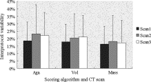
Interprotocol Variability of Coronary Artery Calcium Scoring
Get Radiology Tree app to read full this article<
1/6×[abs(S1−S2)+abs(S1−S3)+abs(S1−S4)+abs(S2−S3)+abs(S2−S4)+abs(S3−S4)]/[1/4×(S1+S2+S3+S4)] 1
/
6
×
[
a
b
s
(
S
1
-
S
2
)
+
a
b
s
(
S
1
-
S
3
)
+
a
b
s
(
S
1
-
S
4
)
+
a
b
s
(
S
2
-
S
3
)
+
a
b
s
(
S
2
-
S
4
)
+
a
b
s
(
S
3
-
S
4
)
]
/
[
1
/
4
×
(
S
1
+
S
2
+
S
3
+
S
4
)
]
where abs is absolute value, and S1, S2, S3, and S4 are CAC scores on the 64-slice prospective, 64-slice retrospective, 16-slice prospective, and 16-slice retrospective, respectively. From the 60 scans, 540 sets of variability (12 CAC materials and three scoring algorithms) data were obtained. The interprotocol variability was compared between the repeated scans and scoring algorithms using two-factor factorial ANOVA.
Get Radiology Tree app to read full this article<
Image Noise
Get Radiology Tree app to read full this article<
Statistical Analyses
Get Radiology Tree app to read full this article<
Radiation Dose
Get Radiology Tree app to read full this article<
DLP(mGy×cm)=CTDIvol(Gy)×12cm. D
L
P
(
m
G
y
×
c
m
)
=
C
T
D
I
v
o
l
(
G
y
)
×
12
c
m
.
A reasonable approximation of the effective dose (E) can be obtained using the equation ( ):
E=k×DLP E
=
k
×
D
L
P
where E is effective dose estimate and k = 0.017 mSv × mGy × cm. This value is applicable to chest scans and is the average between the male and female models.
Get Radiology Tree app to read full this article<
Results
Coronary Artery Calcium Scores
Get Radiology Tree app to read full this article<
Table 1
Agatston, Volume, and Mass Scores on 64-Slice Prospective, 64-Slice Retrospective, 16-Slice Prospective, and 16-Slice Retrospective Scans
64-Slice Prospective 64-Slice Retrospective 16-Slice Prospective 16-Slice Retrospective 1 mm Agatston 27 (37), 1–55 26 (34), 3–47 29 (37), 3–61 25 (31), 1–55 Volume 32 (37), 4–65 35 (45), 9–54 35 (39), 8–61 36 (45), 4–65 Mass 5 (6), 0–8 5 (7), 1–9 6 (7), 1–10 5 (7), 0–10 3 mm Agatston 79 (89), 25–121 80 (97), 22–169 92 (106), 24–187 84 (101), 19–191 Volume 75 (84), 34–106 76 (84), 37–146 87 (93), 35–172 81 (90), 31–170 Mass 15 (18), 5–23 16 (20), 5–24 19 (23), 5–33 16 (18), 4–26 5 mm Agatston 109 (123), 52–161 125 (140), 56–253 143 (159), 49–275 135 (150), 42–273 Volume 97 (96), 62–129 112 (115), 73–216 125 (129), 70–234 125 (129), 66–212 Mass 22 (24), 9–35 26 (30), 11–49 29 (35), 12–53 27 (32), 9–45 10 mm Agatston 242 (263), 120–413 233 (264), 129–395 269 (304), 107–524 260 (295), 93–441 Volume 204 (212), 139–309 199 (210), 141–288 229 (234), 145–422 223 (231), 139–342 Mass 52 (60), 21–76 53 (61), 24–77 59 (68), 24–90 55 (62), 21–85 Overall Agatston 114 (108), 1–413 116 (102), 3–395 133 (117), 3–524 126 (108), 1–411 Volume 102 (87), 1–441 105 (87), 9–288 119 (101), 8–422 116 (96), 4–342 Mass 24 (21), 0–76 25 (22), 1–77 28 (24), 1–90 26 (22), 0–85
64-Slice Prospective: prospective ECG-triggering scan on 64-slice CT; 1 mm: 1 mm-sized coronary artery calcium models (silicone, putty, and Teflon).
Data are expressed as mean (median), range.
Get Radiology Tree app to read full this article<
Interscan Variability of Repeated Coronary Artery Calcium Scoring
Get Radiology Tree app to read full this article<
Get Radiology Tree app to read full this article<
Interprotocol Variability of Coronary Artery Calcium Scoring
Get Radiology Tree app to read full this article<
Get Radiology Tree app to read full this article<
Image Noise
Get Radiology Tree app to read full this article<
Radiation Dose
Get Radiology Tree app to read full this article<
Discussion
Get Radiology Tree app to read full this article<
Get Radiology Tree app to read full this article<
Get Radiology Tree app to read full this article<
Get Radiology Tree app to read full this article<
Get Radiology Tree app to read full this article<
Get Radiology Tree app to read full this article<
Conclusion
Get Radiology Tree app to read full this article<
Get Radiology Tree app to read full this article<
References
1. Callister T.Q., Raggi P., Cooil B., Lippolis N.J., Russo D.J.: Effect of HMG-CoA reductase inhibitors on coronary artery disease as assessed by electron-beam computed tomography. N Engl J Med 1998; 339: pp. 1972-1978.
2. Agatston A.S., Janowitz W.R., Hildner F.J., Zusmer N.R., Viamonte M., Detrano R.: Quantification of coronary calcium using ultrafast computed tomography. J Am Coll Cardiol 1990; 15: pp. 827-832.
3. Callister T.Q., Cooil B., Raya S.P., et. al.: Coronary artery disease: Improved reproducibility of calcium scoring with an electron-beam CT volumetric method. Radiology 1998; 208: pp. 807-814.
4. Yoon H.C., Greaser L.E., Mather R., Sinha S., McNitt-Gray M.F., Goldin J.G.: Coronary artery calcium: Alternate methods for accurate and reproducible quantitation. Acad Radiol 1997; 4: pp. 666-673.
5. Wang S.J., Detrano B.C., Secci A., et. al.: Detection of coronary calcification with electron-beam computed tomography: Evaluation of interexamination reproducibility and comparison of three image-acquisition protocols. Am Heart J 1996; 132: pp. 550-558.
6. Achenbach S., Ropers D., Mohlenkamp S., et. al.: Variability of repeated coronary artery calcium measurements by electron beam tomography. Am J Cardiol 2001; 87: pp. 210-213.
7. Maher J.E., Bielak L.F., Raz J.A., Sheedy P.F., Schwartz R.S., Peyser P.A.: Progression of coronary artery calcification: A pilot study. Mayo Clin Proc 1999; 74: pp. 347-355.
8. Janowitz W.R., Agatston A.S., Viamonte M.: Comparison of serial quantitative evaluation of calcified coronary artery plaque by ultrafast computed tomography in persons with and without obstructive coronary artery disease. Am J Cardiol 1991; 68: pp. 1-6.
9. Fischbach R., Heindel W.: Detection and quantification of coronary calcification: An update. Rofo 2000; 172: pp. 407-414.
10. Ohnesorge B., Flohr T., Fischbach R., et. al.: Reproducibility of coronary calcium quantification in repeat examinations with retrospectively ECG-gated multisection spiral CT. Eur Radiol 2002; 12: pp. 1532-1540.
11. Van Hoe L.R., De Meerleer K.G., Leyman P.P., Vanhoenacker P.K.: Coronary artery calcium scoring using ECG-gated multidetector CT: Effect of individually optimized image-reconstruction windows on image quality and measurement reproducibility. AJR Am J Roentgenol 2003; 181: pp. 1093-1100.
12. Daniell A.L., Wong N.D., Friedman J.D., et. al.: Reproducibility of coronary calcium measurements from multidetector computed tomography. J Am Coll Cardiol 2003; 41: pp. 456A.
13. Horiguchi J., Yamamoto H., Akiyama Y., et. al.: Variability of repeated coronary artery calcium measurements by 16-MDCT with retrospective reconstruction. AJR Am J Roentgenol 2005; 184: pp. 1917-1923.
14. Callister T., Janowitz W., Raggi P.: Sensitivity of two electron beam tomography protocols for the detection and quantification of coronary artery calcium. AJR Am J Roentgneol 2000; 175: pp. 1743-1746.
15. Vliegenthart R., Song B., Hofman A., Witteman J.C.M., Oudkerk M.: Coronary calcification at electron-beam CT: Effect of section thickness on calcium scoring in vitro and in vivo. Radiology 2003; 229: pp. 520-525.
16. Horiguchi J., Matsuura N., Yamamoto H., et. al.: Variability of repeated coronary artery calcium measurements by 1.25-mm- and 2.5-mm-thickness images on prospective electrocardiograph-triggered 64-slice CT. Eur Radiol 2008; 18: pp. 209-216.
17. Horiguchi J., Shen Y., Akiyama Y., et. al.: Electron beam CT versus 16-MDCT on the variability of repeated coronary artery calcium measurements in a variable heart rate phantom. AJR Am J Roentgenol 2005; 185: pp. 995-1000.
18. Horiguchi J., Shen Y., Akiyama Y., et. al.: Electron beam CT versus 16-slice spiral CT: How accurately can we measure coronary artery calcium volume?. Eur Radiol 2006; 16: pp. 374-380.
19. Hong C., Bae K.T., Pilgram T.K., Suh J., Bradley D.: Coronary artery calcium measurement with multi-detector row CT: In vitro assessment of effect of radiation dose. Radiology 2002; 225: pp. 901-906.
20. Hunold P., Vogt F.M., Schmermund A., et. al.: Radiation exposure during cardiac CT: Effective doses at multi-detector CT and electron-beam CT. Radiology 2003; 226: pp. 145-152.
21. Morin R.L., Gerber T.C., McCollough C.H.: Radiation dose in computed tomography of the heart. Circulation 2003; 107: pp. 917-922.
22. Kopp A.F., Ohnesorge B., Becker C., et. al.: Reproducibility and accuracy of coronary calcium measurements with multi–detector row versus electron-beam CT. Radiology 2002; 225: pp. 113-119.
23. Achenbach S., Meissner F., Ropers D., et. al.: Overlapping cross-sections significantly improve the reproducibility of coronary calcium measurements by electron beam tomography: A phantom study. JCAT 2001; 25: pp. 569-573.
24. Muhlenbruch G., Thomas G., Wildberger J.E., et. al.: Effect of varying slice thickness on coronary calcium scoring with multislice computed tomography in vitro and in vivo. Investig Radiol 2005; 40: pp. 695-699.
25. McCollough C.H., Ulzheimer S., Halliburton S.S., et. al.: Coronary artery calcium: A multi-institutional, multimanufacturer international standard for quantification at cardiac CT. Radiology 2007; 243: pp. 527-538.
26. Bielak L.F., Kaufmann R.B., Moll P.P., MacCollough C.H., Schwartz R.S., Sheedy P.F.: Small lesions in the heart identified at electron beam CT: Calcification or noise?. Radiology 1994; 192: pp. 631-636.