
Rationale and Objectives
To optimize visualization of lenticulostriate artery (LSA) by time-of-flight (TOF) magnetic resonance angiography (MRA) with slice-selective off-resonance sinc (SORS) saturation transfer contrast pulses and to compare capability of optimal TOF-MRA and flow-sensitive black-blood (FSBB) MRA to visualize the LSA at 3T.
Materials and Methods
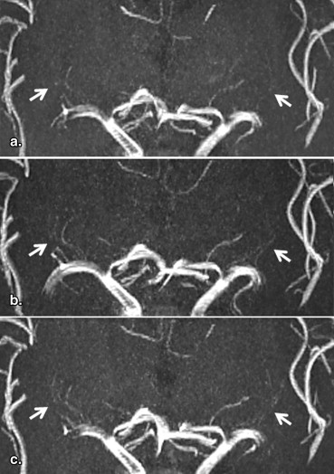
This study was approved by the local ethics committee, and written informed consent was obtained from all the subjects. TOF-MRA was optimized in 20 subjects by comparing SORS pulses of different flip angles: 0, 400°, and 750°. Numbers of LSAs were counted. The optimal TOF-MRA was compared to FSBB-MRA in 21 subjects. Images were evaluated by the numbers and length of visualized LSAs.
Results
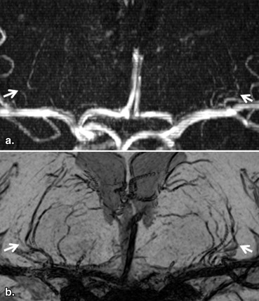
LSAs were significantly more visualized in TOF-MRA with SORS pulses of 400° than others ( P < .003). When the optimal TOF-MRA was compared to FSBB-MRA, the visualization of LSA using FSBB (mean branch numbers 11.1, 95% confidence interval (CI) 10.0–12.1; mean total length 236 mm, 95% CI 210–263 mm) was significantly better than using TOF (4.7, 95% CI 4.1–5.3; 78 mm, 95% CI 67–89 mm) for both numbers and length of the LSA ( P < .0001).
Conclusions
LSA visualization was best with 400° SORS pulses for TOF-MRA but FSBB-MRA was better than TOF-MRA, which indicates its clinical potential to investigate the LSA on a 3T magnetic resonance imaging.
Impairment of the lenticulostriate artery (LSA) often leads to lacunar infarction and cerebral hemorrhage, which is recognized as ‘ small vessels, big problems .’ The LSA branches supply blood to the basal ganglia and its vicinity , and their occlusion results in infarction of these structures . Recently, LSA branches have successfully been visualized using 7T and 3T magnetic resonance imaging (MRI) systems with three-dimensional (3D) time-of-flight (TOF) magnetic resonance angiography (MRA). Whereas at 1.5T, a recent study reported that flow-sensitive black blood (FSBB) MRA, which decreases blood flow signal with weak motion-dephasing gradient, performed better than TOF-MRA for visualization of the LSA . Moreover, FSBB-MRA at 1.5T could detect differences in the LSA between patients with lacunar infarction and/or hypertension and control subjects . However, at 3T, there is no study so far that has compared LSA visualization using TOF-MRA to FSBB-MRA.
In TOF-MRA acquisition, the saturation transfer contrast pulse is often used to reduce background signal, but the blood signal is also reduced, although the blood to background contrast is usually increased. To less reduce the blood signal, slice-selective off-resonance sinc (SORS) pulse can be applied, and enhanced visualization of LSA branches at 1.5T has been achieved .
Get Radiology Tree app to read full this article<
Materials and methods
Get Radiology Tree app to read full this article<
Subjects
Get Radiology Tree app to read full this article<
Image Acquisition
Get Radiology Tree app to read full this article<
Get Radiology Tree app to read full this article<
Get Radiology Tree app to read full this article<
Get Radiology Tree app to read full this article<
Image Analysis
Get Radiology Tree app to read full this article<
Get Radiology Tree app to read full this article<
Get Radiology Tree app to read full this article<
Statistical Analysis
Get Radiology Tree app to read full this article<
Results
Get Radiology Tree app to read full this article<
Get Radiology Tree app to read full this article<
Table 1
Number of Visualized LSA Branches for TOF-MRA
0° 400° 750° Branch numbers 3.7 (2.9–4.4) 5.3 (4.5–6.1) ∗ , † 4.1 (3.2–5.0)
LSA, lenticulostriate artery; MRA, magnetic resonance angiography; TOF, time-of-flight.
The numbers in the parentheses are 95% confidence intervals.
Get Radiology Tree app to read full this article<
Get Radiology Tree app to read full this article<
Get Radiology Tree app to read full this article<
Get Radiology Tree app to read full this article<
Get Radiology Tree app to read full this article<
Table 2
Branch Numbers and Length of Visualized LSA Branches for TOF (400°) and FSBB-MRA
Branch Numbers Length (mm) TOF 4.7 (4.1–5.3) 78 (67–89) FSBB 11.1 (10.0–12.1) 236 (210–263)P value <.0001 <.0001
FSBB, flow-sensitive black blood; LSA, lenticulostriate artery; MRA, magnetic resonance angiography; TOF, time-of-flight.
The numbers in the parentheses are 95% confidence intervals.
Get Radiology Tree app to read full this article<
Discussion
Get Radiology Tree app to read full this article<
Get Radiology Tree app to read full this article<
Get Radiology Tree app to read full this article<
Get Radiology Tree app to read full this article<
Get Radiology Tree app to read full this article<
Get Radiology Tree app to read full this article<
Get Radiology Tree app to read full this article<
Get Radiology Tree app to read full this article<
Acknowledgments
Get Radiology Tree app to read full this article<
Get Radiology Tree app to read full this article<
References
1. Greenberg S.M.: Small vessels, big problems. N Engl J Med 2006; 354: pp. 1451-1453.
2. Marinkovic S.V., Milisavljevic M.M., Kovacevic M.S., et. al.: Perforating branches of the middle cerebral artery. Microanatomy and clinical significance of their intracerebral segments. Stroke 1985; 16: pp. 1022-1029.
3. Marinkovic S., Gibo H., Milisavljevic M., et. al.: Anatomic and clinical correlations of the lenticulostriate arteries. Clin Anat 2001; 14: pp. 190-195.
4. Feekes J.A., Hsu S.W., Chaloupka J.C., et. al.: Tertiary microvascular territories define lacunar infarcts in the basal ganglia. Ann Neurol 2005; 58: pp. 18-30.
5. Feekes J.A., Cassell M.D.: The vascular supply of the functional compartments of the human striatum. Brain 2006; 129: pp. 2189-2201.
6. Cho Z.H., Kang C.K., Han J.Y., et. al.: Observation of the lenticulostriate arteries in the human brain in vivo using 7.0T MR angiography. Stroke 2008; 39: pp. 1604-1606.
7. Kang C.K., Park C.W., Han J.Y., et. al.: Imaging and analysis of lenticulostriate arteries using 7.0-Tesla magnetic resonance angiography. Magn Reson Med 2009; 61: pp. 136-144.
8. Chen Y.C., Li M.H., Li Y.H., et. al.: Analysis of correlation between the number of lenticulostriate arteries and hypertension based on high-resolution MR angiography findings. AJNR Am J Neuroradiol 2011; 32: pp. 1899-1903.
9. Akashi T., Taoka T., Ochi T., et. al.: Branching pattern of lenticulostriate arteries observed by MR angiography at 3.0 T. Jpn J Radiol 2012; 30: pp. 331-335.
10. Gotoh K., Okada T., Miki Y., et. al.: Visualization of the lenticulostriate artery with flow-sensitive black-blood acquisition in comparison with time-of-flight MR angiography. J Magn Reson Imaging 2009; 29: pp. 65-69.
11. Okuchi S., Okada T., Ihara M., et. al.: Visualization of lenticulostriate arteries by flow-sensitive black-blood MR angiography on a 1.5T MRI system: a comparative study between subjects with and without stroke. AJNR Am J Neuroradiol 2013; 34: pp. 780-784.
12. Miyazaki M., Kojima F., Ichinose N., et. al.: A novel saturation transfer contrast method for 3D time-of-flight magnetic resonance angiography: a slice-selective off-resonance sinc pulse (SORS) technique. Magn Reson Med 1994; 32: pp. 52-59.
13. Admiraal-Behloul1 F., Blink1 E., Zhang1 B., et. al.: High resolution time-of flight MRA using slice selective saturation transfer contrast and water excitation technique for the visualization of the lenticulostriate arteries at 1.5T. Proc Intl Soc Mag Reson Med 2011; 19: pp. 363.
14. Okada T, Miyazaki M, Kido A, Visualization of the lenticulostriate arteries at 3T: a comparative study of TOF-MRA with MTC and FSBB-MRA. Proceedings of the 17th Annual Scientific Meeting of KSMRM. 2012:389.
15. Kundel H.L., Polansky M.: Measurement of observer agreement. Radiology 2003; 228: pp. 303-308.
16. Wolff S.D., Balaban R.S.: Magnetization transfer contrast (MTC) and tissue water proton relaxation in vivo. Magn Reson Med 1989; 10: pp. 135-144.
17. Koenig S.H.: Cholesterol of myelin is the determinant of gray-white contrast in MRI of brain. Magn Reson Med 1991; 20: pp. 285-291.
18. Pike G.B., Hu B.S., Glover G.H., et. al.: Magnetization transfer time-of-flight magnetic resonance angiography. Magn Reson Med 1992; 25: pp. 372-379.
19. Bradley W.G., Waluch V.: Blood flow: magnetic resonance imaging. Radiology 1985; 154: pp. 443-450.
20. Edelman R.R., Mattle H.P., Wallner B., et. al.: Extracranial carotid arteries: evaluation with “black blood” MR angiography. Radiology 1990; 177: pp. 45-50.
21. Alexander A.L., Buswell H.R., Sun Y., et. al.: Intracranial black-blood MR angiography with high-resolution 3D fast spin echo. Magn Reson Med 1998; 40: pp. 298-310.
22. Mayo J.R., Culham J.A., MacKay A.L., et. al.: Blood MR signal suppression by preexcitation with inverting pulses. Radiology 1989; 173: pp. 269-271.
23. Parker D.L., Goodrich K.C., Masiker M., et. al.: Improved efficiency in double-inversion fast spin-echo imaging. Magn Reson Med 2002; 47: pp. 1017-1021.
24. Reichenbach J.R., Barth M., Haacke E.M., et. al.: High-resolution MR venography at 3.0 Tesla. J Comput Assist Tomogr 2000; 24: pp. 949-957.
25. Haacke E.M., Xu Y., Cheng Y.C., et. al.: Susceptibility weighted imaging (SWI). Magn Reson Med 2004; 52: pp. 612-618.
26. Le Bihan D., Breton E., Lallemand D., et. al.: Separation of diffusion and perfusion in intravoxel incoherent motion MR imaging. Radiology 1988; 168: pp. 497-505.
27. Kimura T., Ikedo M., Takemoto S.: Hybrid of opposite-contrast MR angiography (HOP-MRA) combining time-of-flight and flow-sensitive black-blood contrasts. Magn Reson Med 2009; 62: pp. 450-458.
28. Kang C.K., Park C.A., Lee H., et. al.: Hypertension correlates with lenticulostriate arteries visualized by 7T magnetic resonance angiography. Hypertension 2009; 54: pp. 1050-1056.
29. Kang C.K., Park C.A., Park C.W., et. al.: Lenticulostriate arteries in chronic stroke patients visualised by 7 T magnetic resonance angiography. Int J Stroke 2010; 5: pp. 374-380.
30. Gotoh K., Okada T., Satogami N., et. al.: Evaluation of CT angiography for visualisation of the lenticulostriate artery: difference between normotensive and hypertensive patients. Br J Radiol 2012; 85: pp. e1004-e1008.
31. Sasaki T., Kodama N., Matsumoto M., et. al.: Blood flow disturbance in perforating arteries attributable to aneurysm surgery. J Neurosurg 2007; 107: pp. 60-67.
32. Park D.H., Kang S.H., Lee J.B., et. al.: Angiographic features, surgical management and outcomes of proximal middle cerebral artery aneurysms. Clin Neurol Neurosurg 2008; 110: pp. 544-551.
33. Lustig M., Donoho D., Pauly J.M.: Sparse MRI: the application of compressed sensing for rapid MR imaging. Magn Reson Med 2007; 58: pp. 1182-1195.