
Rationale and Objectives
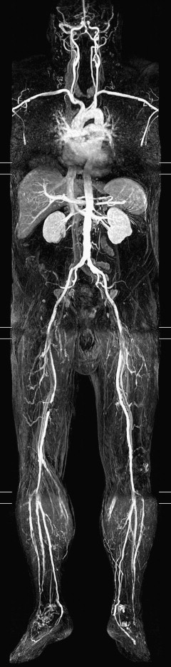
Whole-body magnetic resonance angiography (WB-MRA) at 3 T with body coil acquisition has not previously been investigated. In this study, WB-MRA was performed in this manner using the blood pool contrast agent gadofosveset trisodium.
Materials and Methods
Eleven consecutive patients (five men, six women) with symptomatic peripheral arterial disease (two with critical limb ischemia, nine with claudication) were examined. Conventional digital subtraction angiography (DSA) of the aorta and the inflow and runoff arteries was used as the reference method. WB-MRA was performed using four slightly overlapping stations covering the arteries from the neck to the ankles. The arterial system was divided into 42 segments that were analyzed for the presence of significant arterial disease (≥50% luminal narrowing or occlusion) by two blinded observers.
Results
Sensitivities for detecting a significant arterial lesion with WB-MRA using gadofosveset as the contrast agent were 0.66 (95% confidence interval [CI], 0.49–0.79) and 0.68 (95% CI, 0.52–0.81) for the two observers. Specificities were 0.82 (95% CI, 0.74–0.88) and 0.93 (95% CI, 0.87–0.96), respectively. Intermodality agreement between WB-MRA and DSA was moderate to good, with overall κ values of 0.44 (95% CI, 0.29–0.59) and 0.63 (95% CI, 0.5–0.77) for the two observers. Interobserver agreement for WB-MRA was good, at κ = 0.60 (95% CI, 0.50–0.71).
Conclusion
WB-MRA at 3 T with body coil acquisition in patients with peripheral arterial disease showed good reproducibility but only moderate to good agreement with DSA. Further assessment of the method’s clinical application is warranted.
Exact visualization of the arteries and the extent of atherosclerotic disease is essential before peripheral arterial reconstruction. Today, digital subtraction angiography (DSA) is still the routine modality for lower-limb imaging at most centers. However, DSA is invasive and carries a small but constant risk for major complications of approximately 2% . Therefore, replacing DSA with a noninvasive alternative, such as duplex ultrasound, computed tomographic angiography, or contrast-enhanced magnetic resonance angiography (MRA), seems promising. These techniques focus on a specific part of the arterial system, despite the fact that atherosclerosis is a systemic disease affecting the entire arterial system. In this point of view, imaging procedures examining the whole arterial system are interesting .
Recent developments in magnetic resonance imaging (MRI), including faster gradient systems and extended tabletops, have made contrast-enhanced whole-body MRA (WB-MRA) from head to feet possible . The feasibility and clinical importance of WB-MRA at 1.5 T have been investigated in recent years, leading to acceptance of the method as a valuable diagnostic tool . WB-MRA can be performed with a magnetic resonance system’s built-in body coil or different systems of surface coils, allowing better signal-to-noise ratio (SNR) and higher spatial resolution .
Get Radiology Tree app to read full this article<
Materials and methods
Patients
Get Radiology Tree app to read full this article<
MRA
Get Radiology Tree app to read full this article<
Get Radiology Tree app to read full this article<
Get Radiology Tree app to read full this article<
Get Radiology Tree app to read full this article<
DSA
Get Radiology Tree app to read full this article<
Get Radiology Tree app to read full this article<
Image Evaluation
Get Radiology Tree app to read full this article<
Get Radiology Tree app to read full this article<
Get Radiology Tree app to read full this article<
Get Radiology Tree app to read full this article<
Get Radiology Tree app to read full this article<
Statistical Analysis
Get Radiology Tree app to read full this article<
Results
Get Radiology Tree app to read full this article<
Get Radiology Tree app to read full this article<
Get Radiology Tree app to read full this article<
Table 1
Nondiagnostic Segments on 3-T Whole-body Magnetic Resonance Angiography
Reason Observer 1 Observer 2 Segment partly outside field of view 7 9 Susceptibility artifact 4 7 Bolus/data acquisition mistiming 4 6 Motion artifact 2 5 Overall 17 28
Get Radiology Tree app to read full this article<
Get Radiology Tree app to read full this article<
Get Radiology Tree app to read full this article<
Table 2
Sensitivities and Specificities for the Detection of Significant Arterial Disease on 3-T Whole-body Magnetic Resonance Angiography
Observer 1 Observer 2 Region Sensitivity Specificity Sensitivity Specificity Pelvic ∗ 0.80 (0.51–0.95) 0.88 (0.74–0.96) 0.91 (0.65–1) 0.95 (0.83–0.99) Proximal femur † 0.78 (0.40–0.96) 0.93 (0.76–0.94) 0.80 (0.44–0.96) 0.91 (0.76–0.98) Distal femur ‡ 0.78 (0.40–0.96) 0.94 (0.68–0.94) 0.77 (0.40–0.96) 0.94 (0.69–0.99) Proximal crus § 0.20 (0.01–0.70) 0.80 (0.64–0.90) 0.20 (0.01–0.70) 0.93 (0.76–0.99) Distal crus || 0.50 (0.14–0.86) 0.61 (0.36–0.81) 0.20 (0.01–0.70) 0.91 (0.57–0.99) Overall 0.66 (0.49–0.79) 0.82 (0.74–0.88) 0.68 (0.52–0.81) 0.93 (0.87–0.96)
Values in parentheses are 95% confidence intervals.
Get Radiology Tree app to read full this article<
Get Radiology Tree app to read full this article<
Get Radiology Tree app to read full this article<
Get Radiology Tree app to read full this article<
Get Radiology Tree app to read full this article<
Table 3
Intermodality Agreement (κ) Between Whole-body Magnetic Resonance Angiography and Digital Subtraction Angiography
Region Observer 1 Observer 2 Pelvic ∗ 0.65 (0.44–0.87) 0.84 (0.65–1) Proximal femur † 0.71 (0.44–0.98) 0.69 (0.44–0.94) Distal femur ‡ 0.73 (0.45–1) 0.78 (0.46–1) Proximal crus § 0.22 (0–0.62) 0.16 (0.61) Distal crus || 0.09 (0–0.55) 0.13 (0.77) Overall 0.44 (0.29–0.59) 0.63 (0.5–0.77)
Values in parentheses are 95% confidence intervals.
Get Radiology Tree app to read full this article<
Get Radiology Tree app to read full this article<
Get Radiology Tree app to read full this article<
Get Radiology Tree app to read full this article<
Get Radiology Tree app to read full this article<
Table 4
Interobserver Agreement for 3-T Whole-body Magnetic Resonance Angiography
Region κ Neck ∗ 0.79 (0.38–1) Thorax † 0.84 (0.53–1) Abdomen ‡ 0.70 (0.32–1) Pelvis § 0.70 (0.50–0.91) Proximal femur || 0.59 (0.36–0.82) Distal femur ¶ 0.81 (0.63–0.99) Proximal crus # 0.58 (0.31–0.86) Distal crus ∗∗ 0.57 (0.27–0.88) Overall 0.60 (0.50–0.71)
Values in parentheses are 95% confidence intervals.
Get Radiology Tree app to read full this article<
Get Radiology Tree app to read full this article<
Get Radiology Tree app to read full this article<
Get Radiology Tree app to read full this article<
Get Radiology Tree app to read full this article<
Get Radiology Tree app to read full this article<
Get Radiology Tree app to read full this article<
Get Radiology Tree app to read full this article<
Get Radiology Tree app to read full this article<
Get Radiology Tree app to read full this article<
Table 5
Significant Arterial Stenoses Detected on 3-T Whole-body Magnetic Resonance Angiography
Arterial Segment Observer 1 Observer 2 Internal carotid 3 2 Common carotid 2 1 Subclavian artery 4 3 Renal artery 4 3 Abdominal aorta 1 1
Get Radiology Tree app to read full this article<
Get Radiology Tree app to read full this article<
Discussion
Get Radiology Tree app to read full this article<
Get Radiology Tree app to read full this article<
Get Radiology Tree app to read full this article<
Get Radiology Tree app to read full this article<
Get Radiology Tree app to read full this article<
Get Radiology Tree app to read full this article<
Get Radiology Tree app to read full this article<
Get Radiology Tree app to read full this article<
Get Radiology Tree app to read full this article<
Get Radiology Tree app to read full this article<
Get Radiology Tree app to read full this article<
Get Radiology Tree app to read full this article<
Get Radiology Tree app to read full this article<
Get Radiology Tree app to read full this article<
References
1. Egglin T.K., O’Moore P.V., Feinstein A.R., et. al.: Complications of peripheral arteriography: a new system to identify patients at increased risk. J Vasc Surg 1995; 22: pp. 787-794.
2. Ladd S.C., Ladd M.E.: Perspectives for preventive screening with total body MRI. Eur Radiol 2007; 17: pp. 2889-2897.
3. Ruehm S.G., Goehde S.C., Goyen M.: Whole body MR angiography screening. Int J Cardiovasc Imaging 2004; 20: pp. 587-591.
4. Ruehm S.G., Goyen M., Barkhausen J., et. al.: Rapid magnetic resonance angiography for detection of atherosclerosis. Lancet 2001; 357: pp. 1086-1091.
5. Goehde S.C., Hunold P., Vogt F.M., et. al.: Full-body cardiovascular and tumor MRI for early detection of disease: feasibility and initial experience in 298 subjects. AJR Am J Roentgenol 2005; 184: pp. 598-611.
6. Goyen M., Herborn C.U., Kroger K., et. al.: Detection of atherosclerosis: systemic imaging for systemic disease with whole-body three-dimensional MR angiography—initial experience. Radiology 2003; 227: pp. 277-282.
7. Goyen M., Herborn C.U., Kroger K., et. al.: Total-body 3D magnetic resonance angiography influences the management of patients with peripheral arterial occlusive disease. Eur Radiol 2006; 16: pp. 685-691.
8. Herborn C.U., Goyen M., Quick H.H., et. al.: Whole-body 3D MR angiography of patients with peripheral arterial occlusive disease. AJR Am J Roentgenol 2004; 182: pp. 1427-1434.
9. Ladd S.C., Debatin J.F., Stang A., et. al.: Whole-body MR vascular screening detects unsuspected concomitant vascular disease in coronary heart disease patients. Eur Radiol 2007; 17: pp. 1035-1045.
10. Brennan D.D., Johnston C., O’Brien J., et. al.: Contrast-enhanced bolus-chased whole-body MR angiography using a moving tabletop and quadrature body coil acquisition. AJR Am J Roentgenol 2005; 185: pp. 750-755.
11. Hansen T., Wikstrom J., Eriksson M.O., et. al.: Whole-body magnetic resonance angiography of patients using a standard clinical scanner. Eur Radiol 2006; 16: pp. 147-153.
12. Henness S., Keating G.M.: Gadofosveset. Drugs 2006; 66: pp. 851-857.
13. Klessen C., Hein P.A., Huppertz A., et. al.: First-pass whole-body magnetic resonance angiography (MRA) using the blood-pool contrast medium gadofosveset trisodium: comparison to gadopentetate dimeglumine. Invest Radiol 2007; 42: pp. 659-664.
14. Rutherford R.B., Baker J.D., Ernst C., et. al.: Recommended standards for reports dealing with lower extremity ischemia: revised version. J Vasc Surg 1997; 26: pp. 517-538.
15. Levey A.S., Bosch J.P., Lewis J.B., et. al., Modification of Diet in Renal Disease Study Group: A more accurate method to estimate glomerular filtration rate from serum creatinine: a new prediction equation. Ann Intern Med 1999; 130: pp. 461-470.
16. Gardner M.J., Altman D.G.: Confidence intervals rather than P values.Altman D.G.Machin D.Bryant T.N.Gardner M.J.Statistics with Confidence.2000.Blackwell BMJ BooksOxford, UK:pp. 15-27.
17. Altman D.G.: Diagnostic tests.Altman D.G.Machin D.Bryant T.N.Gardner M.J.Statistics with Confidence.2000.Blackwell BMJ BooksOxford, UK:pp. 105-119.
18. Fenchel M., Scheule A.M., Stauder N.I., et. al.: Atherosclerotic disease: whole-body cardiovascular imaging with MR system with 32 receiver channels and total-body surface coil technology—initial clinical results. Radiology 2006; 238: pp. 280-291.
19. Fenchel M., Requardt M., Tomaschko K., et. al.: Whole-body MR angiography using a novel 32-receiving-channel MR system with surface coil technology: first clinical experience. J Magn Reson Imaging 2005; 21: pp. 596-603.
20. Goyen M., Quick H.H., Debatin J.F., et. al.: Whole-body three-dimensional MR angiography with a rolling table platform: initial clinical experience. Radiology 2002; 224: pp. 270-277.
21. Nael K., Ruehm S.G., Michaely H.J., et. al.: Multistation whole-body high-spatial-resolution MR angiography using a 32-channel MR system. AJR Am J Roentgenol 2007; 188: pp. 529-539.
22. Nael K., Fenchel M., Krishnam M., et. al.: High-spatial-resolution whole-body MR angiography with high-acceleration parallel acquisition and 32-channel 3.0-T unit: initial experience. Radiology 2007; 242: pp. 865-872.
23. Griswold M.A., Jakob P.M., Heidemann R.M., et. al.: Generalized autocalibrating partially parallel acquisitions (GRAPPA). Magn Reson Med 2002; 47: pp. 1202-1210.
24. Pruessmann K.P., Weiger M., Scheidegger M.B., et. al.: SENSE: sensitivity encoding for fast MRI. Magn Reson Med 1999; 42: pp. 952-962.
25. Nael K., Fenchel M.C., Kramer U., et. al.: Whole-body contrast-enhanced magnetic resonance angiography: new advances at 3.0 T. Top Magn Reson Imaging 2007; 18: pp. 127-134.
26. Lohan D.G., Tomasian A., Krishnam M., et. al.: MR angiography of lower extremities at 3 T: presurgical planning of fibular free flap transfer for facial reconstruction. AJR Am J Roentgenol 2008; 190: pp. 770-776.
27. Thurnher S., Miller S., Schneider G., et. al.: Diagnostic performance of gadobenate dimeglumine enhanced MR angiography of the iliofemoral and calf arteries: a large-scale multicenter trial. AJR Am J Roentgenol 2007; 189: pp. 1223-1237.
28. Napoli A., Catalano C., Anzidei M., et. al.: Imaging the whole body atherosclerosis: high resolution magnetic resonance angiography using blood-pool agent. Initial clinical experience. Minerva Cardioangiol 2007; 55: pp. 291-301.
29. Nikolaou K., Kramer H., Grosse C., et. al.: High-spatial-resolution multistation MR angiography with parallel imaging and blood pool contrast agent: initial experience. Radiology 2006; 241: pp. 861-872.
30. Rohrer M., Bauer H., Mintorovitch J., et. al.: Comparison of magnetic properties of MRI contrast media solutions at different magnetic field strengths. Invest Radiol 2005; 40: pp. 715-724.
31. Leiner T., de V.M., Hoogeveen R., et. al.: Contrast-enhanced peripheral MR angiography at 3.0 Tesla: initial experience with a whole-body scanner in healthy volunteers. J Magn Reson Imaging 2003; 17: pp. 609-614.
32. Alexandrova N.A., Gibson W.C., Norris J.W., et. al.: Carotid artery stenosis in peripheral vascular disease. J Vasc Surg 1996; 23: pp. 645-649.
33. Drouet L.: Atherothrombosis as a systemic disease. Cerebrovasc Dis 2002; 13: pp. 1-6.
34. Wachtell K., Ibsen H., Olsen M.H., et. al.: Prevalence of renal artery stenosis in patients with peripheral vascular disease and hypertension. J Hum Hypertens 1996; 10: pp. 83-85.
35. Criqui M.H., Coughlin S.S., Fronek A.: Noninvasively diagnosed peripheral arterial disease as a predictor of mortality: results from a prospective study. Circulation 1985; 72: pp. 768-773.
36. Bilecen D., Jager K.A., Aschwanden M., et. al.: Cuff-compression of the proximal calf to reduce venous contamination in contrast-enhanced stepping-table magnetic Resonance angiography. Acta Radiol 2004; 45: pp. 510-515.
37. Herborn C.U., Vogt F.M., Waltering K.U., et. al.: Optimization of contrast-enhanced peripheral MR angiography with mid-femoral venous compression (VENCO). Rofo 2004; 176: pp. 157-162.
38. Herborn C.U., Ajaj W., Goyen M., et. al.: Peripheral vasculature: whole-body MR angiography with midfemoral venous compression—initial experience. Radiology 2004; 230: pp. 872-878.
39. Eiberg J.P., Madycki G., Hansen M.A., et. al.: Ultrasound imaging of infrainguinal arterial disease has a high interobserver agreement. Eur J Vasc Endovasc Surg 2002; 24: pp. 293-299.