
Rationale and Objectives
To assess how well radiologists visualize relevant features of lesions seen with automated breast volumetric scanning (ABVS) in comparison to hand-held breast ultrasound in women going to breast biopsy.
Materials and Methods
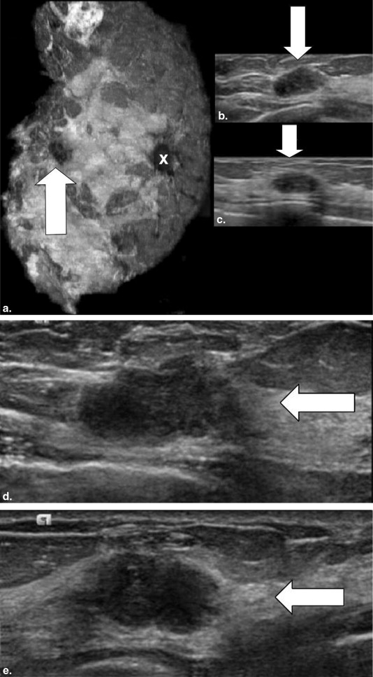
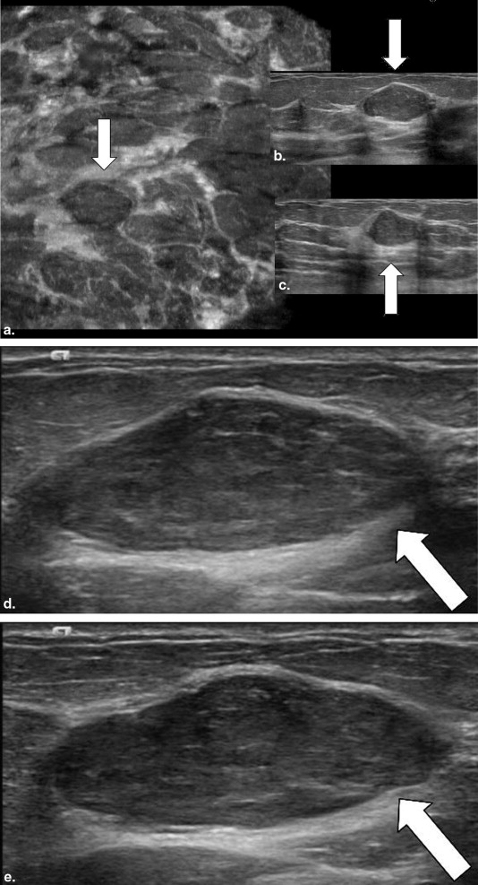
Twenty-five subjects were recruited from women who were scheduled to undergo a breast biopsy for at least one Breast Imaging–Reporting and Data System four or five lesion identified in a diagnostic setting. In this institutional review board–approved study, the subjects underwent imaging of the breast(s) of concern using a dedicated system that allowed both hand-held breast ultrasound and ABVS. Five experienced breast radiologists reviewed the 30 lesions in 25 subjects in a reader study. Each reader was asked to specify the lesion type, size, imaging features, Breast Imaging–Reporting and Data System, and suspicion of malignancy and to compare the lesion characteristics of shape and margins between the two modalities.
Results
Seven (23.3%) masses were malignant and 23 (76.4%) were benign. Across all lesions regardless of size or final pathology, there was no significant difference in sensitivity or specificity ( P > .15) between the two modalities. For malignant lesions, the reader visualization confidence scores between the two ultrasound modalities were not significantly different ( P > .1). However, analysis for nonmalignant cases showed a statistically significant increase in reader visualization confidence in lesion shape and margins ( P < .001).
Conclusions
Radiologists showed increased confidence in visualization of benign masses and equal confidence in suspicious masses with ABVS imaging. This information could help decrease the need for additional hand-held imaging after automated whole breast ultrasound.
Breast cancer is the most commonly diagnosed noncutaneous cancer in American women . Mammography remains the gold standard for screening women for breast cancer, but the major limitation of this technology is decreased sensitivity in women with dense breast tissue . Even with improvements in breast cancer detection using digital mammography, the sensitivity of mammography in women with dense breasts tends to be lower than the reported sensitivity of mammography in women with nondense breasts because of the added challenge of detecting breast cancers that may be obscured by overlapping breast tissue . This concept is known as “masking” . Unfortunately, masking is common in women with dense breasts. Dense breast tissue is found in 40%–60% of all women, of any age, undergoing mammographic imaging . Thus, this is a major contributing factor for the use of an additional screening modality to be used in conjunction with mammography. Ultrasound is one such tool that is being studied as a potential supplemental screening modality .
Ultrasound is an alternative technology that is not limited by breast density and has served as an important adjunct to mammography in the diagnostic evaluation of breast lesions for several decades. Although a highly sensitive imaging modality in breast cancer detection and evaluation, current ultrasound breast scanning technology requires a hand-held transducer. Evaluation is limited by the small hand-held ultrasound transducer size and to areas of the breast that are scanned. Thus, assessment is often deficient because the whole breast may not be evaluated by the operator. In addition, other limitations of conventional ultrasound are primarily because of operator-dependence and poor standardization of technique, which can result in missed cancers and unnecessary biopsies, additional medical expenses, and patient anxiety .
Get Radiology Tree app to read full this article<
Materials and methods
Subjects
Get Radiology Tree app to read full this article<
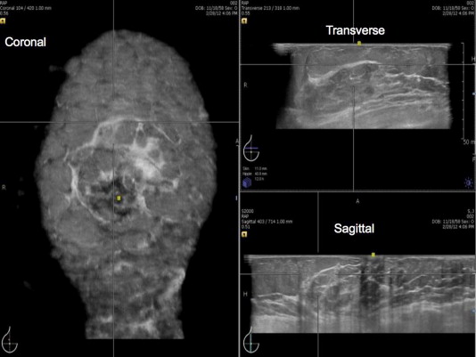
HHBUS and ABVS
Get Radiology Tree app to read full this article<
Get Radiology Tree app to read full this article<
Training and Start Up
Get Radiology Tree app to read full this article<
Image Acquisition
Get Radiology Tree app to read full this article<
Postimaging
Get Radiology Tree app to read full this article<
Reader Study
Get Radiology Tree app to read full this article<
Get Radiology Tree app to read full this article<
Get Radiology Tree app to read full this article<
Get Radiology Tree app to read full this article<
Statistical Analysis
Get Radiology Tree app to read full this article<
Results
Get Radiology Tree app to read full this article<
Table 1
Breast Lesion Pathology and Size
Lesion Number Pathology Largest Size (cm) 1 Intraductal papilloma 2.0 2 Fat necrosis and pseudoangiomatous stromal hyperplasia 2.2 3 Fat necrosis with chronic inflammation 1.6 4 Fibrocystic changes 1.1 5 Focal adenosis 0.5 6 Pseudoangiomatous stromal hyperplasia 1.0 7 Fibrocystic changes 1.6 8 Intraductal papilloma 1.5 9 Invasive ductal carcinoma grade II; DCIS, nuclear grade 2, solid type 1.5 10 Invasive ductal carcinoma with tubular features, grade I 0.6 11 Fibroadenoma 0.6 12 Fibrocystic changes 0.6 13 Fibrocystic changes 0.4 14 Focal chronic inflammation 1.1 15 Fibrocystic changes 0.7 16 Fibrocystic changes 2.0 17 Fibrocystic changes 0.6 18 Invasive ductal carcinoma grade III; DCIS, nuclear grade 3, solid type 2.0 19 Fibrocystic changes 2.0 20 Fibroadenoma 3.4 21 Fibrocystic changes 1.8 22 Fibrocystic changes 0.9 23 Invasive ductal carcinoma, grade III 1.4 24 Invasive lobular carcinoma, grade II 1.0 25 Invasive ductal carcinoma, grade II 1.0 26 Fibroadenoma 2.0 27 Ductal carcinoma in situ, nuclear grade 2, cribriform type 0.8 28 Fat necrosis with dense fibrosis 2.5 29 Fibrocystic changes 1.0 30 Fibroadenoma 1.6
DCIS, ductal carcinoma in situ.
Get Radiology Tree app to read full this article<
Get Radiology Tree app to read full this article<
Get Radiology Tree app to read full this article<
Get Radiology Tree app to read full this article<
Table 2
Sensitivity and Specificity of ABVS and HHBUS Based on Combined Readers’ BI-RADS and Clinical Suspicion of Malignancy Scale
Measures BI-RADS Scale Clinical Suspicion Scale ABVS HHBUS ABVS HHBUS Sensitivity 97.1% 97.1% 94.3% 91.4% Specificity 27.8% 30.4% 36.5% 39.1%P Value >.15 >.15
ABVS, automated breast volumetric scanning; BI-RADS, Breast Imaging–Reporting and Data System; HHBUS, hand-held breast ultrasound.
Get Radiology Tree app to read full this article<
Get Radiology Tree app to read full this article<
Table 3
Mean Reader Confidence Scores of ABVS and HHBUS for the Two Reading Conditions for all Lesions. The Average Confidence Score Scale is −3 to 3
Diagnostic Result Condition I: ABVS vs. HHBUS Condition II: HHBUS vs. ABVS Lesion Shape/Morphology Lesion Margin ABVS Coronal View Only vs. HHBUS Lesion Shape/Morphology ABVS Coronal View Only vs. HHBUS Lesion Margin Lesion Shape/Morphology Lesion Margin HHBUS vs. Coronal View Only ABVS Lesion Shape/Morphology HHBUS vs. ABVS Coronal View Only Lesion Margin Cancer 0.46 0.43 0.63 0.49 0.31 0.34 0.43 0.37 Noncancer 0.70 0.70 1.31 1.23 0.37 0.33 0.70 0.76
ABVS, automated breast volumetric scanning; HHBUS, hand-held breast ultrasound.
Table 4
Mean Reader Confidence Scores of ABVS and HHBUS for the Two Reading Conditions for Lesions in Subjects with Dense Breasts. The Average Confidence Score Scale is −3 to 3
Diagnostic Result Condition I: ABVS vs. HHBUS Condition II: HHBUS vs. ABVS Lesion Shape/Morphology Lesion Margin ABVS Coronal View Only vs. HHBUS Lesion Shape/Morphology ABVS Coronal View Only vs. HHBUS Lesion Margin Lesion Shape/Morphology Lesion Margin HHBUS vs. Coronal View Only ABVS Lesion Shape/Morphology HHBUS vs. ABVS Coronal View Only Lesion Margin Cancer 0.30 0.30 0.35 0.35 0.1 0.05 0.30 0.20 Noncancer 0.58 0.69 1.00 1.00 0.29 0.27 0.51 0.55
ABVS, automated breast volumetric scanning; HHBUS, hand-held breast ultrasound.
Get Radiology Tree app to read full this article<
Get Radiology Tree app to read full this article<
Get Radiology Tree app to read full this article<
Get Radiology Tree app to read full this article<
Get Radiology Tree app to read full this article<
Discussion
Get Radiology Tree app to read full this article<
Get Radiology Tree app to read full this article<
Get Radiology Tree app to read full this article<
Get Radiology Tree app to read full this article<
Get Radiology Tree app to read full this article<
Get Radiology Tree app to read full this article<
Get Radiology Tree app to read full this article<
Conclusions
Get Radiology Tree app to read full this article<
Acknowledgments
Get Radiology Tree app to read full this article<
References
1. American Cancer Society. Breast cancer facts and figures 2013-2014. Available at: http://www.cancer.org/research/cancerfactsstatistics/breast-cancer-facts-figures . Accessed December 10, 2014.
2. Fracheboud J., Otto S.J., van Dijck J.A., et. al.: Decreased rates of advanced breast cancer due to mammography screening in the Netherlands. Br J Cancer 2004; 91: pp. 861-867.
3. Coburn N.G., Chung M.A., Fulton J., et. al.: Decreased breast cancer tumor size, stage, and mortality in Rhode Island: an example of a well-screened population. Cancer Control 2004; 11: pp. 222-230.
4. Jatoi I., Chen B.E., Anderson W., et. al.: Breast cancer mortality trends in the United States according to estrogen receptor status and age at diagnosis. J Clin Oncol 2007; 25: pp. 1683-1690.
5. Pisano E.D., Gatsonis C., Hendrick E., et. al.: Diagnostic performance of digital versus film mammography for breast-cancer screening. N Engl J Med 2005; 353: pp. 1773-1783.
6. Whitehead J., Carlile T., Kopecky K.J., et. al.: Wolfe mammographic parenchymal patterns. A study of the masking hypothesis of Egan and Mosteller. Cancer 1985; 56: pp. 1280-1286.
7. Buist D.S., Porter P.L., Lehman C., et. al.: Factors contributing to mammography failure in women aged 40-49 years. J Natl Cancer Inst 2004; 96: pp. 1432-1440.
8. Carney P.A., Miglioretti D.L., Yankaskas B.C., et. al.: Individual and combined effects of age, breast density, and hormone replacement therapy use on the accuracy of screening mammography. Ann Intern Med 2003; 138: pp. 168-175.
9. Bae M.S., Moon W.K., Chang J.M., et. al.: Breast cancer detected with screening US: reasons for nondetection at mammography. Radiology 2014; 270: pp. 369-377.
10. Stomper P.C., D’Souza D.J., DiNitto P.A., et. al.: Analysis of parenchymal density on mammograms in 1353 women 25-79 years old. AJR Am J Roentgenol 1996; 167: pp. 1261-1265.
11. Crystal P., Strano S.D., Shcharynski S., et. al.: Using sonography to screen women with mammographically dense breasts. AJR Am J Roentgenol 2003; 181: pp. 177-182.
12. Kelly K.M., Dean J., Comulada W.S., et. al.: Breast cancer detection using automated whole breast ultrasound and mammography in radiographically dense breasts. Eur Radiol 2010; 20: pp. 734-742.
13. Berg W.A., Zhang Z., Lehrer D., et. al.: Detection of breast cancer with addition of annual screening ultrasound or a single screening MRI to mammography in women with elevated breast cancer risk. JAMA 2012; 307: pp. 1394-1404.
14. Hooley R.J., Greenberg K.L., Stackhouse R.M., et. al.: Screening US in patients with mammographically dense breasts: initial experience with Connecticut Public Act 09-41. Radiology 2012; 265: pp. 59-69.
15. Weigert J., Steenbergen S.: The Connecticut experience: the role of ultrasound in the screening of women with dense breasts. Breast J 2012; 18: pp. 517-522.
16. Skaane P., Engedal K., Skjennald A.: Interobserver variation in the interpretation of breast imaging. Comparison of mammography, ultrasonography, and both combined in the interpretation of palpable noncalcified breast masses. Acta Radiol 1997; 38: pp. 497-502.
17. Wang H.Y., Jiang Y.X., Zhu Q.L., et. al.: Differentiation of benign and malignant breast lesions: a comparison between automatically generated breast volume scans and handheld ultrasound examinations. Eur J Radiol 2012; 81: pp. 3190-3200.
18. Wang Z.L., Xw J.H., Li J.L., et. al.: Comparison of automated breast volume scanning to hand-held ultrasound and mammography. Radiol Med 2012; 117: pp. 1287-1293.
19. Lin X., Wnag J., Han F., et. al.: Analysis of eighty-one cases with breast lesions using automated breast volume scanner and comparison with handheld ultrasound. Eur J Radiol 2012; 81: pp. 873-878.
20. Sickles E.A., D’Orsi C.J., Bassett L.W., et. al.: Breast imaging reporting and data system, BI-RADS: mammography.2013.American College of RadiologyReston, VA
21. Berg W.A., Zhang Z., Cormack J.B., et. al.: Multiple bilateral circumscribed masses at screening breast US: consider annual follow-up. Radiology 2013; 283: pp. 673-683.
22. Mandelson M.T., Oestreicher N., Porter P.L., et. al.: Breast density as a predictor of mammographic detection: comparison of interval- and screen-detected cancers. J Natl Cancer Inst 2000; 92: pp. 1081-1087.
23. Jackson V.P., Kelly-Fry E., Rothschild P.A., et. al.: Automated breast sonography using a 7.5-MHz PVDF transducer: preliminary clinical evaluation. Radiology 1986; 159: pp. 679-684.
24. Golatta M., Franz D., Harcos A., et. al.: Interobserver reliability of automated breast volume scanner (ABVS) interpretation and agreement of ABVS findings with hand-held breast ultrasound (HHUS), mammography and pathology results. Eur J Radiol 2013; 82: pp. e332-e336.
25. Boyd N.F., Guo H., Martin L.J., et. al.: Mammographic density and the risk and detection of breast cancer. N Engl J Med 2007; 356: pp. 227-236.
26. Kaplan S.S.: Clinical utility of bilateral whole-breast US in the evaluation of women with dense breast tissue. Radiology 2001; 221: pp. 641-649.
27. Kolb T.M., Lichy J., Newhouse J.H.: Comparison of the performance of screening mammography, physical examination, and breast US and evaluation of factors that influence them. Radiology 2002; 225: pp. 165-175.
28. Brem R.F., Tabár L., Duffy S.W., et. al.: Assessing improvement in detection of breast cancer with three-dimensional automated breast US in women with dense breast tissue: The SomoInsight Study. Radiology 2014; 274: pp. 663-673.
29. Berg W., Blume J.D., Cormack J.B., et. al.: Combined screening with ultrasound and mammography vs. mammography alone in women at elevated risk of breast cancer. JAMA 2008; 299: pp. 2151-2163.